Orsi, M.A. 1,2; Doretto Jr, L.3.; Camillo, S.C.A.1; Reischak, D.1; Ribeiro, S.A.M.1; Ramazzoti, A.1; Mendonça, A.O. 1;
Spilki, F.R.4 ; Buzinaro, M.G. 5; Ferreira, H.L.6;Arns, C.W. 2,6*
1
Laboratório Nacional Agropecuário, Ministério da Agricultura, Pecuária e Abastecimento, Campinas, SP, Brasil; 2Faculdade de
Ciências Médicas, Universidade Estadual de Campinas, Campinas, SP, Brasil; 3Centro Brasileiro de Pesquisa Animal, Amparo,
SP, Brasil; 4Instituto de Ciências da Saúde, Centro Universitário Feevale, Novo Hamburgo, RS, Brasil; 5Departamento de Medicina Veterinária Preventiva e Reprodução Animal, Faculdade de Ciências Agrárias e Veterinárias, Universidade Estadual
Paulista, Jaboticabal, SP, Brasil; 6*Laboratório de Virologia, Instituto de Biologia, Universidade Estadual de Campinas, Campinas, SP, Brasil.
Submitted: June 06, 2009; Returned to authors for corrections: July 23, 2009; Approved: February 17, 2010.
ABSTRACT
This study was carried out during 2002/2003, aiming to determine the prevalence of virulent Newcastle
disease virus strains (NDV) in Brazilian commercial poultry farms. Clinical samples were obtained from
the Southeastern, Southern and Central-Western regions, which comprise the main area of the Brazilian
poultry production. Serum samples and tracheal and cloacal swabs of 23,745 broiler chickens from 1,583
flocks, including both vaccinated chickens and those with no vaccination information, were tested for
NDV using a diagnostic ELISA kit. The seropositivity was 39.1%, and the isolation percentage by flock
varied from 1.0 to 7.6%, and by region from 6.5 to 58.4%. Higher isolation rates (74.3-83.3%) were
obtained after three passages in embryonated chicken eggs. All isolates preliminarily identified as NDV
were characterized as nonpathogenic strains, as their Intracerebral Pathogenicity Index (ICPI) was below
0.7. Based on results of this study, Brazil can claim a virulent NDV-free status for commercial flocks.
Key words: Newcastle Disease Virus, NDV-free status, pathogenicity, broiler chickens, biological
characterization.
INTRODUCTION
Avian paramyxovirus-1 (APMV-1), the causative agent of
the Newcastle Disease (NDV), is classified as a member of
genus Avulavirus in the family Paramyxoviridae (14, 15). Newcastle disease (ND), one of the most important viral
diseases in industrial aviculture (3), affects domestic poultry
and wild birds and may cause acute mortality marked by
hemorrhagic lesions, respiratory and apparent or unapparent
enteric infections, among others. Therefore, the etiopathogenic
diagnosis should be conventionally based on isolation and
biological characterization of field samples (29).
NDV detection and pathotyping of avian isolates are
extremely important because the appearance of virulent virus
has significant economic consequences related to vaccination
and eradication, impairing the ability of a given geographic
region to export poultry products (24).
birds and the use of such viruses as live vaccines mean that
isolation of NDV is not enough to confirm a disease diagnosis
and compliance with statutory requirements that may be in
place (8). Viral characterization using the pathogenicity test or
nucleotide sequencing are also required, as the importance and
impact of a given NDV isolate are directly related to its
virulence. Once analysis of a given field disease solely may be
an unreliable measurement of pathogenicity due to the
possibility of concurrent infections and bad technical
management, laboratory assessments of the virus pathogenicity
are necessary. For this purpose, currently three “in vivo” tests are available, which include determination of ICPI
(Intracerebral Pathogenicity Index), IVPI (Intravenous
Pathogenicity Index) and MDT (Mean Death Time) (3).
The World Organisation for Animal Health (OIE) is
recognized by the World Trade Organization for
standardization of matters related to animal health that may
affect international trade. NDV infection is defined as a
notifiable disease if the virus in day-old chick (Gallus gallus) has ICPI of 0.7 or above, or contains certain multiple basic
amino acids at the fusion (F) protein cleavage site (19).
APMV-1 that does not meet the OIE definition for causing ND
is referred to as a low-virulence APMV-1 or NDV.
The first description of this disease in Brazil occurred in
Belém and Macapá in 1953, (22). The outbreak was a
consequence of the importation of NDV-contaminated frozen
chicken carcasses from the United States (28). The first NDV
isolation in Brazil was accomplished by Cunha and Silva (10).
This NDV strain has been designated as M33 and its biological
characterization was performed by Oliveira et al. (20). After
the first impact in the 1950’s decade, the Newcastle disease,
although endemic, has been seen only sporadically, attacking
breeding stocks of small economic expression. Outbreaks have
been quickly controlled by vaccination and complementary
prophylactic measures.
Poultry is the Brazilian leading export product in the meat
sector. The performance of this sector in 2006 consolidated
Brazil in the first-position, conquered in 2004, as the world’s
biggest exporter both in volume and revenue. Behind the
soybean complex, poultry ranks second in the Brazilian
agribusiness exporting rank (2).
The broiler chicken production in the country is
concentrated in the Southern and Southeastern regions (1),
where the main producers and exporters of genetic material
from chickens are located. Lately the Central-Western region
has also experienced a significant expansion.
The nationwide poultry production efficiency makes
Brazil a competitive nation in international markets, even in the
absence of economic subsidies. Aiming to guarantee better
sanitary conditions to Brazilian avian products, an
epidemiological project, in agreement with the National
Program for Poultry Sanity (PNSA), was implemented for the
control of Newcastle Disease in the country.
This study evaluated the prevalence of Newcastle Disease
in commercial birds in poultry producing areas in Brazil and
the occurrence of the virus in this aviary segment. When
pathogenic viruses are absent, the country can be declared free
of virulent Newcastle Disease.
MATERIALS AND METHODS
Sample calculation and source
Samples were collected weekly from apparently healthy
birds in by official service, in slaughterhouses located in
selected areas of nine states of the Southeastern, Southern and
Central-Western states. Blood serum of 15 birds per flock and
pools of eight cloacal swabs and eight trachea swabs were
placed separately in a buffered saline solution (PBS), with
antibiotics (10,000 U/ml penicillin, 10 mg/ml streptomycin,
0.25 mg/ml gentamicin and 5,000 IU/ml nystatin), adjusted to
pH 7.0-7.4, and cold-stored .
Collected samples were sent to a screening centre in each
state to ensure analysis viability, and insertion of data into a
computerized information system. The material was sealed up
and sent in the thermal ice boxes to the National Agricultural
The calculation of the number of samples for the study
was based on the total population of birds in each federative
state, from a total of 410,729,182 birds in the country,
estimated by the Brazilian Ministry of Agriculture. Federative
states were selected based on their importance for the Brazilian
poultry industry, and comprised three regions: Southeastern
(Minas Gerais and São Paulo states) Southern (Paraná, Santa
Catarina and Rio Grande do Sul states) and Central Western
(Goiás, Distrito Federal, Mato Grosso and Mato Grosso do Sul
states).
Assuming a prevalence of at least 1% and a sensitivity of
99% for the detection of at least one infected flock, a minimum
of 485 flocks were planned to be analyzed per region. The
determination of the number of birds to be sampled by flock
took into account the 95% sensitivity of the diagnostic test
(ELISA), with an expected minimum prevalence of 30% of
infected flocks, with confidence degree of 99%, resulting in 15
chickens per flock.
The number of samples in each region was calculated using
the following formula:
[1- (1-C) 1/ (D*SENS)]* [M- (/2 D*SENS-1)], where:
C= reliability degree
M = n. of units (animal/flocks) at risk
D = n. of ill/infected units
SENS = sensitivity test
The calculated number of samples was 23,745 broiler
chickens (1,583 flocks) being 8,880 birds from 592 farms of
the Southeastern region, 7,530 birds from 502 farms in the
Southern region and 7,335 birds from 489 farms in the
Central-Western region. This project was carried out during 2002/2003,
and the period of sample receipt was April 10th to December 30th 2002.
An epidemiological enquiry was performed in all regions
where the viral isolation was made, including the identification
of counties and their properties. An epidemiological survey
was conducted in each positive case to determine the possible
Detection of NDV antibodies
Chicken serum samples were diluted 1:500 and examined
for NDV antibodies by indirect enzyme-linked immunosorbent
assay (ELISA), using a commercial ELISA test kit
(Flockscreen - Guildhay Laboratories Inc., Guilford, England),
run in 96-well microtiter plates containing NDV antigen. The
ELISA test was performed according to the manufacturer’s
recommendations. When at least one bird from a flock was
ELISA positive, the whole flock was considered positive.
Virus isolation
Cloacal and tracheal swabs from all ELISA seropositive
birds and from 2.5% of the seronegative flocks were submitted
to viral isolation. The swabs, stored in transport media
composed of phosphate-buffered saline solution (PBS)
containing antibiotics, were sent to the National Agricultural
Laboratory (Lanagro/SP), Campinas, São Paulo within 48 hrs
after collection, in a refrigerated container (2-8ºC). In the
laboratory the samples were stored at -80ºC until analyzed. The
swabs were pooled and inoculated into five
specific-pathogen-free embryonated chicken eggs (9-11 days old), and
processed according to standard NDV isolation procedures
described by the Regulation #182/94 of the Brazilian Ministry
of Agriculture (5). The samples were submitted to three trials
in embryonated chicken eggs, before considered negative.
Haemagglunation (HA) and haemagglutination inhibition
(HI) tests were carried out according to the technique described
in the Regulation # 182/94 of the Brazilian Ministry of
Agriculture (5).
Viral identification
NDV was determined using reference antisera APMV-1 to
APMV9 by haemagglutination inhibition (HI) test, carried out
according to Allan et al (4) and Regulation # 182/94 of the
Brazilian Ministry of Agriculture (5). As APMV-5 does not
produce haemagglutination, this antiserum was not used in the
analyses. The antisera were produced and kindly provided by
Biological characterization
The pathogenicity of the NDV isolates was determined by
measurement of the Intracerebral Pathogenicity Index (ICPI),
as described in the Regulation # 182/94 (5). According to the
World Organisation for Animal Health (OIE), an IPIC 0.70
indicates that NDV is pathogenic.
Statistical analysis
Statistical analysis was performed using the Chi-square
test or Fisher’s exact test (11,23), Differences were considered
statistically significant when p 0.05.
RESULTS
The highest prevalence of seropositive samples occurred
in Southeastern region (66.4%), followed by Central Western
(23.3%) and Southern (22.3%) regions. In the country, the
prevalence was 39.1%. The seropositivity and frequency of
virus isolation results are shown in Table 1. There was a
significant difference in the percentage of seropositive samples
in the three regions (p= 0.0001). The average percentage of
isolation per flock was 4.9%, being 7.6% in the Southeastern
region, 5.4% in the Southern region and 1.0% in the
Central-Western region. The Southeastern region presented the highest
percentage when compared to the other regions. Considering
the total number of isolates, 58.4% of the flocks were from the
Southeastern region, followed by 35.1% in Southern region and
6.5% in the Central-Western region.
The number of passages in embryonated eggs required for
isolation of NDV is shown in Tables 2 and 3. The isolation
percentage in each passage in relation to the total samples is
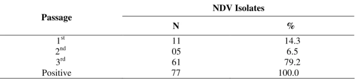
summarized in Table 2, showing that 14.3% of the samples
were positive in the 1st passage, 6.5% in the 2nd passage and 79.2% in the 3rd passage. The isolation percentage in each passage of vaccinated birds in relation to total isolates is
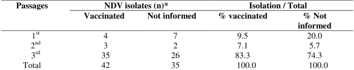
presented in Table 3, showing 9.5% positivity in 1st passage, 7.1% in 2nd passage and 83.3% in 3rd passage. The isolation percentages from flocks with no information on vaccinal status,
in relation to total isolates, were as follows: 20% positivity in
1st passage, 5.7% in 2nd passage and 74.3% in 3rd passage. There was no significant difference in the percentage of
isolation by vaccinal status (p=0.4793).
Table 1. Relationship between seropositivity and NDV isolation
* The percentage of seropositive flocks in the Southeastern region was significantly higher than that in the two other regions
Table 2. NDV isolates in each passage in embryonated chicken eggs (SPF) in relation to the total number of samples
NDV Isolates Passage
N %
1st 11 14.3
2nd 05 6.5
3rd 61 79.2
Positive 77 100.0
Isolation
Region Flocks
(n)
ELISA Seropositive*
flocks % (n) Flocks
%
Region %
Southeastern 592 393 (66.4)* 45 7.6 58.4
Southern 502 112 (22.3) 27 5.4 35.1
Central-Western 489 114 (23.3) 05 1.0 6.5
Table 3. NDV isolates by passages in embryonated chicken eggs, according to vaccination
NDV isolates (n)* Isolation / Total
Passages
Vaccinated Not informed % vaccinated % Not
informed
1st 4 7 9.5 20.0
2nd 3 2 7.1 5.7
3rd 35 26 83.3 74.3
Total 42 35 100.0 100.0
*There was no significant difference in the percentage of isolation by vaccinal status (p=0.4793).
The state with highest viral isolation percentage per flock
was São Paulo (8.8%), followed by Paraná (7.9%), Minas
Gerais (5.3%), Santa Catarina (4.7%), Rio Grande do Sul
(2.8%), Mato Grosso do Sul (1.8%) and Mato Grosso (1.0%).
Among isolates, 44.2% were from São Paulo state, 19.5% from
Paraná, 14.3% from Minas Gerais state, 10.4% from Santa
Catarina, 5.2% from Rio Grande do Sul and Mato Grosso do
Sul, and 1.3% from Mato Grosso. As shown in Table 6, no
virus isolation was obtained in the Federal District and state of
Goiás. Figure 1 shows the isolation of NDV by geographic
region.
In the pathogenicity characterization of NDV isolates
(Table 4), the ICPI ranged between 0 and 0.66. In 41.5% of the
NDV isolates, ICPI varied from 0 to 0.10, in 35.1% varied
from 0.11 to 0.30, in 18.2% varied from 0.31 to 0.50 and in
5.2% varied from 0.51 to 0.66. These data indicate that none of
the isolates was pathogenic, as their ICPI was lower than 0.70.
The correlation between ICPI and samples region was not
significant (p=0.6792), nor was significant the correlation
Table 4. Biological characterization of NDV
between ICPI and birds vaccination status.
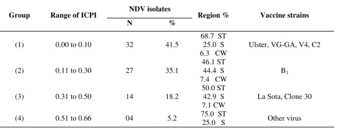
The grouping of the viruses by ICPI and region is shown
in the Table 5. ICPI of vaccine strains are also shown in this
table, for comparison. The 1st group, with 41.5% isolates, presented ICPI between 0 and 0.10, similar to vaccinal strains
Ulster, VG-GA and V4. The isolates in the 1st group were mainly in the Southeastern regions (68.7%), followed by the
Southern (25%) and Central-Western (6.3%) regions. The 2nd group, represented by B1 vaccinal strain (ICPI from 0.11 to
0.30), comprised 35.1% of NDV isolates, being 46.1% in the
Southeastern region, 44.4% in the Southern region and 7.4% in
the Central-Western region. In the 3rd group, represented by the La Sota and Clone 30 vaccinal strains. comprised 18.2% of
isolates (ICPI from 0.31 to 0.50), being 50% in the
Southeastern region, 42,9% in the Southern region and 7.1% in
the Central-Western region. The 4th group, with no vaccine strain used in Brazil, comprised 5.2% of isolates (ICPI from
0.51 to 0.66), being 75% in the Southeastern region and 25% in
the Southern region.
Region Vaccination
status
Number of isolates
Range of ICPI
+ 20 0-0.1
+ 12 0.11 – 0.30
+ 07 0.31-0.50
Southeastern
+ 03 0.51-0.66
- 02 0-0.1
Southeastern
- 01 0.11-0.30
- 08 0-0.1
- 12 0.11-0.30
- 06 0.31-0.50
Southern
- 01 0.51-0.66
- 02 0-0.1
Table 5. Grouping of NDV isolates and vaccine strains by ICPI and region
NDV isolates
Group Range of ICPI
N %
Region % Vaccine strains
(1) 0.00 to 0.10 32 41.5
68.7 ST 25.0 S 6.3 CW
Ulster, VG-GA, V4, C2
(2) 0.11 to 0.30 27 35.1
46.1 ST 44.4 S 7.4 CW
B1
(3) 0.31 to 0.50 14 18.2
50.0 ST 42.9 S 7.1 CW
La Sota, Clone 30
(4) 0.51 to 0.66 04 5.2 75.0 ST
25.0 S Other virus
ST= Southeastern S= Southern CW= Central-Western
Table 6. Percentage of NDV isolates by state
NDV isolates
State Number of flocks
surveyed NDV/flock % n %
Minas Gerais 206 5.3 11 14.3
São Paulo 386 8.8 34 44.2
Paraná 190 7.9 15 19.5
Santa Catarina 171 4,7 08 10.4
Rio Grande do Sul 141 2.8 04 5.2
Mato Grosso do Sul 226 1,8 04 5,2
Mato Grosso 98 1.0 01 1.3
Goiás 108 0 0 0
Distrito Federal 57 0 0 0
Figure1. Number of Newcastle
Disease Virus isolates according
to the state (number of isolates in
DISCUSSION
The main objective of the present study was to investigate
the presence of NDV in commercial healthy birds in the most
important geographic areas for Brazilian poultry production
and export. Birds vaccination efficacy can be monitored by
means of serological tests (18) and a number of serological
techniques can be used to detect specific antibodies to NDV.
The haemagglutination inhibition (HI) test is the method of
choice, but in recent years, several enzyme-linked
immunosorbent assays (ELISA) were developed (16,30). Many
studies on the sensitivity, specificity and correlation between
HI test and ELISA indicate that results may not agree
(9,17,28). Commercially available NDV antibody ELISA kits
are more sensitive than the HI test and, for diagnostic
laboratories, the major advantages of ELISA kits are the assay
standardization, the enhanced effectiveness due to
semi-automation, and the speed for rapid screening for multiples
agents (26).
The percentages of positive samples detected by ELISA
were 66.4%, 23.3% and 22.3% in Southeastern,
Central-Western and Southern regions, respectively. These results
confirm that poultry in the Southeastern region is vaccinated
and that in Minas Gerais and São Paulo states and the northern
region of Paraná state all categories of commercial birds are
vaccinated against Newcastle disease, including broiler
chickens, explaining the large number of sample serology
reagents of this disease in these states. In addition, these results
suggest virus movement from one region to others where no
information on vaccination is available. This movement may
be a consequence of the high density of birds in some regions,
proximity to other species of birds, and also the coexistence of
subsistence farms, side-by-side to well developed poultry
husbandry.
ND vaccination in broiler chicken is not a practice in the
states of Rio Grande do Sul, Santa Catarina, Mato Grosso,
Mato Grosso do Sul, Goiás and the Federal District. However,
in these states, birds for long life purposes, such as
selling maps available in the vaccine producing laboratories
(data not shown), and the amount of vaccines sold for
veterinary stores during the sampling period suggest that
vaccination of broiler chickens was done, including
non-industrial husbandry exploration and subsistence birds.
In this study, the frequency of NDV in healthy birds varied
from 1.0 to 7.6% per flocks, varying from 6.5% to 58.4%
according to the geographic region. The isolation was higher in
regions where vaccination is widely used, confirming results
reported by Alexander (3), who observed that vaccine protects
birds from clinical disease where virus replication and
excretion may occur, even though in low levels. Kapcyznski et
al., (12), studying exotic Newcastle disease (END) viruses that
caused a major outbreak among commercial and backyard
poultry in California (USA), observed that vaccines protected
chickens against morbidity and mortality and significantly
reduced the incidence and viral titers shed from chickens in
comparison with sham controls, but did not prevent infection
and virus shedding. Vaccinated commercial broilers exhibited
66% mortality and shed significantly more virus than broiler
breeders.
A serological study was also conducted in Benin-Africa, in
three different regions (Southern, Central and Northern
regions), and 56%, 75% and 69% of the chickens were
seropositive, respectively, (6, 7). The African results were
similar to those obtained in this work. The presence of virus
already in the first week of life of the birds, observed in the
most regions due to litter reuse, led to the stimulation of the
immune system
The highest virus isolation was observed in Southeastern
region, followed by Southern and Central-Western regions.
This is the first report indicating presence of NDV in regions
where vaccinal status is not informed. Results also indicated a
higher virus circulation in Southern region than in
Central-Western region. The high number of vaccination reported in
the Southeastern region certainly correlates with the high
number of isolates in the area. Similar prevalence, between 5
Seropositivity and virus isolation in states with no
vaccination against ND in broiler chickens can be explained by
the high density of birds in some regions, the proximity with
distinct categories of birds, and the coexistence of subsistence
and low-technology farms along with highly technified poultry
farms.
A better viral isolation was reached when samples were
submitted to three passages in embryonated eggs.
Contradictorily, Kouwenhoven (13) observed that 85% of the
positive samples were positive in the 1st passage and that only 10% needed a second blind passage. In exceptional cases, three
blind passages were needed. The nature of the sample plays an
important role in these tests. The two main sites of NDV
replication in infected poultry appear to be the respiratory and
intestinal tracts; therefore, specimens should always include
cloacal and tracheal swabs (3). As pools from both tracheal and
cloacal samples were taken from healthy birds with no sign of
the disease, it is possible that some harvests occurred during a
period of very small elimination of the virus. This may be
observed by the low number of isolations in the first and
second passages. In most samples, the embryos were not killed
in the initial two passages and no lesions were observed, which
could be due to low virus content of the inoculum. Therefore, a
third passage seems to be necessary for virus adaptation to
embryonated eggs.
In 94.8% of the isolates, the ICPI varied from 0.0 to 0.50,
which is the range where the Ulster 2 C, V4 Queensland, B1
and La Sota vaccine strains are located (4). This explained by
the fact that the most frequently used vaccines in Brazil are La
Sota and B1 (21). Based on these results, the isolated NDV
strains can be classified as avirulent (lentogenic), although the
virus genome has not been sequenced yet.
The isolation of virus with ICPI varying from 0.51 to 0.66
indicates the circulation of non-virulent/apathogenic strains in
regions where vaccination status was not informed.
Yongolo (31) found similar results as the author also
isolated lentogenic strains from healthy carrier birds.
The results in the present study show that the industrial
poultry produced in the nine studied Brazilian states is free of
Newcastle disease, which is in accordance to requirements of
the International Animal Health Code.
ACKNOWLEDGEMENTS
Authors are grateful to the Brazilian Ministry of
Agriculture for providing financial and technical resources and
authorization for the publication of this paper. The authors also
thank Lanagro/SP, in particular the civil servants at the Poultry
Disease Sector and Administration Sectors (computer
assistance). Cleide A. M. Silva and Andrea F. Semolini
(FCM/Unicamp) for their statistical support. C.W.A. and
F.R.S. acknowledge the support from CNPq (National Council
of Scientific and Technological Development, Brazil).
REFERENCES
1. ABEF- Associação Brasileira dos Produtores e Exportadores de Frangos. (2005). Relatório Anual
2. ABEF- Associação Brasileira dos Produtores e Exportadores de Frangos, (2006). Relatório Anual.
3. Alexander, D.J.; Gough, R.E. (2003).Newcastle disease other avian paramyxoviruses, and avian pneumovirus infection. In: Y.M.Saif, H.J. Barnes, A.M. Fadly, J. R. Glisson, L.R. McDougald & D.E. Swayne (Eds), Diseases of poultry 11th , Ames, IA: Iowa State University Press, p.63-99.
4. Allan, W.H.; Lancaster, J.E.; Toth, B. (1978). Newcastle disease vaccines-Their production and use FAO Animal Production and Health Series Nº. 10. FAO: Rome,Italy.
5. BRASIL - Portaria Ministerial nº 182, de 8 de novembro de 1994. Aprova as normas de credenciamento e monitoramento de laboratórios de diagnóstico da doença de Newcastle. Diário Oficial da República do Brasil, Brasília, DF.
6. Bell, J.G. (1991). Vaccination of Africa village poultry against Newcastle disease. In: Demey and Pandey, V.S.(eds.)., Newcastle disease vaccination of village poultry in Africa and Asia. Proceedings of the seminar held on 13-14 February, Antwerp, p.3-8.
7. Bell, J.G. (1992). Newcastle disease in village chickens in North, West and Central Africa. In: Spradbrow P.B. Ed., Newcastle Disease in Village chickens, Control with Thermostable Oral vaccines, Proceedings, International Workshop held in Kaula Lumpur, Malaysia, 6-10 October, 1991. Centre for International Agriculture Research ACIAR, Canberra, p.142-143.
9. Cvelic-Cabrilo, V.; Mazija, H.; Bindin, Z.; Ragland, W.L. (1992). Correlation of haemagglutination inhibition and enzyme-linked immunosorbent assays for antibodies to Newcastle disease virus. Avian Pathol. (21),509-512.
10. Cunha, R.G.; Silva, R.A. (1955).Isolamento e identificação do vírus da doença de Newcastle no Brasil. Soc. Bras. Med Vet. (23)17-33. 11. Fleiss, J.L. (1981). Statistical Methods for Rates and Proportions.2ª ed.
John Wiley & Sons Inc. Nova Iorque.
12. Kaczynski, D.R.; King, D.J. (2005). Protection of chickens against overt clinical disease and determination of viral shedding following vaccination with commercially available Newcastle disease virus vaccines upon challenge with highly virulent virus from the California 2002 exotic Newcastle disease outbreak. 2005. Vaccine 23 (26), 3424-3433.
13. Kouwenhoven, B. (1993). Newcastle disease. Virus Infections of Birds In: McFerran, J.B.; McNulty, M.S. Elsevier, chapter 23, p.350. 14. Mayo, M.A. (2002a). Virus taxonomy-Houston 2002. Arch. Virol.147,
1071-76.
15. Mayo, M.A. (2002b). A summary of taxonomic changes recently approved by ICTV. Arch Virol.(147),1655-1656.
16. Miers. L.A.; Bankowski, R.A.; Zee, Y.C. (1983). Optimizing the enzyme-linked immunosorbent assay for evaluating immunity in chickens to Newcastle disease. Avian Dis. 27,1112-1125.
17. Meulemans. G.; Carlier, M.C.; Gonze, M.; Petit, P.; Halen, P. (1984). Diagnostic serologique de la maladie de Newcastle par les tests d´inhibition de l´hemagglutination et Elisa. Zetralbl Veterinaermed (B) 31:690-700.
18. Office International des Epizooties. (2007). Terrestrial Animal Health Standards Commission Report March 2007, APPENDIX 3.8.X.Guidelines on surveillance for Newcastle disease. Article 3.8.X. 19. Office International des Epizooties. (2000). Newcastle disease,
p.221-232. In Manual of standards for diagnostic tests and vaccines, 4th edition. World Organization for Animal Health, Paris, France.
20. Oliveira, B.O.; Belluci, M.S.P.; Portz, C.; Oliveira, J.R.J.G.; Doretto Jr, L.; Orsi, M.A.; Mazur, C.; Andrade, C.M. (2000). Biological
characterization of M33 field isolate of Newcastle Disease virus. Virus Rev. Res. 05 (2), 56.
21. Orsi, M.A.; Doretto Jr., L.; Albieri, S.C.; Ribeiro, S.AM.; Yoshida, L.T. (2001). Quality control of live vaccines against Newcastle disease in the period 1993 to 2000. Virus Rev. Res., 06(2), 126
22. Santos, J.A. e col., (1954). A ocorrência da doença de Newcastle no Brasil. (Nota Previa). Rev. Prod Animal, (Rio),1 (1): 5-12.
23. SAS System for windows (Statistical Analysis System), versão 9.1.3 Service Pack 3. SAS Institute Inc, 2002-2003, Cary, NC, USA
24. Stram, Y.; Shchori, D.; Chinitch, Y.; David, D.; Molad, T.; Samina, I. (1998). Molecular characterization of an unassigned Israeli Newcastle disease virus isolate. Avian Dis. 42(4), 746-51.
25. Schelling, E.; Thur, B.; Griot, C.; Audige, L. (1999). Epidemiological study of Newcastle disease in backyard poultry and wild bird populations in Switzerland. Avian Pathol. 28(3), 263-272.
26. Snyder, D.B.; Marquardt, W.W.; Mallinson, E.T.; Saveage, P.K.; Allen, D.C. (1984). Rapid serological profiling by enzyme-linked immunosorbent assay. III Simultaneous measurements of antibody titers to infectious Bronchitis virus, infectious bursal disease and Newcastle disease virus in a single serum dilution. Avian Dis. 28,12-24.
27. Thayer, S.G.; Villegas, P.; Fletcher, O.J. (1987). Comparison of two commercial enzyme-linked immunosorbent assays and conventional methods for avian serology. Avian Dis. 31,120-124.
28. Vaitsman, J.; Moussatché, I. (1954). Doença de Newcastle. Boletim. 801 do Serviço de Informação Agrícola, Ministério da Agricultura, 56 p. 29. Vianna, J.S.M.; Mazur, C.; Portz, C.; Ferreira, I.I.; Almeida, C.A.S.;
Galler, R. (2000). Identificação e caracterização biomolecular do vírus da doença de Newcastle pela técnica de RT-PCR. R. Bras. Med. Vet. 22,160-163.
30. Wilson, R.A.; Perrotta, C.; Frey, B.; Eckroade, R.J. (1984). An enzyme– linked immunosorbent assay that measures protective antibody levels to Newcastle disease virus in chickens. Avian Dis. 28, 1079-1085. 31. Yongolo, M.G.S. (1996) Epidemiology of Newcastle Disease in Village