Vol.59: e16160301, January-December 2016 http://dx.doi.org/10.1590/1678-4324-2016160301
ISSN 1678-4324 Online Edition
BRAZILIAN ARCHIVES OF BIOLOGY AND TECHNOLOGY
A N I N T E R N A T I O N A L J O U R N A L
Isolation, Identification and Molecular Characterization of
Highly Pathogenic Newcastle Disease Virus From Field
Outbreaks
$VPD$VKUDI
0XKDPPDG6DODKXG'LQ6KDK
0XGDVVHU+DELE
0XMDKLG
+XVVDLQ
6KDKLG0DKERRE
.KDOLG$O*KDQLP
1Government College University Faisalabad, Faisalabad, Pakistan; 2Nuclear Institute of Agriculture and Biology Faisalabad,
Pakistan; 3King Saud University, Department of Zoology, College of Science, P. O. Box 2455, Riyadh, Saudi Arabia.
ABSTRACT
Newcastle disease (ND) is a major infectious disease of the poultry caused by a virulent strain of Avian Paramyxovirus – 1, that is a single strand non-segmented negative sense RNA virus. ND virus is major threat to the poultry industry in many countries of the world. The study was aimed to isolate and identify Newcastle disease virus (NDV) by using a haemagglutination inhibition (HI) test and reverse transcription-polymerase chain reaction (RT-PCR) assay. A total 100 samples of infected and dead birds were collected from different poultry farms. The weight of the birds was ranged 1000-1200g. The birds were divided into 3 groups. Haemagglutination assay (HA) was performed to detect the presence of NDV in suspension of infected homogenized tissues and it was found that HA is not the best method to detect the virus when it is in trace amounts. RT-PCR using NDV specific primers analyzed different clinical and postmortem samples. Reverse transcriptase polymerase chain reaction and specific primers was used for determining the presence of viruses. It was found that the virus was present in most of the infected samples except the serum of infected birds. During multiple sequence alignment (MSA) it was found that, our isolates have high homology (98%) with other reported NDV isolates. Phylogenetic analysis revealed that our isolate was closely related with viscerotropic velogenic types of NDV, which are highly pathogenic Newcastle disease virus.
Key words: Newcastle disease; epidemic; molecular characterization; avian virus; RT-PCR
1Authors for correspondence: shahidmahboob60@hotmail.com
INTRODUCTION
Newcastle disease (ND) is an important viral disease of poultry and other bird species disregarding of sex and age [1-4]. ND is a major cause of huge economic losses in various parts of the world [5-6]. Newcastle disease virus (NDV) is belong to a group of the avian paramyxovirus - I [7-9]. The disease is characterized by an involvement of digestive, respiratory and nervous systems [10-11]. This disease can vary in nature from mild to severe, depending upon the type of the virus. In non-vaccinated chickens, the morbidity and death rates may be up to 100% each, depending upon the virulent intensity of NDV. In recent years, outbreaks have continuously occurred in Pakistan resulting in huge losses. Infected samples indicated the presence of virus identified by chicken embryonated egg inoculation and haemagglutination assay [2].
NDV has a wide range of hosts range, inclusive of approximately 241 species [12] belongs to 27 orders out of which 50 orders of birds [13]. In various developing countries, ND is an endemic and thus have a high economic impact. Due to this disease, poultry industry is facing losses of billions of Rupees annually in Pakistan [14] and millions of dollars worldwide [15].
ND is a serious threat to the poultry industry [16]. The rate of mortality and morbidity of poultry in unvaccinated flock [2] varies from 90-100%, depending upon the strain of ND virus [10]. The outbreaks of ND are regularly reported from all continents of the world [17]. “An intermittent form of ND present in Pakistan throughout the year, only a limited number of cases are reported annually. A severe outbreak of ND occurred during 2012 at Jallo Wildlife Park in Lahore, Pakistan, caused by APMV- 1 serotype. Within a week, it took the lives of approximately 190 peacocks with a 100% mortality rate and 50% loss of the susceptible birds. Isolation of virus and serological diagnostics, such as HI Test, ELISA and molecular diagnostic tests like real time PCR confirmed the presence of velogenic Newcastle disease virus” [18]. The study was aimed to isolate and identify Newcastle disease virus (NDV) by using a haemagglutination inhibition (HI) test and reverse transcription-polymerase chain reaction (RT-PCR) assay from the dead birds from the poultry farms of Punjab
MATERIALS AND METHODS
Collection of infected samples:
The samples were collected during 2013 from the poultry farms of different areas of Punjab province, Pakistan. A total 100 samples of infected and dead birds were collected from different poultry farms. The weight of the birds was ranged 1000-1200g. The birds were divided into 3 groups. Post-mortems were conducted and infected tissues, i.e. intestine, proventriculus, spleen, lungs and trachea were collected from the dead chickens. After collection, the samples were transported on ice packs and stored at -20 o C to -25 o C for further processing.
Virus isolation in embryonated eggs:
“Virus isolation was carried out as described by [19-20]. Eggs from healthy and ND-seronegative chickens were used which were conventionally raised. Tissue was homogenized as a 10% (w/v) suspension in PBS containing antibiotics (streptomycin, penicillin). After centrifugation, 0.1 ml supernatant was inoculated into the allantoic cavity of 9- to 11-day-old embryonated chicken eggs. Allantoic fluid from dead embryos or in live embryos after 72 hours of incubation was collected and NDV was detected by HA test”.
Haemagglutination assay (HA):
The titer of the virus in allantoic fluid or tissue homogenate was determined by hemagglutination assay as described by [20-22].
Reverse transcription-polymerase chain reaction: Allantoic fluid was used for RNA extraction by following the protocol of Favor Prep. TM Viral Nucleic Acid Extraction mini kit. RNA extracted by using 150µl of allontoic fluid, 570µl of VNE Buffer, 570µl of ethanol, 500µl Wash Buffer 1 and 750µl of wash buffer. Each RNA sample was dissolved in 40 µl sterile RNAse-free water and stored at –70°C.
centrifuged and then was incubated for 60 min at 42ºC”. The reaction terminated after heating it at 70ºC for 5 minutes, then after briefly spin the tube before cDNA was used for PCR amplification. RT-PCR was performed as described by [25]. NDV-F /NDV-R primers were selected to amplify a 202 bp fragment of the F gene including the cleavage site. “Primer sequences are shown in Table-1. PCR was carried out in a 50 μl reaction containing 5.0 μl10X buffer, 2.0 μl 25 mM MgCl2, 2.0 μl 10 mM dNTP, 0.2 μl Taq, 0.8 μl NDV-F
primer (100 pmol), 0.8 μl NDV-R primer (100 pmol), 1.5 μl cDNA, and 37.7 μl DEPC was added to each tube. The amplification profile started with one cycle at 94°C for 2 min. followed by 35 cycles of 94°C for 15 sec, 48°C for 30 sec and 72°C for 30 sec and final extension of 72°C for 10 min. Sterile RNAse free water or tissue samples from animals slaughtered on day 0, were used as negative controls”.
Table 1: Specific primers used for RT-PCR
Sr.
No. Gene Primer Primer Sequence
PCR
Product Reference
i. F NDV-F 5’-GGTGAGTCTATCCGGARGATACAAG-3’
202bp
Creelan et.
al., (2002) ii.
F NDV-R 5’-TCATTGGTTGCRGCAATGCTCT -3’
Agarose Gel Electrophoresis:
PCR products were subjected to agarose gel electrophoresis. For this 10 μl of PCR product along with 2 μl of 6X loading dye were mixed and loaded on 1.5% agarose gel along with 100bp ladder. The gel was run in 1X TAE buffer till the dye reach near other end. At the end, the gel was stained with ethidium bromide and observed under UV light.
Nucleotide sequence analysis:
The PCR product was purified and its nucleotide sequence was determined using both forward and reverse primers. The nucleotide sequence was analyzed using BLAST software and its homology was searched against available nucleotide sequences from GenBank. Phylogenetic analysis was performed using a partial nucleotide sequence of the fusion protein gene and phylogenetic tree was drawn on the basis of observed divergence using software DNAMAN by Lynnon Biosoft, Canada.
RESULTS AND DISCUSSION
Clinical signs and symptoms:
The infected broilers showed clinical symptoms of depression, dizziness, gasping, paralysis of neck, legs or wings and loss of appetite. Swelling of the eyes and discharge from eyes were also observed. Greenish yellow colored diarrhea was very prominent. Similar signs and symptoms were also reported by [1, 11].
Postmortem lesions:
Fig 1:(a) Typical conjunctivitis (b) Postmortem examination showing hemorrhages of intestine
(c) Enlarged spleen and hemorrhages in mucosa of proventriculus, along with normal organs for comparison.
(a)
(b)
Detection of NDV by Haemagglutination Assay: Haemagglutination assay (HA) was performed to detect the presence of NDV in suspension of infected homogenized tissues. Results are shown in Table 2. HA is not the best method to detect the virus when it is in trace amounts. When the
homogenized viral suspension was allowed to multiply in allantoic fluid of embryonated eggs, then HA can detect successfully the virus in allantoic fluid. [5] also found that HA is one of the rapid and successful techniques to detect the NDV.
Table 2: Comparison of results of Haemagglutination assay (HA) and RT-PCR
Test
Haemagglutination Assay
RT-PCR
Type of samples
Direct tissue
suspension
Allentoic
Fluid
Direct tissue
suspension
Allentoic
Fluid
Clinical
samples
Tracheal swabs
-
+
+
+
Serum
-
-
-
-
Fecal
-
+
+
+
Proventriculus
+
+
+
+
Spleen
-
+
+
+
L 1 2 3 4 5 6
7
1000b p
100bp 200bp 500bp
Fig 2: Agarose gel electrophoresis showing RT-PCR results.
Lane L: 100bp ladder
Lane 1,3,4,6,7: Positive results, showing 202 bp PCR product
Post-mortem
samples
Lungs
-
-
+
+
Intestine
+
+
+
+
Confirmation of NDV by RT-PCR:
RT-PCR using NDV specific primers analyzed different clinical and postmortem samples. Results of comparison of both techniques are shown in Table 2. It was found that the virus was present in most of the infected samples except the serum of infected birds. It might be because virus presence in the blood is for short periods during infection. RT-PCR is very sensitive technique to detect the presence of NDV in different tissue samples, even if the virus was present in minute quantity [2, 24]. Moreover, [25] “ reported one-step RT-PCR test coupled with restriction enzyme assay (REA) as fast and specific method for the detection and typing of APMV-1 from field samples”.
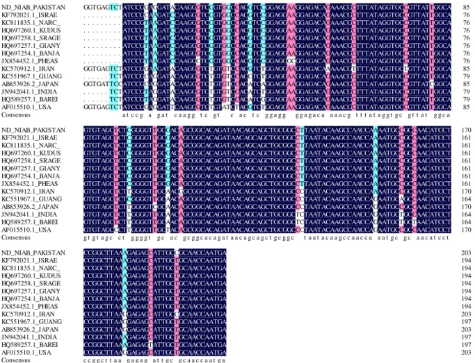
Nucleotide sequence analysis
During multiple sequence alignment (MSA) it was found that, our isolates have high homology (98%)
Fig 4:
Multiple sequence alignment of partial fusion gene (203bp) of different reported NDV isolates along with NDV isolate of Pakistan (ND-NIAB-Pak).CONCLUSION
It was concluded the virus was present in most of the infected samples except the serum of infected birds. The multiple sequence alignment (MSA) exhibited, that these isolates have, high homology (98%) with other reported NDV isolates.
Phylogenetic analysis revealed that our isolate is closely related with viscerotropic velogenic types of NDV, which are highly pathogenic Newcastle disease virus.
ACKNOWLEDGMENTS
The authors would like to express their sincere appreciation to the Deanship of Scientific Research at King Saud University for its funding of this research through the Research Group Project No. RG-1435-012.
REFERENCES
[1] Alexander DJ (2003). Newcastle disease, other avian paramyxoviruses and pneumovirus infections. J Diseas Poultry 11: 63-99.
[2] Haque MH, Hossain MT, Islam MT, Zinnah, MA, Khan MSR, Islam MA. (2010). Isolation and Detection of Newcastle disease Virus from field outbreaks in broiler and layer chickens by reverse transcription–polymerase chain reaction. Bangla J Vet Med 8(2): 87-92.
[3] Orsi MA, Doretto JrL, Camillo SCA, Reischak D, Ribeiro SAM, Ramazzoti A, Mendonca AO, Spilki FR, Buzinaro MG, Ferreira HL, Arns CW. (2010). Prevalence of Newcastle disease virus in broiler chickens (Gallus gallus) in Brazil. Brazil J Microbiol 41: 349-357.
paramyxovirus type 1 (Newcastle disease virus) isolates by phylogenetic analysis of a partial nucleotide sequence of the fusion protein gene. Avian Pathol32: 239-256.
[6] Diel DJ, Susta L, Garcia SC, Killian ML, Brown CC, Miller PJ, Afonso CL, (2012). Complete Genome and Clinicopathological Characterization of a Virulent Newcastle Disease Virus Isolate from South America. J Clini Microbiol50(2): 378-387
[7] Yu L, Wang Z, Jiang Y, Chang L, Kwang J (2001). Characterization of Newly Emerging Newcastle Disease Virus Isolates from the People’s Republic of China and Taiwan. J Clin Microbiol 39(10): 3512-3519.
[8] Choi KS, Lee EK, Jeon WJ, Kwon JH. (2010). Antigenic and immunogenic investigation of the virulence motif of the Newcastle disease virus fusion protein. J Vet Sci11(3): 205-211.
[9] Qin Z M, Tan LT, Xu HY, Ma BC, Wang YL, Yuan XY, Liu WJ (2008). Pathotypical Characterization and Molecular Epidemiology of Newcastle Disease Virus Isolates from Different Hosts in China from 1996 to 2005. J Clin Microbiol 46(2): 601-611. [10] Nanthakumar T, Kataria RS, Tiwari AK, Butchaiah
G, Kataria JM (2000). Pathotyping of Newcastle Disease Viruses by RT-PCR and Restriction Enzyme Analysis. J Vet Res Commun 24: 275-286.
[11] Tiwari AK, Katari RS, Nanthakumar T, Dash BB, Desai G (2004). Differential detection of Newcastle disease virus strains by degenerate primers based RT-PCR. J Comp Immunol Microbiol Infect Diseas 27:163-169.
[12] Madadgar O, Karimi V, Nazaktabar A, Kazemimanesh M, Ghafari MM, Dezfouli SMA, Hojjati P (2013). A study of Newcastle disease virus obtained from exotic caged birds in Tehran between 2009 and 2010. Avian Pathol 42(1): 27-31.
[13] Carrasco AOT, Rodrigues JNM, Seki MC, Moraes FE, Silva JR, Durigon EL, Pinto AA (2013). Use of reverse transcriptase polymerase chain reaction (RT-PCR) in molecular screening of Newcastle disease virus in poultry and free-living bird populations. Trop Anim Heal Prod 45:569-576.
[14] Waheed U, Siddique M, Arshad M, Ali M, Saeed A (2013). Preparation of Newcastle Disease Vaccine from VG/GA Strain and its Evaluation in Commercial Broiler Chicks. Pak J Zool 45(2): 339-344.
[15] Susta, L., Miller, P. J., Afonso, C. L., & Brown, C. C. (2010). Clinicopathological Characterization in Poultry of Three Strains of Newcastle Disease Virus Isolated From Recent Outbreaks. J Vet Pathol48(2): 349-360.
[16] Narayanan MS, Parthiban M, Sathiya P, Kumanan K (2010). Molecular detection of Newcastle disease virus using Flinders Molecular detection of Newcastle disease virus using Flinders Technology
Associates-PCR Technology Associates-PCR. J Veterin Arhiv 80(1): 51-60.
[17] Munir M, Shabbir MZ, Yaqub T, Shabbir MAB, Mukhtar N, Khan MR, Berga M. (2012). Complete Genome Sequence of a Velogenic Neurotropic Avian Paramyxovirus 1 Isolated from Peacocks (Pavo
cristatus) in a Wildlife Park in Pakistan. J Virol 86(23): 13113-13114.
[18] virus isolated from outbreaks in Egypt during 2006. Virol J 8(237): 01-04.
Munir S, Hussain M, Farooq U, ZabidUllah JQ, Afreen M, Bano K, Khan J, Ayaz S, Kim KY, Anees, M (2012). Quantification of antibodies against poultry haemagglutinating viruses by haemagglutination inhibition test in Lahore. Afr J Microbiol Res 6(21), 4614-4619.
[19] Mayr A, Bachmann PA, Bibrack B, Wittmann G (1974). Virologische Arbeitsmethoden Band 1, 1st edn (pp. 37–39). Stuttgart: Gustav Fischer Verlag. [20] OIE (1996). Newcastle disease. OIE Manual of
Standards forDiagnostic Tests and Vaccines (pp. 161–169). Paris: Office International des Epizooties. [21] Hierholz JC, Suggs MT, Hall EC (1969). Standardized viral hemagglutination and hemagglutination-inhibition tests. Description and statistical evaluation. Appl Microbiol 18:824–833 [22] Nadeem Y, Chaudhary TM, Shah M S, Ashraf A
(2004). Oil adjuvanted newcastle disease vaccine production using local viral isolates. Proceedings of 24th PakiCong Zool pp: 51- 56.
[23] Ashraf A, Shah MS (2014). Newcastle Disease: Present status and future challenges for developing countries. Afr J Microbiol Res 8(5): 411-416. [24] Yi J, Liu C (2011). Detecting Newcastle disease
virus in combination of RT-PCR with red blood cell absorption. Virol J 8: 202-208.
[25] Creelan JL, David AG, Samuel JMc (2002). Detection and differentiation of pathogenicity of avian paramyxovirus serotype 1 from field cases using one-step reverse transcriptase-polymerase chain reaction, Avian Pathol 31:5, 493-499
[26] Mohamed HA, Kumar S, Paldurai A, Samal SK (2011). Sequence analysis of fusion protein gene of Newcastle disease
[27] Zhang S, Wang X, Zhao C, Liu D, Hu Y, Zhao J, Zhang G (2011). Phylogenetic and Pathotypical Analysis of Two Virulent Newcastle Disease Viruses Isolated from Domestic Ducks in China. PLOS ONE 6(9):1-9.
Erratum
In Article
“
Isolation, Identification and Molecular Characterization of Highly Pathogenic
that read:
“
Asma Ashraf
1; Mohammad Slah U Din
2; Muhammad Habib
1; Mujahid Hussain
2;
Shahid Mahboob
1,3*; Khalid Al-Ghanim
3;
1Government College University Faisalabad, Faisalabad, Pakistan; 2Nuclear Institute of Agriculture and
Biology Faisalabad, Pakistan; 3King Saud University, Department of Zoology, College of Science, P. O. Box
2455, Riyadh, Saudi Arabia.”
Read:
“Asma Ashraf
1; Muhammad Salah-ud-Din Shah
2; Mudasser Habib
2; Mujahid
Hussain
2; Shahid Mahboob
2,3*; Khalid Al-Ghanim
3;
1Government College University Faisalabad, Faisalabad, Pakistan; 2Nuclear Institute of Agriculture and
Biology Faisalabad, Pakistan; 3King Saud University, Department of Zoology, College of Science, P. O. Box
2455, Riyadh, Saudi Arabia.”