First Characterization of
Candida albicans
by Random Amplified
Polymorphic DNA Method in Nicaragua and Comparison of the
Diagnosis Methods for Vaginal Candidiasis in Nicaraguan Women
Martha Darce Bello/*/
+, Alcides Gonzalez*, Christian Barnabé**, Georges Larrouy
Laboratoire de Parasitologie Médicale et d’Ecologie Humaine, 37, Allées Jules-Guesde 31062 Toulouse Cedex, France *Centro Nacional de Diagnostico y Referencia, Ministerio de Salud de Nicaragua, Managua, Nicaragua **Unité de Recherche:
“Génétique des Maladies Infectieuses”, Centre National de la Recherche Scientifique/Institut de Recherche pour le Développement, Montepellier, France
A total of 106 women with vaginitis in Nicaragua were studied. The positive rate for the identification of Candida species was 41% (44 positive cultures out of 106 women with vaginitis). The sensitivity of microscopic examination of wet mount with the potassium hydroxide (KOH) was 61% and 70% with Gram’s stain when using the culture of vaginal fluid as gold standard for diagnosis of candidiasis. Among the 44 positives cultures, isolated species of yeast from vaginal swabs were C. albicans (59%), C. tropicalis (23%), C. glabrata (14%) and C. krusei (4%). This study reports the first characterization of 26 C. albicans stocks from Nicaragua by the random amplified polymorphic DNA method. The genetic analysis in this small C. albicans population showed the existence of linkage disequilibrium, which is consistent with the hypothesis that C. albicans undergoes a clonal propagation.
Key words: vulvovaginal candidiasis - diagnosis - Candida albicans molecular typing random amplified polymorphic DNA -population structure - Nicaragua
The genital candidiasis is one of the pathogenic dem-onstrations of yeast. Candida albicans is the most fre-quent species; it is usually isolated in 85 to 90% from the vaginal mycoses (Odds et al. 1988).
Vaginal candidiasis affects females at least once dur-ing their lifetime, at an estimated rate of 70 to 75%, of whom 40 to 50% will experience a recurrence (Sobel 1999). In Nicaragua, we know very little about the preva-lence and incidence of vaginal candidiasis and no study of the biology of C. albicans has been carried out. In this country, diagnosis of vaginal candidiasis is mainly based on the clinical presentation. Laboratories of the hospitals and health centres (peripheral laboratories) carry out only the microscopic diagnosis from the vaginal fluid. In the laboratory of the National Centre of Diagnosis and Refer-ence of Nicaragua (CNDR), the yeast identification is based on the observation of the microscopic aspects, culture and biochemical tests.
Most of the genetic studies revealed that C. albicans is predominantly clonal (Pujol et al. 1993, Helstein et al. 1993, Lockhart et al. 1995, Xu et al. 1999). Some authors have proposed that clonal propagation with a remaining capacity of recombination, shape the population struc-ture of C. albicans (Caugant & Sandven 1993, Gräser et al. 1996, Tibayrenc 1997).
Pujol et al. (1997) demonstrated that the fingerprinting of C. albicans randon amplified polymorphic DNA (RAPD), multilocus enzyme electrophoresis (MLEE) and
+Corresponding author. Fax: +33-5-61-14.5979. E-mail:
[email protected] Received 7 November 2001 Accepted 29 July 2002
Southern blot hybridization with the moderately repeti-tive DNA Ca3 probe, not only clustered moderately re-lated isolates in a similar fashion but also afforded similar levels of resolution of microevolution within a clonal popu-lation.
The goal of this study was to use the RAPD method to examine the patterns of yeast genetic diversity among women with vaginitis from a single geographic area. We were specifically interested to know the frequency of yeast in vulvovaginal secretions. We also compared the con-ventional methods of yeast diagnosis from vaginal samples used in Nicaragua and yeast culture method.
MATERIALS AND METHODS
Sabouraud glucose agar was used to distinguish between C. albicans and C. dublinensis.
We compared the microscopy and KOH test with the culture (chosen as gold standard), which is at present considered as the most sensitive method (Sobel 1999).
Sensitivity and specificity of each technique were esti-mated with the culture results with the following formulas: Sensitivity = True positives/True positives + False negatives Specificity = True negatives/True negatives + False positives
The identified C. albicans stocks were used to study the genetic structure of this population. The stocks were stored in 1.5 ml of a stationary phase culture (until reach-ing a density of 2 x 10⁸ cells:ml), mixed with 0.1 ml of dimethyl sulfoxide and frozen at -70°C until use. Four reference C. albicans strains of the American Type Cul-ture Collection (ATCC) were used as control strains: 90028, 64548, 64550 and 64551. Six stocks belonging to other species from the strains collection of the Parasitology Laboratory of the Purpan Hospital of Toulouse, France, were used as outgroups for phylogenetic analysis (C. krusei, C. tropicalis, C. glabrata, C. parapsilopsis, Sac-charomyces cerevisiae and Geotrichum candidum).
DNA extraction- A modification of procedure previ-ously applied to S. cerevisiae was used (Philippsen et al. 1991). Briefly, the yeast colonies were taken from each agar plate and were inoculated in 15 ml of YPD (2% yeast extract, 1% peptone, 2% dextrose). Yeast were grown over-night at 37°C to a cell density of approximately 2x10⁸ cells/ ml (early stationary phase). The yeast were collected by centrifugation at 3200 g for 5 min, washed in 15 ml of water at 1700 g for 5 min and suspended once again in 1.5 ml of a solution (pH 7.5) of 0.9 M sorbitol, 0.1 M EDTA, 50 mM dithiothreitol plus 0.5 mg of Zymoliase 20T (ICN, biomedicals inc, UK). After 1 h of incubation at 37°C, the cells were centrifuged at 1700 g for 5 min and suspended in 0.5 ml of lysis buffer: 50 mM Tris HCl pH 8.0, 10 mM EDTA, 1% SDS (W/V), plus 5 µl of proteinase K 20 µg/ml. The mixture was incubated at 56°C for 2 h. Eight µl of RNAse A (100 µg/ml) were then added, followed by mix-ing and incubation at 37°C for 30 min. DNA purification was carried out by classical phenol-chloroform extraction (Sambrook et al. 1989). The DNA was precipitated with cold ethanol at -70°C for 1 h and centrifuged at 5000 g for 30 min. The pellet was washed twice with 70% ethanol. The precipitate was dried and suspended in 100 µl of pu-rified water.
RAPD - Conditions for the RAPD have been previ-ously described (Williams et al. 1990). Briefly, a polymerase chain reaction was performed in 0.5 ml microcentrifuge tubes in a final reaction mixture (60 µl) containing 20 ng of C. albicans DNA, 6 µl of 10X buffer for Taq DNA poly-merase, 0.9 U of Taq DNA polymerase (Boehringer Mannheim, Meylan, France), 100 µM each dATP, dCTP, dGTP and dTTP (Boehringer Mannheim), and 200 nM of the primers (Operon Technologies, Alameda, USA). The decameric primers used were A2 (5’ CAGGCCCTTC 3’); A3 (5’ AGTCAGCCAC 3’); A5 (5’ AGGGGTCTTG 3’); A9 (5’ GGGTAACGCC 3’); A12 (5’ TCGGCGATAG 3’); A15 (5’ TTCCGAACCC 3’); A18 (5’ AGGTGACCGT 3’) and A20 (5’ GTTGCGATCC 3’). Amplifications were performed
in a thermal cycler PTC-100 (MJ Research Inc., USA) pro-grammed for 45 cycles of 1 min at 94°C (denaturation), 1 min at 36°C (annealing) and extension at 72°C for 2 min, with a final 10 min extension at 72°C for the last cycle. Amplified DNA fragments were analyzed by electrophore-sis on 1.6 % agarose gels stained with ethidium bromide. Genetic analysis - Genetic relationships among the stocks were estimated by the Jaccard’s genetic distance (Jaccard 1908). Each RAPD band was coded with a num-ber, starting with 1 for the slowest band. The distance was estimated based on the following formula:
D = 1 – [a/(a +b +c)]
a = number of bands that are common to the two com-pared genotypes
b = number of bands present in the 1st genotype and absent in the 2nd
c = number of bands absent in the 1st genotype and present in the 2nd
The UPGMA method [Unweighted Pair-Group Method with Arithmetic Averages, Sneath and Sokal (1973)] was used to cluster the genotypes together ac-cording to their Jaccard’s distances.
Population genetic analysis was based on linkage dis-equilibrium statistics, with random mating as null hypoth-esis. The following four probability values proposed by Tibayrenc et al. (1990) were used: d1: the combinatorial probability of sampling the most frequent genotype as often as or more often than actually observed if there were random recombination; d2: the probability of ob-serving any genotype as often as or more often than the most common genotype actually observed if there were random recombination; e: the probability of observing as few or fewer genotypes than actually observed if there were random recombination; and f: it gives the probabil-ity of observing linkage disequilibrium as high, or higher than actually observed in the sample if there were random recombination. If a probability is non significant (p > 5 X 10⁻²), random recombination can not be rejected, but it is significant (p < 5 X 10⁻²), it supports the nonrandom as-sociation of loci. The f test is based on Montecarlo simu-lations with 104 iterations. A level of significance < 10-4 means that no case was observed out of 104 iterations (Tibayrenc et al. 1991).
RESULTS
Frequency of candidiasis- Yeast were isolated in 44 out of 106 women (42%), while no trace of yeast was found in 62 women (58%). C. albicans was the most frequently isolated species accounting for 26 (59%), followed by the next most frequent yeast species, C. tropicalis, which was isolated from 10 (23%) of the women. Other species of Candida were also cultured: C. glabrata from 6 (14%) and C. krusei from 2 (4%).
The filamentation test was positive for all the C. albicans stocks and negative for the other species. The RAT con-firmed the presence of chlamydospore of C. albicans, whereas for the other species, we observed only the pres-ence of yeast.
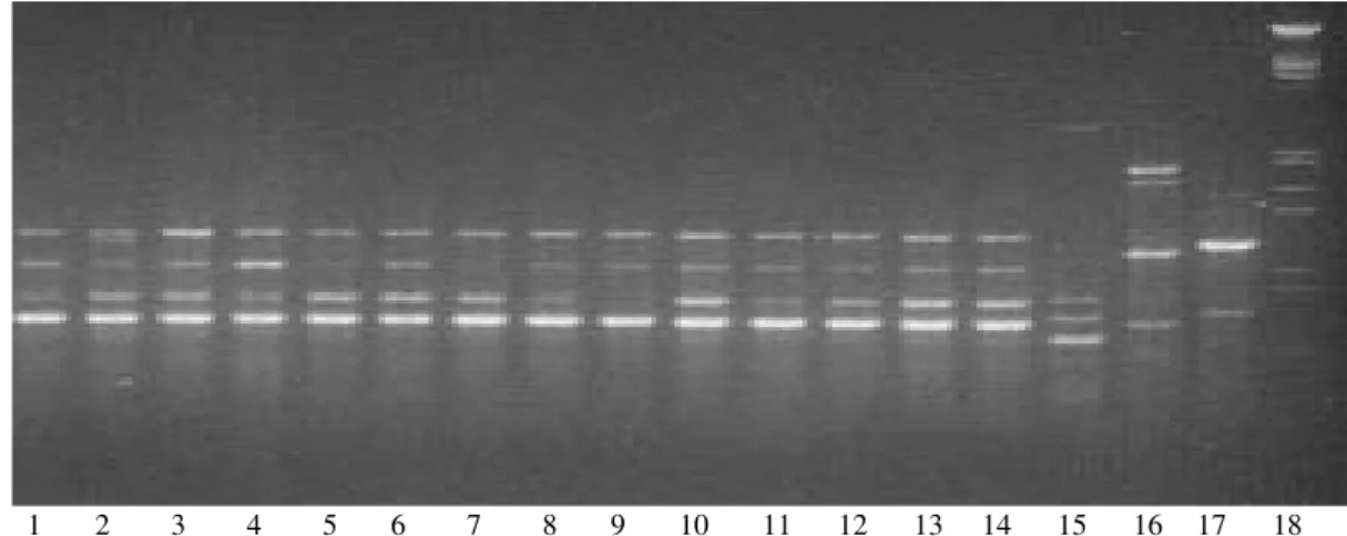
RAPD - The 26 C. albicans stocks collected in this study were analyzed with 8 individual primers. These prim-ers were selected from 20 primprim-ers tested for their capaci-ties to discriminate variability and reproducibility. Eight primers for the C. albicans stocks generated 37 bands. From the total samples (C. albicans and the others spe-cies) the number of bands was 78. RAPD profiles were close to the four reference stocks. The RAPD profiles obtained with the A3 and A20 primers were better for differentiating the yeast stocks (Fig. 1).
Genetic diversity - All primers tested showed the poly-morphic bands. The level of RAPD resolution was high: among 26 stocks, 23 different rapdemes were observed (genotype diversity = 0.88). All the primers exhibit vari-ability.
Phylogenetic clustering- The UPGMA tree derived from RAPD data showed that all C. albicans fell into a single cluster, in which Jaccard’s genetic distances were fairly low, while the other non-C. albicans stocks fell apart
in a sharply distinct cluster (Fig. 2). The most important genetic distance among the stocks of our sample was 0.49, with an average of 0.26 ± 0.1, theoretical maximum is 1.0. The level of polymorphism was 0.75 and the mean genetic diversity was 0.45.
Genetic population tests - The linkage disequilibrium tests (d1, e, and f) were significant within the C. albicans group with all stocks, but when repeated genotypes were removed, the f test only showed a borderline p value (0.06).
DISCUSSION
Laboratory diagnosis- When we used the culture as gold standard for detecting Candida sp. the sensitivity of microscopic examination by Gram’s stain was more ef-fective than the KOH test. These results are similar to a study made by Geiger et al. (1995). A positive culture does not necessarily indicate that the yeast is responsible for the vaginal symptoms. The diagnosis of Candida vagini-tis requires a correlation between clinical conditions, and laboratory results.
Studies indicate that Candida sp. may be isolated from lower genital tract of approximately 20% (occasional stud-ies set the upper limit at 55%) of asymptomatic healthy
TABLE I
Results of diagnosis of Candida sp.by microscopy and polassium hydroxide (KOH) test on 44 culture positive vaginal fluid samples from Nicaraguan vaginitis patients
Microscopy KOH test
Positive Negative Positive Negative Total
Isolates a N % N % N % N % N %
C. albicans 19 73 7 27 16 61 10 39 26 59
C. tropicalis 6 60 4 40 5 50 5 50 10 23
C. glabrata 4 67 2 33 4 67 2 33 6 14
C. krusei 2 100 0 0 2 100 2 100 2 4
Total 31 70 13 30 27 61 17 39 44 100
a: isolates were typed based on the chromagar test and confirmed with ID 32C identification kits; N: number of corresponding samples
1 2 3 4 5 6 7 8 9 10 11 12 13 14 15 16 17 18
TABLE II
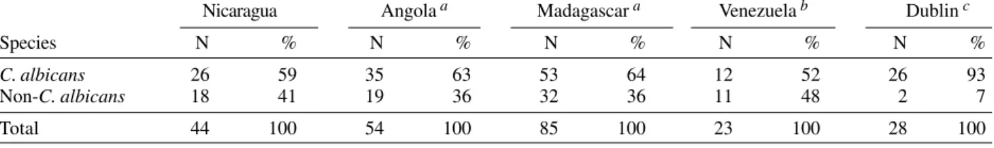
Distribution of Candida albicans and non-C. albicans species in vaginal samples from patients with vaginitis in five countries Nicaragua Angola a Madagascar a Venezuela b Dublin c
Species N % N % N % N % N %
C. albicans 26 59 35 63 53 64 12 52 26 93
Non-C. albicans 18 41 19 36 32 36 11 48 2 7
Total 44 100 54 100 85 100 23 100 28 100
a: Tietz et al. (1995); b: Mendoza et al. (1999); c: Al-Rawi et al. (1999)
CAN02
CAN23
CAN22
CAN19
CAN20
CAN03
CAN07
CAN09
CAN13
CAN05
CAN10
CAN06
CAN11
CAN17
ATCC 64550
CAN18
CAN04
CAN08
CAN15
CAN21
CAN14
CAN12
CAN24
CAN29
CAN28
ATCC 900028
CAN01
ATCC 64548
CAN16
ATCC 64551
C. krusei
C. parapsilopsis
Geotrichum candidum
C. glabrata
C. tropicalis
Sacharomyces cereviseae
Fig. 2:UPGMA dendrogram derived from Jaccard’s distances after randon amplified polymorphic DNA data of the 26 Candidaalbicans isolates from Nicaragua and reference stocks.
C. albicans
women (Drake & Maibach 1973). Our results in symptom-atic women showed that yeast were present in 41% of the vaginal specimens. This study was based on the isola-tion of agent from vaginal fluid in culture, which does not allow to differentiate pathogenic from saprophytic Can-dida sp. As these microorganisms are common colonisers of the female genital tract, it would be useful to have a gold standard for identification of Candida sp. able to distinguish pathogenic forms from saprophytic ones.
As previously observed in others studies (Tietz et al.1995, Mendoza et al. 1999),
C. albicans was the most frequent specie of yeast isolated from these vaginal samples from Nicaragua, with a total of 59%.
When we compared ours results with the data from others tropical countries (Venezuela, Angola, and Mada-gascar), no statistical difference in the C. albicans/non-C. albicans distribution was observed (P = 0.75). How-ever, compared to results from Dublin (Al-Rawi et al. 1999), a statistical difference was observed (P < 0.002), with a higher frequency of C. albicans in Dublin (Table II). Re-cruitment strategy of each study could have modified these results, but they suggest the possibility of differ-ences linked to climate or to socio-economic development, further studies on this thematic are necessary.
Genetic and phylogenetic diversity of C. albicans -The present study confirmed early results and demon-strated that stocks attributed to C. albicans by classical morphological and biological criteria display a genetic and phylogenetic diversity. The results recorded here confirm a tendency, already noted by Schmid et al. (1993), in a population of C. albicans strains from vaginal samples using the DNA fingerprints with the moderately repeti-tive sequence Ca3, observing that the majority of the stocks of C. albicans are in a relatively homogeneous group.A high genetic similarity was also reported, using the same Ca3 probe, in strains isolated from women with vaginal candidiasis, from a same geographical region (Schmid et al. 1999).
Population genetic analysis -Like many other patho-gens, the question of the population structure of C. albicans has been the subject of intense debates (Tibayrenc 1997, Vilgalys et al. 1997). Early studies ad-dressing this question (Tibayrenc et al. 1991, Caugant & Sandven 1993) have recorded low levels of linkage dis-equilibrium (nonrandom association of genotypes occuring at different loci), by comparison with other patho-gens such as Trypanosoma or Leishmania (Tibayrenc et al. 1990). By constrast, other authors (Hellstein et al. 1993, Pujol et al. 1993, Xu et al. 1999, Arnavielhe et al. 2000) have found considerable levels of linkage in differents C. albicans populations and have concluded that these populations propagate clonally. Tibayrenc (1997) has pro-posed that the relevant boundary is not between clonal and sexual species, but rather between species that are structured into stable evolutionary units (DTUs), and species in which genetic exchange is frequent enough to render impossible that maintaining of such stable subdi-visions.
the repeated genotypes were removed, only a limited, non-significant, linkage disequilibrium was found. A statisti-cal type II error (lack of power of the test used in our small sample) could explain this fact. This population genetic analysis indicates that the two fundamental consequences of sexual reproduction (segregation and recombination) are apparently absent or rare in this C. albicans popula-tion. Now, it is impossible to identify clear-cut subdivi-sions within the C. albicans cluster (Fig. 2). It is worth noting that for another pathogen, T. cruzi, the agent of Chagas disease, clear subdivisions could be individual-ized by both isoenzyme electrophoresis and RAPD typ-ing with an even lower set of primers (Tibayrenc et al. 1993). This result has been fully confirmed by a an other study involving a broader range of primers (Brisse et al. 1998). The results obtained here: significant linkage dis-equilibrium with apparent lack of stable and clear-cut sub-divisions, are consistent with the proposition that the propagation of C. albicans is mainly clonal.
Nevertheless, complementary studies with other mo-lecular markers and with other C. albicans populations from Nicaragua are necessary to ascertain the C. albicans population structure in this country. In any case, con-frontation of clinical and therapeutic data with set of mo-lecular data, such as RAPD, would be useful for a better understanding of the epidemiological aspects of the can-didiasis infections.
ACKNOWLEDGEMENTS
To Michel Tibayrenc for his support in setting up our col-laboration. To Rafaela Ruiz, Julissa Avila, Justo Reyes, Sergio Lopez, and Brigitte Gras for providing laboratory assistance, Joaquin V Martinez-Suarez for supplying the reference stocks, Pierre-Yves Bello for critically reading the manuscript, Nikki Wilkinson Rodriguez and Ana Cristi Martinez for the English-language revision.
REFERENCES
Al-Rawi N, Kavanagh K 1999. Characterisation of yeasts im-plicated in vulvovaginal candidosis in Irish women. Br J Biomed Sci 56: 99-104.
Arnavielhe S, De Meeus T, Blancard A, Mallie M, Renaud F, Bastide JM 2000. Multicentric genetic study of Candida albicans isolates from non-neutropenic patients using MLEE typing: population structure and mode of reproduc-tion. Mycoses 43: 109-117.
Brisse S, Barnabé C, Tibayrenc M 1998. Trypanosoma cruzi: how many relevant phylogenetic subdivisions are there?
Parasitol Today 14: 178-179.
Caugant DA, Sandven P 1993. Epidemiological analysis of Can-dida albicans strains by multilocus enzyme electrophore-sis. J Clin Microbiol 31: 215-220.
Drake TE, Maibach HI 1973. Candida and candidiasis. 1. Cul-tural conditions, epidemiology and pathogenesis. Postgrad Med J 53: 83-87.
Geiger AM, Foxman B, Sobel JD 1995. Chronic vulvovaginal candidiasis: characteristics of women with Candida albicans,
C. glabrata and no Candida. Genitourin Med 71: 304-307. Gräser YM, Volovsek J, Arrington G, Schönian W, Presber TG, Mitchell Vilgalys R 1996. Molecular markers reveal that population structure of the human pathogen Candida albicans exhibits both clonality and recombination. Proc Nat Acad Sci USA 93: 12473-12477.
Hellstein J, Vawter-Hugart H, Fotos P, Schmid J, Soll DR 1993.
Genetic similarity and phenotypic diversity of commensal and pathogenic strains of Candida albicans isolated from the oral cavity. J Clin Microbiol 31: 3190-3199.
Jaccard P 1908. Nouvelles recherches sur la distribution florale.
Bull Soc Vaudoise Sci Nat 44: 223-270.
Lockhart S, Fritch J, Meier A, Schröppel K, Srikantha T, Galask R, Soll D 1995. Colonizing populations of Candida albicans
are clonal in origin but undergo microevolution through C1 fragment reorganization as demonstrated by DNA finger-printing and C1 sequencing. J Clin Microbiol 33: 1501-1509.
Mendoza M, Gonzalez I, Bellorin EJ, Salazar W, Mendoza L, Zambrano EA, De Albornoz MC 1999. Isolation, identifi-cation and serotyping of yeasts obtained from the vaginal fluid in patients with clinical vaginitis. Invest Clin 40: 25-36.
Odds FC 1988. Ecology of Candida and Epidemiology of Candidosis, Bailliere Tindall, London, p. 124-125. Philippsen P, Stotz A, Scherf C 1991. DNA of Saccharomyces
Cerevisiae. Meth Enzymol 194: 169-173.
Pujol C, Renaud F, Mallie M, de Meeus T, Bastide JM 1997. Atypical strains of Candida albicans recovered from AIDS patients. J Med Vet Mycol 35: 115-121.
Pujol C, Reynes J, Renaud F, Raymond M, Tibayrenc M, Ayala FJ, Janbon F, Mallie M, Bastide JM 1993. The yeast Can-dida albicans has a clonal mode of reproduction in a popu-lation of infected human immunodeficiency virus-positive patients. Proc Natl Acad Sci USA 90: 9456-9459. Sambrook J, Fritsch EF Maniatis T 1989. Molecular Cloning.
A Laboratory Manual, Cold Spring Harbor Laboratory Press, NY.
Schmid J, Rotman M, Reed B, Pierson CL, Soll DR 1993. Ge-netic similarity of Candida albicans strains from vaginitis patients and their partners. J Clin Microbiol 31: 39-46. Schmid J, Scott H, Hunter PR, Cannon RD, Salleh Y, Samad S,
Carr M, Parr D, McKinney W, Schousboe M 1999. Evi-dence for a general-purpose genotype in Candida albicans,
higly prevalent in multiple geographical regions, patients types and types of infection. Microbiology 145: 2405-2413. Sneath PHA Sokal RR 1973. Numerical taxonomy. In DAP Kennedy, The Principle and Practice of Numerical Classifi-cation, Freeman, San Francisco, 537 pp.
Sobel J 1999. Vulvovaginal candidiasis. In KK Holmes, P-A Mardh, PF Sparlinget (eds), Sexually Transmitted Diseases, Mc Graw Hill, UK, p. 629-639.
Tibayrenc M 1997. Are Candida albicans natural populations subdivided? Trends Microbiol 5: 253-257.
Tibayrenc M, Kjellberg F, Arnaud J, Oury B, Brenière SF, Dardé ML, Ayala FJ 1991. Are eukaryotic microorganisms clonal or sexual? A population genetics advantage. Proc Natl Acad Sci USA 88: 5129-5133.
Tibayrenc M, Kjellberg F, Ayala FJ 1990. A clonal theory of parasitic protozoa: the population structure of Entamoeba, Giardia, Leishmania, Naegleria, Plasmodium, Trichomo-nas, and Trypanosoma and their medical and taxonomical consequences. Proc Natl Acad Sci USA 87: 2414-2418. Tietz H-JA, Kussnner M, Thanos M, Pinto de Andrade M,
Presber W, Shonian G 1995. Phenotypic and genotypic characterization of unusual vaginal isolates of Candida albicans from Africa. J Clin Microbiol 33: 2462-2465. Williams JGK, Kubelik AR, Livak KJ, Rafalski JA, Tingey SV
1990. DNA polymorphisms by arbitrary primers are use-ful as genetic markers. Nucleic Acids Res 18: 6531-6535. Xu J, Boyd CM, Livingston E, Meyer W, Madden JF, Mitchell