Differentiation of
Candida
species obtained from nosocomial
candidemia using RAPD-PCR technique
Diferenciação de espécies de
Candida
obtidas de candidemia
nosocomial pela técnica de RAPD-PCR
Henrique Maia Valério
1, Rita de Cássia Botelho Weikert-Oliveira
1and Maria Aparecida de Resende
1ABSTRACT
Thirteen strains of the genus Candida were isolated from catheter, urine and surgical wounds from individual patients of the Santa Casa de Misericórdia, Belo Horizonte, MG, Brazil. Ten strains were characterized as Candida albicans, two as Candida glabrata, and one as Candida parapsilosis. Isolates were evaluated for molecular relatedness by random amplified polymorphic DNA technique using 15 primers. The analysis of the genomic DNA obtained revealed a low intraspecific polymorphism and did not allow for the differentiation between strains of the same species obtained from distinct clinical sources (catheter, urine and surgical wounds). The RAPD profiles generated were able to differentiate among the species of Candida albicans,
Candida parapsilosis and Candida glabrata strains isolated in this study.
Key-words: Candida spp. Random amplified polymorphic DNA. Polymorphism. Nosocomial infection.
RESUMO
Treze amostras de leveduras do gênero Candida foram isoladas de catéter, urina e feridas cirúrgicas de pacientes da Santa Casa de Misericórida de Belo Horizonte, MG, Brasil. Dez amostras foram identificadas como Candida albicans, duas como
Candida glabrata e uma como Candida parapsilosis. Os isolados foram avaliados quanto ao perfil molecular pela técnica de amplificação aleatória de DNA polimórfico utilizando 15 iniciadores. A análise do DNA genômico obtido revelou um baixo polimorfismo intraespecífico e não permitiu a diferenciação entre amostras da mesma espécie obtidas a partir de diferentes espécimes clínicos (catéter, urina e feridas cirúrgicas). Os perfis de RAPD obtidos foram capazes de diferenciar entre as espécies Candida albicans, Candida parapsilosis e Candida glabrata isoladas neste estudo.
Palavras-chaves:Candida spp. Amplificação aleatória de DNA polimórfico. Polimorfismo. Infecção nosocomial.
1. Departamento de Microbiologia do Instituto de Ciências Biológicas da Universidade Federal de Minas Gerais, Belo Horizonte, MG, Brasil.
Address to: Dra Maria Aparecida de Resende. Depto Microbiologia/ICB/UFMG. Av. Antonio Carlos 6627, 31270-901 Belo Horizonte, MG, Brasil.
Tel: 55 31 3499-2760; Fax: 55 31 3499-2730 e-mail: [email protected]
Recebido para publicação em 3/12/2004 Aceito em 25/1/2006
The anamorphic yeast of the genus Candida contains an assemblage of microorganisms that have been placed into different species primarily on the basis on their physiological, biochemical and morphological characteristics. Typing physiological characteristics include those associated with a variety of compounds that are used as the sole sources of carbon or nitrogen11. Diagnostic kits, based on physiological characteristics, have been developed to facilitate the accurate identification of yeast from clinical specimens. However, certain tests results may be borderline, such that the same isolate may show a positive response on one occasion and a negative response on another17.
Other typing methods, which were found to be suitable for genus Candida strain delineation, have relied on biotyping, enzyme profiles, susceptibility to killer toxin, streak morphology, serological agglutination reactions, comparison of the hydrophobic properties of Candida albicans and
Valério HM et al
The random amplified polymorphic DNA (RAPD) technique23 25 relies on the use of arbitrary primers which are annealed to genomic DNA using low temperature conditions. Priming at a number of closely adjacent complementary sites allows the subsequent amplification of dispersed genomic sequences by
taq DNA polymerase enzyme. This technique detects genetic polymorphisms and does not depend on prior knowledge of species-specific sequences. Some authors have demonstrated that distinctive RAPD and RFLP patterns can be obtained from
C. albicans, C. lusitaniae, C. tropicalis, C. parapsilosis, C. krusei, C. haemulonii and C. glabrata strains1 3 12 13 16.
In a pilot study, we detected the difference between several clinical isolates of Candida species based on RAPD profiles. Here we report on the use of RAPD-PCR assays as a molecular typing to differentiate Candida species obtained from distinct clinical sources of isolation.
MATERIAL AND METHODS
Candidaisolates. Thirteen strains of genus Candida were isolated from catheter, urine, and surgical wounds from patients hospitalized in the Santa Casa de Misericórdia, Belo Horizonte, Minas Gerais State, Brazil (Table 1). The strains were identified by germ tube and chlamydoconidia formation and subjected to confirmation using methods described by van der Walt and Yarrow20 and Kurtzman and Fell7. Ten of these strains were characterized as C. albicans, two as C. glabrata and one as C. parapsilosis. The one reference strain of C. albicans (The London School of Hyg. & Trop. Med. 3153) was evaluated for molecular relatedness using RAPD technique.
Table 1 - Sources of strain isolation.
Species Sources of isolation Isolate Lanes number Figure 1 Figure 2 Candida albicans catheter 14 1 1
catheter 68 2 2 catheter 94 3 3 catheter 123 5 5 surgical wounds 115 7 7 surgical wounds 157 8 8 surgical wounds 166 10 10 urine 72 11 11 urine 153 12 12 urine 164 13 NC Candida parapsilosis catheter 107 4 4 Candida glabrata surgical wounds 44 6 6 surgical wounds 162 9 9 NC: negative control
Acquisition of genomic DNA. The strains were grown on Sabouraud Dextrose Agar (SDA; Difco) for 48 hours at 28°C as preinoculum. After this period, 3 x 10⁸ cells.ml-1 were inoculated on 50ml of SDA for 48 hours at 28°C. The cellular biomasses were separated by centrifugation at 10,000 x g and resuspended in 5ml of 0.1M natrium citrate/1.1M sorbitol buffer (pH 5.5) containing glucanase enzyme at 5mg.ml-1 and incubated at 33°C for 3 hours in a shaking water bath. The protoplasts obtained were transferred to 5ml of
lysing buffer (0.04M Tris HCl, pH 8.0; 0.20M NaCl,SDSand 0.01M Na2 EDTA) and washed three times with 5ml of phenol-chloroform and then precipitated with absolute EtOH and 0.3M NaCl. The precipitate obtained was centrifuged and washed twice with 70% ethanol, dried and resuspended in 100ml of 0.10mM Tris HCl (pH 7.5). DNA aliquots were diluted to 50ng.ml-¹ for the RAPD reaction.
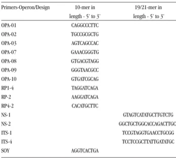
RAPD profiles. The RAPD reactions were conducted in a 30µl volume containing buffer 1X (Promega), 0.2mM each of dATP, dGTP, dCTP and dTTP (Promega), 50ng of genomic-DNA, 2mM of MgCl2 (Promega), 160nM of primer (Operon) and 1 unit of Taq thermostable DNA polymerase (Promega). The amplification parameters consisted of 35 cycles of denaturation at 95°C for 45 sec, primer annealing at 36°C for 2 min, and extension at 72°C for 2 min. In the first cycle, the denaturing step was 5 min and in the final cycle the final extension step was 7 min. The reactions were performed using decamer primers of the OPERON kit (OPA 01, 02, 03, 07, 08, 09 and 10), and the arbitrary primers SOY, RP1-4, RP-2, RP4-28. Amplification using the ribosomal primers NS1, NS2, ITS1 and ITS-424 was performed increasing the melting temperature to 45°C. The primer sequences are shown in Table 2.
Table 2 - Primers included in this study.
Primers-Operon/Design 10-mer in 19/21-mer in length - 5’ to 3’ length - 5’ to 3’ OPA-01 CAGGCCCTTC OPA-02 TGCCGCGCTG OPA-03 AGTCAGCCAC OPA-07 GAAACGGGTG OPA-08 GTGACGTAGG OPA-09 GGGTAACGCC OPA-10 GTGATCGCAG RP1-4 TAGGATCAGA RP-2 AAGGATCAGA RP4-2 CACATGCTTC NS-1 GTAGTCATATGCTTGTCTG NS-2 GGCTGCTGGCACCAGACTTGC ITS-1 TCCGTAGGTGAACCTGCGG ITS-4 TCCTCCGCTTATTGATATGC SOY AGGTCACTGA
Fingerprints were produced by electrophoresis of 10µl aliquots of reaction in 1.5% agarose gels run in TBE (0.45M Tris borate, 0.001M EDTA) buffer at 120V for 90 minutes. The gels were stained with 1µg.ml-¹ ethidium bromide, photographed under UV light using a Polaroid camera (Model DS-34) with a black and white film (Type 667, Polaroid Corp). For each experiment, the base pair sizes were measured from size markers included in every gel (DNA lambda/Hind III or 100-bp ladder, Gibco-BRL).
RESULTS
amplified sample. The RAPD profiles using the OPA kit, R P 1 - 4, RP-2, RP4-2 and SOY primers were able to characterize interspecific polymorphisms among the Candida
species. The OPA 02 and NS2 primers gave representative profiles for the species clusters (Figure 1).
1 2 3 4 5 6 7 8 9 10 11 12 13 14 15
23Kb
2,2-
1,2-
1,0Kb-Figure 1 - Genetic variation within Candida albicans, Candida glabrata and Candida parapsilosis determined by RAPD analysis using primer NS2; lane 14, positive control; lane 15, molecular weight lambda EcoRI/ Hind III (lanes are according to Table1).
Among the C. albicans isolates no polymorphic traits were detected, even though the isolates were obtained from different yeast reservoirs (catheter, surgical wounds and urine). Similar RAPD profiles of C. albicans and C. parapsilosis strains were generated using the primers ITS-1 and ITS-4. However, this set of primers generated a different RAPD profile when C. glabrata DNA was used as a template (Figure 2). For all the primers tested, the RAPD profile of C. albicans type strain was differentiated from other strains of this same species by some polymorphic amplicons (Figure 1).
1 2 3 4 5 6 7 8 9 10 11 12 NC CA MW
23Kb
1Kb 0,9 0,8 0,7 0,5 0,4
Figure 2 - Genetic variation within Candida albicans, Candida glabrata and Candida parapsilosis determined by RAPD analysis using primer OPA2; NC, negative control; CA, Candida albicans reference strain; MW, molecular weight: ladder 100bp (lanes are according to Table 1).
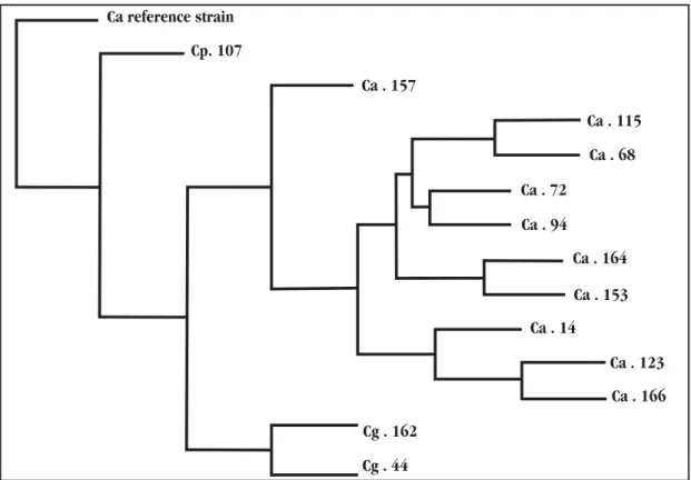
The phenogram grouped the strains of Candida albicans,
Candida parapsilosis and C. glabrata by the similarity in the genotypic characteristics obtained from the RAPD reactions5. The type strain of C. tropicalis was positioned on the phenogram separately, as shown Figure 3, where the clustering of most of the C. albicans strains was relatively differentiated.
The two isolates 44 and 162, belonging to the species
C. glabrata, were originally misidentified as C. albicans. These results suggest that their original presumptive identification by germ tube and chlamydoconidia formation was unreliable. Therefore, the new identities were correctly confirmed by additional biochemical and physiological tests realized in this work, using previously established techniques7 20.
Figure 3 - Phenogram of isolates based on RAPD profiles. The consensual phenogram was constructed using the Wagner parsimony option within the computer program Phylip v.3,572c. Branch lengths and scale bar correspond to the distance values among these Candida albicans (Ca), Candida parapsilosis (Cp), and Candida glabrata (Cg) strains.
Ca reference strain
Cp. 107
Ca . 157
Ca . 115
Ca . 68
Ca . 72
Ca . 94
Ca . 164
Ca . 153
Ca . 14
Ca . 123
Ca . 166
Cg . 162
DISCUSSION
Candida spp has recently emerged as an important cause of nosocomial infection through cross-transmission, particularly in intensive care units14. Fungemia caused by this yeast are reported to be related to previous digestive tract colonization, mucosal surfaces of mammalian bodies, more specifically in human mucosa, or exogenous contamination16 21. These emerging threats of fungal infection require standardized methods for strain characterization in order to identify hospital clusters15. RAPD assays may be an important tool for Candida species identification, increasing the capability of previously established traditional methods. In this work the applicability of this technique was demonstrated, using the primers OPA2 and NS2 to differentiate between C. albicans, C. glabrata and C. parapsilosis.
The RAPD profiles generated showed few differences between strains of the same species. This result demonstrates that it is difficult to identify primers able to detect intraspecific polymorphism in these clonal species26.
The misidentification of strains 44 and 162 demonstrated that the patterns provided with biochemical, physiological and micromorphological tests, widely used for strain identification, can sometimes be unclear and obscure differences in the biochemical tests realized, thus masking the incorrect identification of yeasts from nosocomial candidemia9.
The clinical value of a single procedure, such as RAPD analysis, for determining both species and biotype is most clear in the area of molecular epidemiology18. According to some authors, given the increase in nosocomial infections caused by
Candida species, there is an urgent need for a rapid and simple procedure which would allow for the analysis of both the outbreaks and the incidence of person-to-person transmission associated with these organisms2 13 16. As such, more profound posterior epidemiological analyses are required to more accurately clarify whether there are simply genetic similarities in a characteristic cloned population among these isolates or whether hospital procedures helped disseminate the agents among these patients through cross-infections.
ACKNOWLEDGMENTS
Henrique Maia Valério was a recipient of a Brazilian Government fellowship from the CNPq (Conselho Nacional de Desenvolvimento Científico e Tecnológico).
REFERENCES
1. Bautista-Muñoz C, Boldo XM, Villa-Tanaca L, Hernández-Rodríguez C. Identification of Candida spp by randomly amplified polymorphic DNA analysis and differentiation between Candida albicans and Candida dubliniensis by direct PCR methods. Journal of Clinical Microbiology 41: 414-420, 2003. 2. Boldo XM, Villa-Tanaca L, Zuniga G, Hernandez-Rodriguez C. Genetic
diversity among clinical isolates of Candida glabrata analyzed by randomly amplified polymorphic DNA and multilocus enzyme electrophoresis analyses. Journal of Clinical Microbiology 41: 4799-4804, 2003.
3. Cresti S, Posteraro B, Sanguinetti, Guglielmetti P, Rossolini GM, Morace G, Fadda G. Molecular typing of Candida spp by random amplification of polymorphic DNA and analysis of restriction fragment length polymorphism of ribosomal DNA repeats. New Microbiology 22: 41-52, 1999. 4. Del Castilho L, Bikandi J, Almuneda N, Quindós G, Sentandreu R, Pontón
J. Comparision of morphotypic and genotypic methods for strain delineation in Candida. Mycoses 40: 445-450, 1997.
5. Felsenstein J, Phylip (Phylogeny Inference Package) Department of Genetics, University of Washington, Seattle, WA, v. 3.572c, 1993. 6. Hazen KC, Wu JG, Masuoka J. Comparison of the Hydrophobic Properties
of Candida albicans and Candida dubliniensis. Infection Immunology 69: 779-786, 2001.
7. Kurtzman CP, Fell JW. The yeasts, a taxonomic study. 4th edition. Elsevier,
Amsterdam, 1998.
8. Lehman, PF, Lin D, Lasker BA. Genotypic identification and characterization of species and strains within the genus Candida by using random amplified polymorphic DNA. Journal of Clinical Microbiology 30: 3249-3254, 1992.
9. Marot-Leblond A, Grimaud L, David S, Sullivan DJ, Coleman DC, Ponton J, Robert R. Evaluation of a rapid immunochromatographic assay for identification of Candida albicans and Candida dubliniensis. Journal of Clinical Microbiology 42: 4956-4960, 2004.
10. Marot-Leblond A, Grimaud L, Nail S, Bouterige S, Apaire-Marchais V, Sullivan DJ, Robert R. New Monoclonal Antibody Specific for Candida albicans Germ Tube. Journal of Clinical Microbiology 38: 61-67, 2000. 11. Peman J, Aparisi N, Garcia-Esteban C, Gobernado M. Rapid identification
of Candida glabrata using a new comercial kit. Revista Iberoamericana de Micologia 21: 82-84, 2004.
12. Pinto PM, Resende MA, Koga-Ito CY, Ferreira JA, Tendler M. rDNA-RFLP identification of Candida species in immunocompromised and seriously diseased patients. Canadian Journal of Microbiology 50: 514-520, 2004. 13. Pinto PM, Resende MA, Koga-Ito CY, Ferreira JA, Tendler M. Genetic variability analysis among clinical Candida spp. isolates using random amplified polymorphic DNA. Memórias do Instituto Oswaldo Cruz 99: 147-152, 2004.
14. Reef SE, Lasker BA, Butcher DS, McNeil MM, Pruitt R, Keyserling H, Jarvis WR. Nonperinatal nosocomial transmission of Candida albicans in a Neonatal Intensive Care Unit: Prospective Study. Journal of Clinical Microbiology 36:1255-1259, 1998.
15. Reiss E, Tanaka K, Bruker G, Chazalet V, Coleman D, Debeaupuis JP, Hanazawa R, Latge JP, Lortholary J, Makimura K, Morrison CJ, Murayama SY, Naoe S, Paris S, Sarfati J, Shibuya K, Sullivan D, Uchida K, Yamaguchi H. Molecular diagnosis and epidemiology of fungal infections. Medical Mycology 36: 249-257, 1998.
16. Resende JC, Resende MA, Saliba JL. Prevalence of Candida spp in hospitalized patients and their risk factors. Mycoses 45:306-312, 2002. 17. Riederer KM, Ramanathan J, Barczak J, Baran Jr J, Khatib R. Utility of a
pre-optimized kit for random amplified polymorphic DNA in typing Candida albicans. Canadian Journal of Microbiology 48: 369-373, 2002. 18. Tamura M, Watanabe K, Mikami Y, Yazawa K, Nishimura K. Molecular characterization of new clinical isolates of Candida albicans and C. dubliniensis in Japan: Analysis reveals a new genotype of C. albicans with group I Intron. Journal of Clinical Microbiology 39: 4309-4315, 2001.
19. Tamura NK, Gasparetto A, Svidzinski TI. Evaluation of the adherence of Candida species to urinary catheters. Mycopathologia 156: 269-272, 2003. 20. Van Der Walt JP, Yarrow D. Methods for the isolation, maintenance, classification and identification of yeasts. In: Kurtzman CP, Fell JW (eds) The Yeasts. Elsevier Publishers, Amsterdam, p. 45-104, 1984. 21. Vrioni G, Matsiota-Bernard P. Molecular typing of Candida isolates from
patients hospitalized in an intensive care unit. Journal of Infection 42: 50-56, 2001.
22. Wahyuningsih R, Freisleben HJ, Sonntag HG, Schnitzler P. Simple and rapid detection of Candida albicans DNA in serum by PCR for diagnosis of invasive candidiasis. Journal of Clinical Microbiology 38:3016-3021, 2000.
23. Welsh J, McCleland M. Genomic Fingerprinting Using Arbitrarily Primed PCR and A Matrix of Pairwise Combinations of Primers. Nucleic Acids Research 19: 5275-5279, 1991.
24. White TJ, Bruns T, Lee S, Taylor J. Amplification and direct sequencing of fungal ribosomal RNA genes for phylogenetics. In: Innis MA, Gelfand DH, Sninsky JJ, White TJ (eds) PCR protocols. Academic Press, San Diego, CA: p. 315-322, 1990.
25. Williams JGK, Kubelik AR, Livak KJ, Rafalski JA. DNA Polymorfisms Amplified by Arbitrary Primers are Useful as Genetic Markers.Nucleic Acids Research 18: 6531-6535, 1990.