Schistosomiasis Mansoni in Low Transmission Areas.
Abdominal Ultrasound
R Ruiz, P Candia*, M Garassini*, C Tombazzi*, G Certad, AC Bruces, O Noya,
B Alarcón de Noya/
+Sección de Biohelmintiasis, Instituto de Medicina Tropical and Cátedra de Parasitologia *Cátedra de Gastroenterologia, Escuela de Medicina “Luis Razetti”, Facultad de Medicina, Universidad Central de Venezuela, Apartado Postal 47706, Los Chaguaramos,
Caracas 1041-A, Venezuela
In endemic areas with low prevalence and low intensity of infection, the diagnosis of hepatic pathology due to the Schistosoma mansoni infection is very difficult. In order to establish the hepatic morbidity, a double-blind study was achieved in Venezuelan endemic areas, with one group of patients with schistosomiasis and the other one of non-infected people, that were evaluated clinically and by abdominal ultrasound using the Cairo classification. Schistosomiasis diagnosis was established based on parasitologic and serological tests. The increase of the hepatic size at midclavicular and midsternal lines (in hepatometry) and the hard liver consistency were the clinical param-eters able to differentiate infected persons from non infected ones, as well as the presence of left lobe hepatomegaly detected by abdominal ultrasound. The periportal thickening, especially the mild form, was frequent in all age groups in both infected and uninfected patients. There was not correlation between the intensity of infection and ultrasound under the current circumstances. Our data suggest that in Venezuela, a low endemic area of transmission of schistosomiasis, the hepatic morbidity is mild and uncommon. The Cairo classification seems to overestimate the prevalence of periportal pathology. The specificity of the method must be improved, especially for the recognition of precocious pathology. Other causes of hepatopathies must be investigated.
Key word: ultrasound - schistosomiasis - low transmission - Venezuela
The diagnosis of schistosomiasis in Venezuela is very difficult due to the low prevalence, low intensity of the infection and to the unspecificity of clinical signs usually associated with this disease (Ruiz et al. 1999). For this reason, the Venezuelan Schistosomiasis Research Group has recommended the employment of immunological tech-niques for the diagnosis of schistosomiasis (Alarcón de Noya et al. 1992).
Moreover, the prevalence of hepatic morbidity in schis-tosomiasis is difficult to determine because some of the methods that could be used for diagnosis such as the biopsy, or those that evaluate the hemodynamic alterations of the liver, are dangerous and invasive procedures that can not be performed on patients under field conditions. Periportal fibrosis is one of the most characteristic al-teration in the liver of infected patients with schistoso-miasis (Prata 1987) and it is considered, the most frequent cause of hepatic fibrosis worldwide (Warren 1984). Ab-dominal ultrasound has shown to be an alternative method for diagnosis, when the liver biopsy is contraindicated or
impracticable (El-Rooby 1985). This method is relatively inexpensive, rapid, portable, causes no biological hazards to the patients and its sensitivity and specificity in the recognition of periportal fibrosis, is comparable with the one reported for hepatic biopsy (Abdel-Wahab et al. 1989, Cerri et al. 1984, Homeida et al. 1988), percutaneous transhepatic portography, angiography (Hatz et al. 1992a), and clinical examination (Kardorff et al. 1997). It reflects the dynamic changes produced by portal hypertension, since it measures the portal vein diameter and the pres-ence of systemic collateral blood vessels that are corre-lated with esophageal varices (Abdel-Latif et al. 1981, Davidson et al. 1991, Abdel-Wahab et al. 1993, Richter et al. 1998). However, this tool requires well-trained physi-cians, and its standardization is still a matter of debate after two WHO workshops (Cairo Working Group 1992, Niamey Working Group 2000).
Some studies have reported that ultrasound could improve the accuracy of clinical examination in endemic areas of schistosomiasis (Lambertucci et al. 2000). More-over, it has been shown that ultrasound could be an ex-cellent indicator for diagnosis, and very useful for plan-ning and monitoring control programs in areas of differ-ent endemicity (Hatz et al. 1990, 1992b, Lambertucci et al. 2000).
Almost all investigations on abdominal ultrasound have been done in hospitalized patients or in endemic areas with high schistosomiasis prevalence and intensity of infection. The aim of this work was to evaluate the use of abdominal ultrasound using the Cairo classification for the schistosomiasis hepatic fibrosis diagnosis in Venezu-ela, a low transmission endemic area with low prevalence and morbidity.
This work was financed by the “Programa de Control de Enfermedades Endémicas” of Malariología-World Bank (PCEE/ PNDU) and partially by the “Consejo de Desarrollo Científico y Humanístico” de la Universidad Central de Venezuela for facilitating travel expenses for congress communications. +Corresponding author. Fax:+ 58-212-6053563. E-mail:
MATERIALS AND METHODS
Study area and population - This transverse and double blind study was carried out between 1998 and 2001 in three villages situated in the Venezuelan endemic area for schistosomiasis: Caraballeda, La Curía and Belén. Par-ticipants were considered cases of schistosomiasis ac-cording to a recent proposal of our group (Ruiz et al. 1999) and described below. Simultaneously, we selected a group of non infected persons from endemic areas to match them according to sex and age with the infected ones. These persons did not have antecedents of schistosomiasis and all the laboratorial tests for this disease were negatives.
Stool evaluation and serologic tests - Stool samples were collected and examined for the presence of S. mansoni eggs by the Kato Katz technique (Katz et al. 1972). For the serological diagnosis of schistosomiasis different immu-nological tests were performed: Enzyme Linked Immuno Absorbent Assay with Sodium Metaperiodate (SMP-ELISA) (Alarcón de Noya et al. 2000), Circumoval Precip-itin Test (COPT) (Spencer et al. 1991) and Alkaline Phos-phatase Immunoassay (APIA) (Pujol & Cesari 1990).
Clinical evaluation - This evaluation included a medi-cal history, epidemiologimedi-cal data, current symptoms and a physical examination. Informed consent was obtained from each patient or representing in the case of children, and only volunteers were admitted in this study. Experienced observers carried out an abdominal ultrasound employ-ing a portable Toshiba equipment with curved 3.75 MHz transducer. The echographers make first diagnostic im-pression and cataloged as normal, periportal fibrosis, hepatomegaly and hepatic steatosis according the obser-vations during the exam. Thereafter, the ecographical evaluation was made following the standardized Cairo clas-sification (Cairo Working Group 1992), and measures were done for final ultrasound diagnosis. All schistosomiasis patients were treated with praziquantel, 40 mg/kg in single oral dose.
Definitions - We consider “cases” of schistosomiasis (Ruiz et al. 1999) those people with one of the following criteria: (1) eggs of S. mansoni in stools; these patients have positive COPT, SMP-ELISA, and APIA (Criterion I); (2) persons without S. mansoni eggs in stools but with positive COPT, who have not received previous anti-S.
mansoni chemotherapy in the last 12 months (Criterion II); (3) persons without S. mansoni eggs in stools, with nega-tive COPT, but with both SMP-ELISA and APIA (immu-noassay tests) positive simultaneously and without previ-ous chemotherapy against schistosomiasis (Criterion III). Hepatomegaly was considered when the liver sur-passed the costal margin in persons older than 5 years-old. The right lobe was measured at the anterior axilar line and the left lobe at the line passing by the xyphoid appen-dix. In those persons with palpable liver below the costal margin, it was determined: liver consistence (soft, firm or hard), hepatic surface (smooth or nodular), left lobe promi-nence (when it was proportionally larger than the right lobe) (Prata & Bina 1968). Another hepatomegaly criteria was hepatometry with anterior axilar line (AAL) > 9 cm, midclavicular line (MCL) > 12 cm and midsternal line (MSL) > 9 cm. Splenomegaly was diagnosed when spleen surpassed the costal margin (Prata 1970).
The following findings were considered as indicators of periportal fibrosis: (1) hepatic left lobe in longitudinal section larger than 70 mm; (2) portal vein diameter supe-rior to 12 mm; (3) mean diameter of three peripheral portal vein branches superior to 3 mm. Periportal thickening in turn, was classified in Grade I: 3-5 mm (mild), Grade II: > 5-7 mm (moderate) and Grade III: > 5-7mm (severe); (4) mesen-teric vein diameter above 11 mm; (5) splenic vein superior to 12 mm; (6) longitudinal diameter of spleen superior to 120 mm; (7) gallbladder wall superior to 5 mm; (8) pres-ence of collateral vessels or ascitis.
Statistical analysis - The Chi-square test was used to evaluate differences between proportions (p < 0.05).
RESULTS
In total, 175 S. mansoni infected patients and 87 non-infected were evaluated. Out of the 175 cases of schisto-somiasis, 96 (54.9%) persons were diagnosed by stool examination and 79 (45.1%) individuals by serology. The median of eliminated S. mansoni eggs was 122 per/g of feces (range: 24-1928). According to the elimination of eggs, 65 (67.7%) of them had mild infection, 28 (29.2%) moderate, and 3 (3.1%) severe.
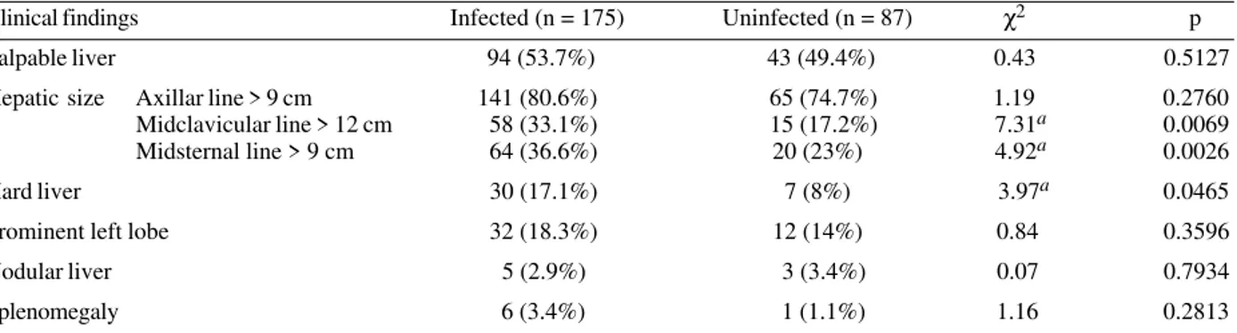
Table I shows clinical findings in infected and non-infected patients. By hepatic percussion the midclavicular and midsternal line values (> 12 cm and > 9 cm
respec-TABLE I
Clinical findings in infected and uninfected persons in the Venezuelan schistosomiasis area, 1998-2001
Clinical findings Infected (n = 175) Uninfected (n = 87) χ2 p
Palpable liver 94 (53.7%) 43 (49.4%) 0.43 0.5127
Hepatic size Axillar line > 9 cm 141 (80.6%) 65 (74.7%) 1.19 0.2760 Midclavicular line > 12 cm 58 (33.1%) 15 (17.2%) 7.31a 0.0069
Midsternal line > 9 cm 64 (36.6%) 20 (23%) 4.92a 0.0026
Hard liver 30 (17.1%) 7 (8%) 3.97a 0.0465
Prominent left lobe 32 (18.3%) 12 (14%) 0.84 0.3596
Nodular liver 5 (2.9%) 3 (3.4%) 0.07 0.7934
Splenomegaly 6 (3.4%) 1 (1.1%) 1.16 0.2813
tively) as well as hard liver, were significantly associated with schistosomiasis. There were not statistically signifi-cant differences between the rest of the variables.
The association between ultrasound findings and in-fection is shown in Table II. It was found that left lobe was hypertrophied in 160 of infected patients (91.4%) and in 72 of uninfected (82.8%), with a statistical difference between both groups. Periportal thickening was found in 159 infected persons (90.9%) and in 73 (83.9%) non-in-fected, this difference was important but it was not statis-tically significant. Other features such as splenomegaly, portal and mesenteric vein dilatation, are also shown in Table II.
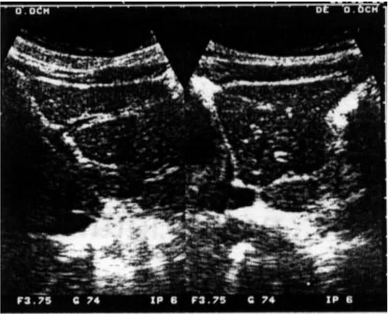
When comparisons were made according to ultrasono-graphical findings and age, it was found that the frequency of left lobe hepatomegaly in infected and non-infected people was similar in all age groups (data not shown). This frequency was 100% among infected persons older than 50 years. There were not statistically significant dif-ferences among these groups (data not shown). The pres-ence of periportal thickening among the infected and non-infected persons was similar in all ranges of age, also, without statistically significant differences. Typical ultra-sound findings, left lobe hepatomegaly, splenomegaly, portal vein dilatation, periportal thickening, in Venezu-elan schistosomiasis patients are shown in Fig. 1. The
TABLE II
Ultrasound findings in schistosomiasis patients and uninfected persons, Venezuela, 1998-2001
Ultrasound findings Infected (n = 175) Uninfected (n = 87) χ2 P
Left lobe > 70 mm 160 (91.4%) 72 (82.8%) 4.31 a 0.0379
Portal vein > 12 mm 6 (3.4%) 1 (1.1%) 1.17 0.2785
Mesenteric vein > 11 mm 9 (5.1%) 1 (1.1%) 2.52 0.1121
Spleen > 120 mm 8 (4.6%) 1 (1.1%) 2.05 0.1521
Periportal thickness (PT) 159 (90.9%) 73 (83.9%) 2.77 0.0962
PT 3-5 mm 128 64 – –
PT > 5-7 mm 30 8 – –
PT > 7 mm 1 1 – –
a: statistically significant
Fig. 2 demonstrates hepatic lesions suggestive of peri-portal thickening in a non-infected person.
A summary of the results obtained by ultrasound ac-cording to the criteria used for diagnosis of schistoso-miasis is presented in Table III. In both groups of pa-tients, the left lobe hepatomegaly was a common finding, 90.6% and 92.4% from those diagnosed by coprology or serology respectively (persons with Criteria II or III). Also, the periportal thickening was found in 90 (93.8%) persons with fecal S. mansoni eggs and in 69 (87.3%) diagnosed only by serology. The rest of the evaluated parameters were less frequent in both groups. In any case, there was not statistical significant difference when ultrasound find-ings were compared according to the criteria used for the diagnosis of schistosomiasis.
The association between the intensity of infection and ultrasound findings is shown in Table IV. The Chi-Square test did not detect statistical differences between these variables.
Some relevant antecedents and first ultrasonographic reports in non-infected persons are shown in people with periportal thickening and left lobe hepatomegaly demon-strable after measures (Table V).
DISCUSSION
Usually, hepatomegaly and intensity of infection are employed as the classic morbidity markers in schistoso-miasis (Arap-Siongok et al. 1976, Barreto & Loureiro 1984, Gryseels 1992). In our study, hepatomegaly below the costal margin was detected in similar percentages in
in-TABLE III
Ultrasound findings according to schistosomiasis diagnosis criteria, Venezuela, 1998-2001
Ultrasound findings Coprology (n = 96) Serology (n = 79) χ2 p
Left lobe > 70 mm 87 (90.6%) 73 (92.4%) < 0.1 0.08810
Portal vein > 12 mm 3 (3.1%) 3 (3.8%) < 0.1 0.8078
Mesenteric vein > 11 mm 5 (5.2%) 4 (5%) 0.0 0.9655
Spleen > 120 mm 5 (5.2%) 3 (3.8%) 0.2 0.6566
Periportal thickness (PT) 90 (93.8%) 69 (87.3%) 2.1 0.1433
PT 3-5 mm 71 57 – –
PT > 5-7 mm 18 12 – –
PT > 7 mm 1 0 – –
fected and uninfected people. However, it is complicated to compare our results with those obtained in other epi-demiological settings, because the diagnosis of clinical hepatomegaly has not been standardized (Cook et al. 1974, Lehman et al. 1976, Kardorff et al. 1997). Moreover, it is possible that not all cases of clinical hepatomegaly found in this work could be explained by schistosomiasis. For that reason, other causes of liver disease should be in-vestigated.
In contrast, the increase of the liver size at midclavicular and midsternal line and liver consistency was statistically superior in schistosomiasis patients when compared to uninfected persons. The values that reflect left lobe hepatomegaly are relevant because they could be used as markers for schistosomiasis, especially in some endemic areas in Venezuela where the recognition of this disease is difficult to asses due to the low intensity of infection (Ruiz et al. 1999). Nevertheless, in the present work, children were evaluated with the same hepatometry parameters used for adults and this could cause loss of invaluable information in the pediatric age, for this rea-son, it is necessary to establish the normal reference val-ues of hepatometry for children.
This classification does not take into consideration the age or height of the patients (Burchard et al. 1998). It is possible that adjusting a body-height dependent refer-ence value, as proposed by the Niamey Working Group (2000), the ultrasound specificity could be improved.
Nodular liver, left lobe prominence, and splenomegaly had low frequency in Venezuelan patients in comparison
TABLE IV
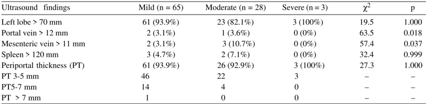
Ultrasound findings according to egg output, Venezuela, 1998-2001
Ultrasound findings Mild (n = 65) Moderate (n = 28) Severe (n = 3) χ2 p
Left lobe > 70 mm 61 (93.9%) 23 (82.1%) 3 (100%) 19.5 1.000
Portal vein > 12 mm 2 (3.1%) 1 (3.6%) 0 (0%) 63.5 0.018
Mesenteric vein > 11 mm 2 (3.1%) 3 (10.7%) 0 (0%) 57.4 0.037
Spleen > 120 mm 3 (4.7%) 2 (7.1%) 0 (0%) 32.4 0.999
Periportal thickness (PT) 61 (93.9%) 26 (92.9%) 3 (100%) 27.3 1.000
PT 3-5 mm 46 22 3 – –
PT5-7 mm 14 4 0 – –
PT > 7 mm 1 0 0 – –
TABLE V
Relevant antecedents and first ultrasonographical findings in non-infected people with definitive periportal thickening and left lobe hepatomegaly at ultrasound, Venezuela, 1998-2001
Final ultrasound diagnosis
Periportal thickening (n = 74) Left lobe hepatomegaly (n = 72)
First ultrasonographic report Normal 57 (77%) 52 (72%)
Periportal fibrosis 7 (9.5%) 7 (9.7%)
Hepatomegaly 7 (9.5%) 8 (11.1%)
Hepatic steatosis 4 (5.4%) 5 (6.9%)
Antecedents Hepatitis 6 (8.1%) 8 (11.1%)
Alcoholic intake 28 (37.8%) 27 (37.5%)
Included in the 72 patients with left hepatomegaly there are 60 persons who have periportal thickening simultaneously.
to the same signs reported for Brazilian patients with schis-tosomiasis (Prata & Bina 1968, Prata 1987). In spite of the small number of infected persons with splenomegaly, our results are similar to those reported by Dietze (1983) in a Brazilian area with high endemicity.
In addition, it was found that using the Cairo Working Group (1992) classification was possible to identify in-fected from non-inin-fected patients based only on the pres-ence of left lobe hepatomegaly, without differpres-ences ac-cording to the age. If hepatomegaly is used as an indica-tor of hepatic morbidity, fibrosis prevalence could be over-estimated in adults with left lobe > 70 mm, and underesti-mated in children with left lobe < 70 mm, since variables like height, weight or corporal surface that could influ-ence liver size, are not considered.
locali-ties (Doehring-Schwedtfeger et al. 1989, Abdel-Wahab et al. 1990) preventing the evolution toward hepatosplenic schistosomiasis (Bina & Prata 1981, 1983).
On the other hand, advanced S. mansoni disease shows less problems for the diagnosis by the Cairo clas-sification (Hatz et al. 1992b). However, the low frequency of portal hypertension signs found by ultrasound, sug-gests that in Venezuela the hepatic morbidity is uncom-mon.
Due to the presence of ultrasonographical abnormali-ties and clinical findings in some uninfected persons, we suggest that some other factors must be influencing the hepatic architecture in these groups. Some of the infor-mation that must be obtained in the clinical history in order to make a differential diagnosis should include: pre-vious schistosomiasis, alcohol consumption, exposition to pesticides, viral hepatitis, other parasitic diseases with hepatic compromise (visceral larva migrans, visceral leish-maniasis).
It was detected that ultrasound parameters did not correlate with laboratory diagnosis, the criteria either para-sitologic or serologic in the case of S. mansoni infection. This is consistent with previous studies that showed that in Venezuelan endemic areas, the serologic assays are of greater value for the schistosomiasis diagnosis (Alarcón de Noya et al. 1992).
It was also found no differences in ultrasound find-ings according to the intensity of infection, but it is im-portant to consider that the universe of individuals with heavy infections was low, and this could influence the obtained results.
As it has been proposed previously for other investi-gations, the validity of the ultrasound for the evaluation and for monitoring the morbidity of the control programs depends on the predictive potential of the indicators of pathology in a given population (Hatz et al. 1992b). For that reason, the standardization of ultrasound must be carried out, especially when ecosonographic is used in areas where the morbidity is difficult to asses and where control measures have been implemented. Only with a correct diagnosis it is possible to evaluate the impact of those measures.
In summary, we found that the diagnosis of hepatic alterations by ultrasound according to the parameters de-scribed above, looks more specific than the quantitative Cairo classification. However, these results must be inter-preted with caution because in this work we have not stan-dardized the qualitative presence or absence of fibrosis.
ACKNOWLEDGMENTS
To the population of Caraballeda, La Curía and Belén by their invaluable cooperation. To Dr Aluizio Prata for the revi-sion of the manuscript. To Lic. Cecilia Colmenares and Lic. Sandra Losada for their collaborative support with the manu-script.
REFERENCES
Abdel Latif Z, Abdel-Wahab F, El-Kady NM 1981. Evaluation of portal hypertension in cases of hepatosplenic schistoso-miasis using ultrasound. J Clin Ultrasound 9: 409-412. Abdel-Wahab MF, Esmat G, Farrag A, El-Boraey Y, Strickland
GT 1993. Ultrasonographic prediction of esophageal
va-rices in schistosomiasis mansoni. Am J Trop Med Hyg 88: 560-563.
Abdel-Wahab MF, Esmat G, Milad M, Abdel-Razek S, Strickland GT 1989. Characteristic sonographic pattern of schistosomal hepatic fibrosis. Am J Trop Med Hyg 40: 72-76.
Abdel-Wahab MF, Esmat G, Narooz SI, Yosery A, Struewing JP, Strickland T 1990. Sonographics studies of schoolchil-dren in a village endemic for Schistosoma mansoni. Trans R Soc Trop Med Hyg 84: 69-73.
Alarcón de Noya B, Colmenares C, Lanz, H, Caracciolo, MA, Losada, S, Noya O 2000. Schistosoma mansoni: immu-nodiagnosis is improved by sodium metaperiodate which reduces cross-reactivity due to glycosylated epitopes of soluble egg antigen. Exp Parasitol 95:106-112.
Alarcón de Noya B, Noya O, Balzán C, Cesari IM 1992. New approaches for the control and eradication of schistosomia-sis in Venezuela. Mem Inst Oswaldo Cruz 87: 227-231. Arap-Siongok TK, Mahmoud AAF, Ouma JH, Warren KS,
Muller AS, Handa AK, Houser, HB 1976. Morbidity in schistosomiasis mansoni in relation to intensity of infec-tion: study of a community in Machakos, Kenia. Am J Trop Med Hyg 25: 273-284.
Barreto ML, Loureiro S 1984. The effect of Schistosoma mansoni infection on child morbidity in the State of Bahia, Brazil. I. Analysis at the ecological level. Rev Inst Med Trop São Paulo 26: 230-235.
Bina JC, Prata A 1981. A possibilidade de prevenção das formas graves da esquistossomose mansoni: papel da terapêutica específica. In Situação e Perspectiva do Controle das Doenças Infecciosas e Parasitárias. Um Seminário na Universidade de Brasília, Editora Universidade Nacional de Brasília, Brasília, p. 45-56.
Bina JC, Prata A 1983. Regressão da hepatosplenomegalia pelo tratamento específico da esquistossomose. Rev Soc Bras Med Trop 16: 213-218.
Boisier P, Ramarokoto CE, Ravaoalimalala VE, Rabarijaona L, Serieye J, Roux J, Esterre P 1998. Reversibility of Schisto-soma mansoni-associated morbidity after yearly mass praziquantel therapy: ultrasonographic assessment. Trans R Soc Trop Med Hyg 92: 451-453.
Boisier P, Serieye J, Ravaolimalala VE, Roux J, Esterre P 1995. Ultrasonographical assessment of morbidity in schistoso-miasis mansoni in Madagascar: a community-based study in a rural population. Trans R Soc Trop Med Hyg 89: 208-212.
Burchard GD, Guissé-Sow F, Diop M, Ly A, Lanuit R, Gryseels B, Gressner AM 1998. Schistosoma mansoni infection in a recently exposed community in Senegal: lack of correlation between liver morphology in ultrasound and connective tis-sue metabolites in serum. Trop Med Intern Health 3: 234-241.
Cairo Working Group 1992. The use of diagnostic ultrasound in schistosomiasis – Attempts at standardization of method-ology. Acta Trop 51: 45-63.
Cerri GG, Alves VAF, Magalhães A 1984. Hepatosplenic schis-tosomiasis mansoni: ultrasound manifestations. Radiology 153: 777-780.
Cook JA, Baker ST, Warren KS, Jordan PA 1974. A controlled study of morbidity of schistosomiasis mansoni in St. Lucian children based on quantitative egg excretion. Am J Trop Med Hyg 23: 625-633.
Davidson RN, Houston S, Kiire F 1991. Schistosomal peripor-tal fibrosis in Zimbabwe: use of ultrasound in patients with esophageal varices. Trans R Soc Trop Med Hyg 85: 380-382.
Medidas Integradas de Controle em uma Área Hi-perendêmica, MSc Thesis, Universidade Nacional de Brasília, p. 139
Doehring-Schwedtfeger E, Ali QM, Abdel-Rahim IM, Kardorff R, Franke D, Kaiser C, Elsheikh M, Ehrich JH 1989. Sonomorphological abnormalities in Sudanese children with Schistosoma mansoni infection: a proposed staging-sys-tem for field diagnosis of periportal fibrosis. Am J Trop Med Hyg 41: 63-69.
El-Rooby A 1985. Management of hepatic schistosomiasis. Semin Liver Dis 5: 263-276.
Gryseels B 1992. Morbidity due to infection with Schistosoma mansoni: an update. Trop Geogr Med 44: 189-200. Hatz C, Jenkins JM, Ali QM, Abdel-Wahab MF, Cerri GG,
Tanner M 1992a. A review of the literature on the use of ultrasonography in schistosomiasis with special reference to its use in fields studies. 2. Schistosoma mansoni. Acta Trop 51: 15-28.
Hatz C, Jenkins JM, Morrow RH, Tanner M 1992b. Ultra-sound in schistosomiasis – A critical look at methodologi-cal issues and potential applications. Acta Trop 51: 89-97. Hatz C, Savioli L, Mayobana C, Dhunputh J, Kisumku UM, Tanner M 1990. Measurement of schistosomiasis-related morbidity at community level in areas of different endemic-ity. Bull WHO 68: 777-787.
Homeida M, Abdel-Gadir AF, Cheever AW, Bennett JL, Arbab BMO, Ibrahium SZ, Abdel-Salam IM, Dafalla AA, Nash TE 1988. Diagnosis of pathologically confirmed Symmers’ periportal fibrosis by ultrasonography: a prospective blinded study. Am J Trop Med Hyg 38: 86-91.
Katz N, Chaves A, Pellegrino J 1972. A simple device for quan-titative stool thick-smear technique in schistosomiasis mansoni. Rev Inst Med Trop São Paulo 14: 397-400. Kardorff R, Gabone RM, Mugashe C, Obiga D, Ramarokoto
CE, Mahlert C, Spannbrucker N, Lang A, Günzler V, Gryseels B, Ehrich JHH, Doehring E 1997. Schistosoma mansoni-related morbidity on Ukerewe Island, Tanzania: clinical, ultrasonographical and biochemical parameters. Trop Med Intern Health 2: 230-239.
Lambertucci JR, Serufo JC, Gerspacher-Lara R, Rayes AAM, Teixeira R, Nobre V, Antunes CMF 2000. Schistosoma mansoni: assessment of morbidity before and after control. Acta Trop 77: 101-109.
Lehman JS, Mott KE, Morrow RH, Muniz, TM, Boyer, MH 1976. The intensity and effect of infection with Schisto-soma mansoni in a rural community in northeast Brazil. Am J Trop Med Hyg 25: 285-294.
Niamey Working Group 2000. Ultrasound in schistosomiasis. A practical guide to standardized use of ultrasonography for assessment of schistosomiasis-related morbidity. Docu-ment no. TDR/STR/SCH/00.1. Available from TDR on request and to download at: www.who.int/tdr/publications/ publications/ultrasound.htm
Nooman ZM, Hassan AH, Mishrirky AM, Ragheb M, Abu-Saif AN, Abaza SM, Serwah AA, Kamal M, Fouad M 1995. The use and limitations of ultrasonography in the diagnosis of liver morbidity attributable to Schistosoma mansoni infection in community-based surveys. Mem Inst Oswaldo Cruz 90: 147-154.
Prata A 1987. Schistosomiasis mansoni in Brazil. Baillieres Clin Trop Med Comm Dis 2: 349-369.
Prata A 1970. Como caracterizar a forma hepatosplênica da esquistossomose. InII Simpósio sobre Esquistossomose, Salvador, p. 179-184.
Prata A, Bina JA 1968. Development of the hepatosplenic form of schistosomiasis. Gaz Med Bahia 68: 49-60.
Pujol FH, Cesari IM 1990. Immunogenicity of adults Schisto-soma mansoni alkaline phosphatase. Parasite Immunol 12: 189-198.
Richter J 2000. Evolution of schistosomiasis-induced pathol-ogy after therapy and interruption of exposure to schisto-somes: a review of ultrasonographic studies. Acta Trop 77: 111-131.
Richter J, Correia Dacal AR, Verguetti Siqueira JG, Poggensee G, Mannsmann U, Deelder A, Feldmeier H 1998. Sonographic prediction of variceal bleeding in patients with liver fibrosis due Schistosoma mansoni. Trop Med Intern Health 3: 728-735.
Ruiz R, Alarcón de Noya B, Colmenares C, Losada S, Contreras R, Cesari IM, Zerpa B, Utrera E, Sierra C, Sojo J, Noya O 1999. El diagnóstico clínico y de laboratorio como criterios en la definición de “casos” de esquistosomiasis en áreas de baja transmisión. Acta Cient Venez 50: 346.
Spencer L, Alarcón de Noya B, Noya O, Masroua O 1991. Análisis comparativo entre la prueba de precipitación cir-cumoval y ELISA con antígenos crudos para el diagnóstico de la esquistosomiasis en Venezuela. GEN 45: 77-83. Thomas AK, Ditrich M, Kardorff R, Talla I, Mbaye A, Sow S,
Niang M, Yazdanpanah Y, Stelma FF, Gryseels B, Doehring E 1997. Evaluation of ultrasonographic staging systems for the assessment of Schistosoma mansoni induced hepatic involvement. Acta Trop 68: 347-356.