Evaluation by ELISA of
Anisakis simplex
Larval Antigen Purified by
Affinity Chromatography
M Rodero, A Jiménez, C Cuéllar
+Departamento de Parasitología, Facultad de Farmacia, Universidad Complutense, 28040 Madrid, España
In order to improve the specificity and sensitivity of the techniques for the human anisakidosis diagnosis, a method of affinity chromatography for the purification of species-specific antigens from Anisakis simplex third-stage larvae (L3) has been developed. New Zealand rabbits were immunized with A. simplex or Ascaris suum antigens or inoculated with Toxocara canis embryonated eggs. The IgG specific antibodies were isolated by means of protein A-Sepharose CL-4B beads columns. IgG anti-A. simplex and -A. suum were coupled to CNBr-activated Sepharose 4B. For the purification of the larval A. simplex antigens, these were loaded into the anti-A. simplex column and bound antigens eluted. For the elimination of the epitopes responsible for the cross-reactions, the A. simplex specific proteins were loaded into the anti-A. suum column. To prove the specificity of the isolated proteins, immunochemi-cal analyses by polyacrylamide gel electrophoresis were carried out. Further, we studied the different responses by ELISA to the different antigenic preparations of A. simplex used, observing their capability of discriminating among the different antisera raised in rabbits (anti-A. simplex, anti-A. suum, anti-T. canis). The discriminatory capability with the anti-T. canis antisera was good using the larval A. simplex crude extract (CE) antigen. When larval A.
simplex CE antigen was loaded into a CNBr-activated Sepharose 4B coupled to IgG from rabbits immunized with A.
simplex CE antigen, its capability for discriminate between A. simplex and A. suum was improved, increasing in the case of T. canis. The best results were obtained using larval A. simplex CE antigen loaded into a CNBr-activated Sepharose 4B coupled to IgG from rabbits immunized with adult A. suum CE antigen. When we compared the different serum dilution and antigenic concentration, we selected the working serum dilution of 1/400 and 1 µg/ml of antigenic concentration.
Key words: Anisakis simplex - antigen purification - affinity chromatography - ELISA - rabbit antisera
The diagnosis of human anisakidosis can be made from the history of eating raw fish, and a final diagnosis is not easy even with the development of gastrofiberscopical examination of patients (Ishikura et al. 1993), and in most cases it is only correctly made after post-operative patho-logical examination of the affected part of the gut. In in-testinal anisakiasis post-operative diagnosis is made by identification of whole larvae or cross sections of larvae in histological sections (Smith & Wootten 1978). Conse-quently, anisakidosis has often been misdiagnosed as appendicits, acute abdomen, gastric tumour or cancer, ile-itis, cholecystile-itis, diverticulile-itis, tuberculous peritonile-itis, cancer of the pancreas, or Crohn’s disease (Sakanari & McKerrow 1989). Immunodiagnostic methods have been developed, but several cross-reactivities have appeared. The earliest method for immunodiagnosis of anisakiasis was a complement fixation text (Daniels 1962). A immun-ofluorescence test was more sensitive than complement
fixation, but cross-reactions with sera from toxocariasis patients can occur (Ruitemberg 1970). Immunoelectro-phoresis on starch demonstrated cross-reactivity to anti-gens from both Toxocara and Ascaris (Suzuki 1968).
Iglesias et al. (1996) observed that the murine sera raised by immunization with Ascaris suum, Toxocara canis and Hys-terothylacium aduncum somatic antigens reacted
even more strongly with the Anisakis simplex antigen
than with the corresponding homologous by ELISA and immunoblotting. In fact, extensive homology between both somatic and excretory-secretory antigens of A. sim-plex and other ascaridoid nematodes mainly A. suum, A. lumbricoides and T. canis has been reported (Kennedy et
al. 1988).
In this work the specificity and sensitivity of A. sim-plex antigens prepared by affinity chromatography were
assayed by ELISA using sera from immunized animals.
MATERIALS AND METHODS
Parasites - A. simplex L3 were picked up manually
from the viscera, flesh and body cavities of naturally in-fected blue whiting (Micromesistius poutassou) and
ex-haustively washed in water (Perteguer & Cuéllar 1998).A. suum and T. canis were obtained from natural infections
of swine or dogs, respectively. T. canis eggs were
ob-tained by dissection of gravid females and, after their pu-rification, they were embryonated under sterile conditions and preserved at 4ºC until use (Guillén et al. 1986).
Antigens - For preparation of the crude extract (CE) of A. simplex (A. simplex CE antigen), L3 were placed at 4ºC
in PBS. This material was homogenized in a
hand-oper-This research was supported by SAF98-0072 (Comisión Interministerial de Ciencia y Tecnología). Marta Rodero is sup-ported by a fellowship from International Pharmaceutical Im-munology, Atache, Smaller, Alacan, Centrum, Agrupación de Interés Económico.
+Corresponding author. Fax: +34-91-394.1815. E-mail: cuellarh@farm.ucm.es
ated glass tissue grinder at 4oC, followed by sonication
for 10 s with a Virsonic 5 (Virtis, NY, USA) set at 70% output power. The homogenate was extracted in PBS at 4oC overnight and, subsequently, delipidized with
n-hex-ane and then centrifuged at 8,497 g for 30 min at 4oC
(Biofuge 17RS: Heraeus Sepatech, Gmb, Osterode, Den-mark). The supernatant was dialysed overnight at 4oC in
PBS. Protein content of the extract was estimated and the extract was frozen at -20oC until used (Perteguer & Cuéllar
1998). CE antigen from A. suum adults (A. suum CE
anti-gen) was obtained using a modification of the Welch et al. (1983) method by homogenization and extraction in PBS at 4oC overnight (instead of ultrasonic burst). Its protein
content was estimated by the Bradford (1976) method, and then the antigen frozen at -20oC until use (Águila et
al. 1987, Cuéllar et al. 1990). T. canis eggs were obtained
by dissection of gravid females, resulting from a natural infection. After purification, they were embryonated un-der sterile conditions at 37oC and preserved at 4oC until
use (Guillén et al. 1986, Fenoy et al. 1987, 1988).
Hyperimmune sera - New Zealand rabbits of about 3
kg body weight were immunized with larval A. simplex or
adult A. suum CE antigen as multiple doses of 3 ml of
antigen (1,000 µg/ml in final volume) in Freund’s Com-plete Adjuvant (FCA) intramuscularly given weekly for 3 weeks. Other New Zealand rabbits were inoculated with multiple doses of 2,000 embryonated eggs of T. canis
weekly, during 4 weeks, by oral administration with gas-tric tubing. Animals were bled weekly postimmunization (p.i.) after the 1st inoculation (week 0) (Cuéllar et al. 1990, García et al. 1996).
Purification of A. simplex CE products by affinity chro-matography - Protein A-Sepharose CL-4B beads
(Pharmacia Biotech) columns were prepared according to the manufacturer’s instructions. Rabbit anti-A. simplex,
or -A. suum antibodies, in sample buffer [0.05 M Tris, 0.5
M NaCl (pH 8.0)] were loaded into the columns. Fractions of 1 ml were then collected. Unbound immunoglobulins were washed with washing buffer (0.05 M Tris, 0.5 M NaCl). Bound immunoglobulins then were eluted with gly-cine buffer (0.2 M glygly-cine, 0.5 M NaCl, pH 2.8). Fractions were collected onto 100 µl of collection buffer (Tris-base 1 M, pH 8.5) and read on a spectrophotometer at A280 for calculating IgG concentration. A column was prepared with Protein A affinity isolated IgG anti-A. simplex, at a
concentration of 5 mg/ml in NaHCO3 0.1 M with NaCl 0.5 M (pH 8.5) coupled to CNBr-activated Sepharose 4B ac-cording to the manufacturer’s instructions (Pharmacia Biotech). A. simplex CE antigen in sample buffer was
loaded into the column and incubated 3 h at room tem-perature. Fractions of 1 ml were then collected. Unbound antigens were washed with washing buffer and bound antigens were then eluted with glycine buffer followed by 50 mM diethylamine in saline, pH 11.5 and collected into glycine to neutralise the eluted fractions. Fractions were read at A280. This antigen was named as A. simplex PAK
antigen. The same procedure was carried out using col-umns prepared with rabbit IgG anti-A. suum, obtaining the A. simplex PAS antigen and the A. simplex EAS antigen. SDS-PAGE - Sodium dodecylsulphate-polyacrilamide
gel electrophoresis (SDS-PAGE) was carried out as
de-scribed by Laemmli (1970) and revised by Hames (1986) using a Mini Protean® II cell (Bio Rad). The gels con-sisted of a 4% stacking gel and a 5-20% gradient separat-ing gel. Samples were dissolved in a sample buffer (50 mM Tris-HCl buffer, pH 8.6, containing 2% SDS, 20% glyc-erol and 0.02% bromophenol blue) diluted 1:1 in electrode buffer (25 mM Tris, 192 mM glycine, pH 8.3), containing 1% SDS. Electrophoresis was performed for 2 h at a con-stant 100 V in Tris-glycine electrode buffer (see above). Broad range molecular weight markers (6500-205,000 or 7200-209,000 Da, Bio Rad) were incorporated into each electrophoretic run. Gels were stained with silver.
Determination of specific antibody levels - The 96
well microtitre plates (Nunc-Immuno Plate PolySorpTM) were sensitized overnight at 4oC by the addition of 100 µl/
well of antigen diluted at 0.5 or 1 µg/ml in a 0.1 M carbon-ate buffer, pH 9.6. After washing three times with 0.05% PBS-Tween 20 (PBS-Tween), wells were blocked by the addition of 200 µl per well of 0.1% BSA in PBS, for 1 h at 37oC. After washing, 100 ml of duplicate dilutions of rab-bit sera at 1/100, 1/200 or 1/400 in PBS-Tween, containing 0.1% BSA were added and incubated at 37oC for 2 h.
Once the plates were washed, 100 µl per well of affinity isolated, peroxidase conjugated, goat anti-rabbit IgG (Caltag Laboratories, San Francisco, CA, USA), at the appropriate dilution in PBS-Tween, 0.1% BSA, were incu-bated for 1 h at 37oC. After adding substrated
(phosphate-citrate buffer, containing 0.04% H2O2 and 0.04% o-phe-nylenediamine) the reaction was stopped with 3N sulfuric acid and the plates were read at 490 nm (García et al. 1996). Results were expressed as O.D.p-O.D.c indexes by sub-tracting the mean optical density (O.D.) of the control from the mean O.D. of the test sera once the non-specific reaction with the BSA used in the blocking was subtracted.
RESULTS
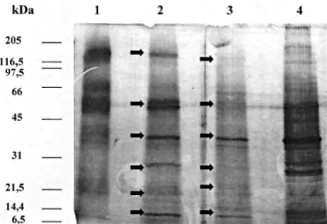
The electrophoresis comparative study between the
A. simplex CE antigen and the one represented by the
mixed fractions eluted with glycine buffer followed by di-ethylamine from the Sepharose 4B-rabbit IgG anti-A. sim-plex column (A. simplex PAK antigen) indicates that both
preparations are very similar (Fig. 1).
Likewise, A. simplex PAK antigen was loaded into the
column prepared with anti-A. suum rabbit IgG. Unbound
(A. simplex free A. suum antigens) antigens were washed
and bound (cross-reacting A. suum antigens) antigens
eluted. After passing across the anti-Ascaris column, the
proteins of A. simplex PAK antigen were maintained but
increased in intensity the bands of 40 and 14 kDa. With A. simplex PAK antigen a doublet around 60 kDa appeared.
With the A. simplex PAS antigen, 60 kDa was maintained
with a high intensity. Bright bands of high molecular weight of about 209 and 150 kDa were seen. In Fig. 1 electrophoretic patterns obtained with the above men-tioned antigens by SDS-PAGE are shown. In the A. sim-plex PAK the previously mentioned doublet is shown. In
the A. simplex PAS antigen the protein of 120 kDa
in-creased in concentration, as well as, a doublet of about 66-45 kDa, the protein of 40 kDa was maintained but in-creased in intensity the protein of 25 kDa, and a doublet between 21.5 and 14 kDa. In the A. simplex EAS antigen
debris of rabbits IgG were observed at 97-66 kDa. Like-wise, very bright proteins of about 35 kDa and 7 kDa were present.
Further, the ELISA method has been carried out in order to determine the IgG levels in sera from rabbits ex-perimentally immunized with larval A. simplex or adult A. suum CE antigen as multiple doses in FCA
intramuscu-larly or multiple doses of T. canis embryonated eggs.
After they were bled weekly p.i., the sera with strong re-actions were selected by ELISA against their homologous antigens. The selected rabbit antisera were tested at the 1/100, 1/200 or 1/400 dilutions and the antigens at 1 or 0.5 µg/ml. The antigenic preparations used were as follows: larval A. sim-plex CE antigen (A. simplex CE antigen); A. simplex CE
antigen after loading into a CNBr-activated Sepharose 4B coupled to IgG from rabbits immunized with A. simplex CE
antigen (A. simplex PAK antigen); A. simplex PAK
anti-gen after loading into a CNBr-activated Sepahrose 4B coupled to IgG from rabbits immunized with adult A. suum
CE antigen (A. simplex PAS antigen); A. simplex PAK
antigen eluted from the anti-A. suum column (A. simplex
EAS antigen) and adult A. suum CE antigen (A. suum CE
antigen). Results were expressed as O.D.p-O.D.c indexes by substracting the mean optical density of the test sera once the non-specific reaction with the BSA used in the blocking was substracted (Figs 2, 3).
When the capability of the different antigens to dis-criminate among the different antisera raised in rabbits was studied, the minimal responses were observed using sera from rabbits inoculated per os with T. canis
embryo-nated eggs against all the antigenic preparations used. The highest responses were obtained when the sera from rabbits immunized with larval A. simplex or adult A. suum
CE were tested against their corresponding homologous and heterologous CE from A. simplex or A. suum. In order
to study the different antigenic preparations of A. simplex
used we have obtained an index as the ratio between the optical density resulting from the anti-A. simplex
antise-rum and the optical density of the anti-A. suum or anti-T. canis heterologous antisera once their corresponding
non-specific reaction with the BSA used in the postcoating
were substracted, as well as, the optical density of the negative controls. In the case of the A. simplex CE
anti-gen those indexes were between 0.7 (serum dilution 1/ 100-antigen concentration 0.5 µg/ml) and 1.7 (serum dilu-tion 1/200 and 1/400-antigen concentradilu-tion 0.5 µg/ml) us-ing anti-A. suum antisera. On the contrary, the
discrimina-tory capability with anti-T. canis antisera was good with
indexes situated between 5.1 (serum dilution 1/100-anti-gen concentration 1 µg/ml) and 14 (serum dilution 1/200-antigen concentra-tion 0.5 µg/ml). When A. simplex PAK antigen was tested,
its capability for discriminate between A. simplex and A. suum was improved with indexes situated between 1.7
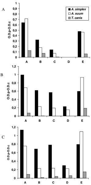
Fig. 2: ELISA IgG responses expressed as O.D.p-O.D.c indexes with serum samples. A: at 1/100; B: at 1/200 and C: at 1/400, of rabbits respectively immunized with Anisakis simplex crude extract (CE) antigen, Ascaris suum CE antigen, or Toxocara canis embryonated
eggs, against the following antigens diluted at 1 µg/ml: larval A. simplex CE antigen (A. simplex CE antigen); A. simplex CE antigen
treated in a IgG anti-A. simplex CE column (A. simplex PAK anti-gen); A. simplex PAK antigen treated in a IgG immunized anti-A. suum CE column (A. simplex PAS antigen); A. simplex PAK anti-gen eluted from the A. suum column (A. simplex EAS antigen);
adult A. suum antigen (A. suum CE antigen)
A
B
C
0 0,1 0,2 0,3 0,4 0,5 0,6 0,7 0,8 0,9 1
A B C D E
O.D.p-O.D.c
A. simplex A. suum
T. canis
0 0,2 0,4 0,6 0,8 1 1,2
A B C D E
O.D.p-O.D.c
0 0,2 0,4 0,6 0,8 1 1,2
A B C D E
(serum dilution 1/100-antigen concentration 1 µg/ml) and 3 (serum dilution 1/400-antigen concentration 1 µg/ml), increasing in the case of T. canis from 4.3 (serum dilution
1/100-antigen concentration 1 µg/ml) to 103.3 (serum dilu-tion 1/200-antigen concentradilu-tion 1 µg/ml).
Using A. simplex PAS antigen, with anti-A. suum
anti-sera, we obtained values from 1.8 (serum dilution 1/100-antigen concentration 1 µg/ml) to 3.3 (serum dilution 1/400-antigen concentration 1 µg/ml; serum dilution 1/100-antigen concentration 0.5 µg/ml). In the case of T. canis
with the A. simplex PAS antigen the values were from 4.6
(serum dilution 1/100-antigen concentration 1 µg/ml) to
571 (serum dilution 1/200-antigen concentration 1 µg/ml). In summary, to eliminate the cross-reactivity between A. simplex and T. canis and A. suum it is necessary a
sequen-tial step affinity chromatography using IgG from rabbits immunized with A. simplex and A. suum.
DISCUSSION
The aim of this work was to assay by ELISA the speci-ficity and sensitivity of larval A. simplex CE antigen
puri-fied by means of affinity chromatography using sera from rabbits experimentally immunized.
The first purification step consisted in binding the larval A. simplex antigen to homologous antibodies from
rabbits experimentally immunized with this antigen and eluted from the column in order to eliminate the major cross-reactivity molecules. The SDS-PAGE patterns of A. simplex CE antigen showed proteins of 205, 120, 66-45, 40,
31-21 and 14 kDa. In the A. simplex PAK antigen we
ob-served the same proteins but several were in different proportions. The molecular mass of ABA-1, the Ascaris
nematode polyprotein, is controversial, it has been previ-ously estimated at 14,000 Da (Christie et al. 1990), but mass spectrometry analysis indicated that there were five componentes of similar size, with the major species being 14,643.2 ± 1.4 Da with a high degree of similarity amongst ascaridid parasites (Christie et al. 1993). Yahiro et al. (1998) cloned the cDNA of TBA-1, the nematode polyprotein allergen of T. canis, and on the basis of the amino acid
sequence found it to be most similar to ABA-1. They ob-served a transient TBA-1 IgG antibody response during the infection that could explain the failure of Kennedy et al. (1989) in the aim of finding anti-TBA-1 antibodies in animals infected with T. canis for a prolonged period. Also
different forms of ABA-1 have been reported by Kato and Komatsu (1996), which could explain the different an-tibody recognition such, also two different forms of TBA-1 may be expressed in a stage specific manner. Also Kennedyet al. (1988) observed evidence that a Mr 14,000 component of A. simplex has a homologue in A. suum, A. lumbricoides and T. canis, but did not elicit an antibody
response in anisakiasis.
Zarnowska and Jastrzebska (1994) by SDS-PAGE of larval ES products from T. canis showed polypeptides of
molecular weights ranging from 19 to 200 kDa. However, an additional polypeptide, not observed on stained gels, and resolving at 14 kDa, was detected by immunoblotting. Sera from patients with A. lumbricoides recognized
polypeptides of 74, 75 and 160 kDa. Iglesias et al. (1996) confirmed by immunoblotting the high degree of cross-reactivity between the somatic antigens of A. simplex and
somatic antigens of the ascaridoids A. suum, T. canis and H. aduncum, although several A. simplex components in
the 11-18 kDa range were only recognized by sera from mice infected with A. simplex. Tanaka et al. (1983)
devel-oped a radioimmunoassay for A. suum protein and
ob-served that T. canis had a high concentration of a
sub-stance partially cross-reactive with A. suum protein. Also,
small amounts of subtance cross-reacting with A. suum
protein was also exhibited by Anisakis larvae. High
con-centrations of A. suum protein were observed in sera from
patients with ascariasis (64.5 ± 18.8 ng/ml), anisakiasis
A
B
0 0,1 0,2 0,3 0,4 0,5 0,6 0,7 0,8 0,9
A B C D E
O.D.p-O.D.c
A. simplex A. suum T. canis
0 0,2 0,4 0,6 0,8 1 1,2
A B C D E
O.D.p-O.D.c
0 0,2 0,4 0,6 0,8 1 1,2
A B C D E
O.D.p-O.D.c
C
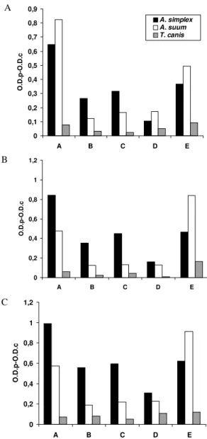
Fig. 3: ELISA IgG responses expressed as O.D.p-O.D.c indexes with serum samples. A: at 1/100; B: at 1/200 and C: at 1/400, of rabbits respectively immunized with Anisakis simplex crude extract (CE)
antigen, Ascaris suum CE antigen, or Toxocara canis embryonated eggs, against the following antigens diluted at 0.5 µg/ml: larval A. simplex CE antigen (A. simplex CE antigen); A. simplex CE antigen treated in a IgG anti-A. simplex CE column (A. simplex PAK
anti-gen); A. simplex PAK antigen treated in a IgG immunized anti-A. suum CE column (A. simplex PAS antigen); A. simplex PAK
(75.2 ± 28.0 ng/ml) and toxocariasis (78.4 ± 31.3 ng/ml). Nunes et al. (1997) detected at least one band with mo-lecular weight around 55-66 kDa that seems to be respon-sible for the cross-reactivity between T. canis and A. suum
once it disappears when previous absorption of serum samples with A. suum antigens was performed. Kennedy
et al. (1989) observed, using radioimmunoprecipitation and
SDS-PAGE, that there was a significant antigenic similar-ity between the antigens of A. suum and T. canis. Among
the cross-reactive components, these authors found a 14 kDa internal protein which has a homologue in the two parasites, observing that was the subject of an IgG anti-body response in Ascaris infection, but there was not
measurable response to it in toxocariasis. McWilliams et al. (1987) observed that A. suum cross-reacted allergically
with T. canis and that the cross-reacting allergens were
predominatly of high molecular weight.
A. simplex PAK antigen was purified from the anti-A. suum rabbit IgG column. In the A. simplex PAS antigen
the proteins of A. simplex PAK antigen were maintained
but increased in intensity the bands of 40 and 14 kDa. In the A. simplex PAS antigen, the proteins were of 120,
66-45, 40, 31-21 and 14 kDa with elevated concentrations of these specific proteins that those observed in the non-purified samples except in the case of the 14 kDa protein. The eluted fractions which contained the cross-reacting antigens with A. suum showed bands in the ranges of
molecular weight situated at 40 and 14 kDa, observing in the latter higher concentrations than in the specific anti-gen. In our experimental conditions, the minimal responses were observed by ELISA using sera from rabbits inocu-lated per os with T. canis embryonated eggs against all
the antigenic preparations used. Conversely, the highest responses were obtained when the sera from rabbits im-munized with larval A. simplex or adult A. suum CE were
tested against their corresponding homologous and het-erologous CE from A. simplex or A. suum. Further, we
stud-ied the different responses to the different antigenic prepa-rations of A. simplex used, observing their capability of
discriminating among the different antisera raised in rab-bits. The discriminatory capability with the anti-T. canis
antisera was good using the larval A. simplex CE antigen.
When A. simplex PAK antigen was used, its capability for
discriminate between A. simplex and A. suum was improved,
increasing in the case of T. canis. The best results were
obtained using A. simplex PAS antigen. When we
com-pared the different serum dilution and antigenic concen-tration, we selected the working serum dilution of 1/400 and 1 µg/ml of antigenic concentration. These conditions insure the maximal differences among the different sera using small quantities of the samples. Works are in progress to study the specificity and sensitivity of A. sim-plex antigens prepared by affinity chromatography using
sera from human patients diagnosed of anisakidosis, ascariosis and visceral larva migrans by ELISA and west-ern-blot techniques.
REFERENCES
Águila C del, Cuéllar C, Fenoy S, Guillén JL 1987. Compara-tive study of assay detecting circulating immuno-complexes and specific antibodies in patients infected with Toxocara
canis. J Helminthol 61: 196-202.
Bradford M 1976. A rapid a sensitive method for the quantitation of microgram quantities of protein utilizing the principle of protein-dye-binding. Ann Biochem72: 248.
Cuéllar C, Fenoy S, del Águila C, Guillén JL 1990. Evaluation of chemotherapy in experimental toxocarosis by determi-nation of specific immune complexes. J Helminthol64:
279-289.
Christie JF, Dunbar B, Davidson I, Kennedy MW 1990. N-terminal amino acid sequence identity between a major al-lergen of Ascaris lumbricoides and Ascaris suum, and
MHC-restricted IgE responses to it. Immunology69: 596-602. Christie JF, Dunbar B, Kennedy MW 1993. The ABA-1
aller-gen of the nematode Ascaris suum: epitope stability, mass
spectrometry, and N-terminal sequence comparison with its homologue in Toxocara canis. Clin Exp Immunol92:
125-132.
Daniels JJHM 1962. De eosinofile flegmone van het maagdarmkanaal veroorzaakt door de haringworm. Ned Tijdschr Geneeskd 106: 131-132.
Fenoy S, Cuéllar C, Guillén JL 1987. Estudio comparativo de la influencia de la luz en el embrionamiento experimental de los huevos de Toxocara canis y Toxascaris leonina. Rev Ibér Parasitol Vol Ext: 173-177.
Fenoy S, Cuéllar C, Guillén JL 1988. Estudio comparativo de la influencia de la luz en el embrionamiento experimental de huevos de Toxocara canis, Toxascaris leonina y Ascaris suum. Rev Ibér Parasitol48: 395-401.
García L, González ML, Esteban MI, Mirabent E, Perteguer MJ Cuéllar C 1996. Enzyme-linked immunosorbent assay, immunoblot analysis and RAST fluoroimmunoassay analy-sis of serum responses against crude larval antigens of
Anisakis simplex in a Spanish random population. J Helminthol70: 281-289.
Guillén JL, Cuéllar C, del Águila C 1986. Fotodependencia del desarrollo embrionario de Toxocara canis (Werner, 1782)
Stiles, 1905. Rev Ibér Parasitol46: 67-74.
Hames BD 1986. An introduction to polyacrilamide gel elec-trophoresis. In BD Hames, D Rickwood (eds), Gel Electro-phoresis in Proteins, IRL Press, Oxford.
Iglesias R, Leiro J, Ubeira FM, Santamarina MT, Navarrete I, Sanmartín ML 1996. Antigenic cross-reactivity in mice be-tween third-stage larvae of Anisakis simplex and other
nema-todes. Parasitol Res82: 378-381.
Ishikura H, Kikuchi K, Nagasawa K, Ooiwa T, Takamiya H, Sato N, Sugane K 1993. Anisakidae and anisakidosis. In T Sun, Progress in Clinical Parasitology, Vol. III,
Springer-Verlag, New York.
Kato Y, Komatsu S 1996. ASABF, a novel cysteine-rich anti-bacterial peptide isolated from the nematode Ascaris suum.
Purification, primary structure, and molecular cloning of cDNA. J Biol Chem271: 30493-30498.
Kennedy MW, Qureshi F, Fraser EM, Haswell-Elkins MR, Elkins OB, Smith HV 1989. Antigenic relationships be-tween the surface-exposed, secreted and somatic materials of the nematode parasites Ascaris lumbricoides, Ascaris suum and Toxocara canis. Clin Exp Immunol75: 493-500.
Kennedy MW, Tierney J, Ye P, McMoanagle FA, McIntosh A, McLaughlin D, Smith JW 1988. The secreted and somatic antigens of the third stage larva of Anisakis simplex, and
antigenic relationship with Ascaris suum, Ascaris lumbricoides, and Toxocara canis. Mol Biochem Parasitol 31: 35-46.
Laemmli UK 1970. Cleavage of structural proteins during the assembly of the head of bacteriophage T4. Nature 227: 680.
immuno-logical investigation of the allergens from Ascaris suumperienteric fluid. Cross-reactivity, molecular weight
distribution and correlation with phosphorilcholine-contain-ing components. Int Arch Allergy Appl Immunol 82:
125-132.
Nunes CM, Tundisi RN, García JF, Heinemann MB, Ogassawara S, Richtzenhain LJ 1997. Cross-reactions between Toxo-cara canis and Ascaris suum in the diagnosis of visceral
larva migrans by western blotting technique. Rev Inst Med Trop São Paulo39: 253-256.
Perteguer MJ, Cuéllar C 1998. Isotype-specific immune re-sponses in murine experimental anisakiasis. J Vet Med B45:
603-610.
Ruitemberg EJ 1970. Anisakiasis: Pathogenesis, Serodiagno-sis and Prevention, Thesis, University of Utrecht,
Nether-lands.
Sakanari JA, McKerrow JH 1989. Anisakiasis. Clin Microbiol Rev2: 278-284.
Smith JW, Wootten R 1978. Anisakis andAnisakiasis. In WHR
Lumsden, K Muller, JK Baker (eds), Advances in Parasi-tology, Vol. 16, Academis Press, London.
Suzuki T 1968. Studies on the immunological diagnosis of anisakiasis. Japan J Parasitol17: 213-220.
Tanaka K, Kawamura H, Tohgi N, Tsuji M, Miyachi Y, Miyoshi A 1983. The measurement of Ascaris suum protein by
ra-dioimmunoassay in sera from patients with helminthiasis and with gastrointestinal diseases. Parasitology86:
291-300.
Welch JS, Symons MH, Dobson C 1983. Immunodiagnosis of parasitic zoonoses: purification of Toxocara canis antigens
by affinity chromatography. Int J Parasitol13: 171-178.
Yahiro S, Cain G, Butler JE 1998. Identification, characteriza-tion and expression of Toxocara canis nematode polyprotein
allergen TBA-1. Parasite Immunol20: 351-357.
Zarnowska H, Jartrzebska M 1994. Excretory-secretory larval antigens of Toxocara canis: physico-chemical