293 293 293 293 293 Mem Inst Oswaldo Cruz, Rio de Janeiro, Vol. 100(3): 293-301, M ay 2005
Enzyme-linked immunosorbent assay and Western blot antibody
determination in sera from patients diagnosed with different
helminthic infections with
Anisakis simplex
antigen purified by
affinity chromatography
M Rodero, T Chivato* , A M uro* * , C Cuéllar/+
Departamento de Parasitología, Facultad de Farmacia, Universidad Complutense, 28040 Madrid, España *Servicio de Alergia e Inmunología, Hospital del Aire, Madrid, España **Laboratorio de Parasitología, Facultad de Farmacia, Universidad de Salamanca,
Salamanca, España
An evaluation of the sensitivity and the specificity of the Anisakis simplex antigens purified by affinity chroma-tography was performed using sera from patients diagnosed with Anisakis sensitisation and sera from patients previously diagnosed with different helminthic infections. Only the sera of the patients diagnosed with Schistosoma
mansoni or Onchocerca volvulus parasitic infections were negative against the A. simplex antigen and its purified
fractions (PAK antigen: A. simplex antigen purified using columns prepared with anti-A. simplex rabbit IgG and PAS antigen: PAK antigen purified using columns prepared with anti-Ascaris suum rabbit IgG). However all the sera were positive against the A. suum antigen. In all the sera from the patients diagnosed with Anisakis sensitisation, the antibody levels detected using the purified antigens (PAK and PAS antigens) were lower than the observed using
the A. simplex crude extract with the highest diminution in the case of the IgG. When these same sera were tested
against the A. simplex crude extract by Western blot, several bands of high molecular masses were observed as well as, intense bands at 60 and/or 40 kDa. A concentration of these last proteins was observed in the PAK and the PAS antigens. When the sensitivity and the specificity determinations were performed, only seven of the 38 patients diagnosed of Anisakis sensitisation were positive, as well as, the sera from the patients diagnosed with parasitisms
by Echinococcus granulosus or Fasciola hepatica.
Key words:Anisakis simplex - enzyme-linked immunosorbent assay - Western blot - helminthoses - cross-reactivity
Anisakidosis is a human disease caused by the ingtion of larval nematodes from the family Anisakidae,
es-pecially Anisakis simplex. The infection is acquired by
eating raw seafood or undercooked fish and squid (Sakanari & McKerrow 1989). Anisakidosis is divided into gastric, intestinal, and ectopic (Ishikura et al. 1993). Due to the vagueness of the symptoms, this disease is often misdiagnosed as appendicitis, acute abdomen, gastric tumor or cancer, ileitis, cholecystitis, diverticulitis, tuber-culous peritonitis, cancer of the pancreas or Chrohn’s dis-ease (Sakanari & McKerrow 1989).
Immunodiagnostic methods have been developed, but several cross-reactivities have been observed. Although, the introduction of sensitive techniques, such as enzyme-linked immunosorbent assay (ELISA) did not avoid the presence of antibodies that provoke cross-reactivities with Ascaris and Toxocara (Kennedy et al. 1988) when the A. simplex larval crude extract was used as reactive for the determination of specific IgE levels in patients with sus-pected anisakidosis, so by fluoro enzymo immuno assay
(FEIA), immunoblot or prick test, due to the high
propor-tion of false negative results detected (Lorenzo et al. 2000).
+Corresponding author. E-mail: [email protected] Received 14 June 2004
Accepted 14 March 2005
In fact, extensive homology between both somatic and
excretory-secretory antigens of A. simplex and other
ascaridoid nematodes mainly A. suum, A. lumbricoides,
and T. canis has been reported (Kennedy et al. 1988). For these reasons, in this work, an evaluation of the
sensitivity and the specificity of the A. simplex antigen
purified by affinity chromatography (Rodero et al. 2001, 2002) was carried out using sera from patients previously
diagnosed with Anisakis sensitisation and different
hel-minthic infections.
MATERIALS AND METHODS
Parasites - A. simplex L3 were picked up manually from the viscera, flesh, and body cavities of naturally
in-fected blue whiting (Micromesistius poutassou) and
ex-haustively washed in water (Perteguer & Cuéllar 1998). A.
suum were obtained from swine natural infections (Guillén
et al. 1986).
Antigens - For the preparation of the crude extract
(CE) of A. simplex (A. simplex CE antigen or AK antigen),
294 294 294 294
294 Purified antigen to identify anisakidosis • M Rodero et al.
estimated and the extract was frozen at 20ºC until used
(Perteguer & Cuéllar 1998). The antigen from A. suum adults
(A. suum CE antigen or AS antigen) was obtained using a modification of the Welch et al. (1983) method by homo-genisation and extraction in PBS at 4ºC overnight (instead of ultrasonic burst). Its protein content was estimated by the Bradford (1976) method, and then the antigen was frozen at 20ºC until use (Águila et al. 1987, Cuéllar et al. 1990).
Hyperimmune sera - New Zealand rabbits of about 3
kg body weight were immunised with 1 mg of larval A.
simplex or adult A. suum CE antigens by the intramuscu-lar route. The animals were bled weekly postimmunization (p.i.) after the first inoculation (week 0) (García-Palacios et al. 1996). The experiments were carried out according to the European Council on applied animals experiments, published in the Guidelines 86/609ED, and controlled in Spain by Royal Decree 223/1988 of 14 March, on the pro-tection of animals used for research and other scientific ends.
Human sera - Thirty-eight human anti-Anisakis sera were obtained from the Servicio de Alergia del Hospital del Aire de Madrid. The concentration of circulating anti-Anisakis IgE antibodies was measured by means of the Pharmacia CAP System RAST FEIA (Pharmacia AB, Uppsala, Sweden). The results of this fluoro immuno as-say were reported in kU/l and converted to CAP “scores” of 0 (< 0.35), 1 (0.35-0.7), 2 (0.7 - 3.5), 3 (3.5 - 17.5), 4 (17.5 -50), 5 (50 - 100) and 6 (> 100). All the sera showed positive
CAP values to Anisakis by FEIA, which varied from
val-ues of CAP = 1 (3%), CAP = 2 (26%), CAP = 3 (47%), CAP = 4 (10%), CAP = 5 (8%) and to CAP = 6 (5%). Nine sera
from patients diagnosed with Loa loa, Onchocerca
vol-vulus, A. lumbricoides, Trichuris trichiura, T. canis, S. mansoni, S. intercalatum, Echinococcus granulosus and Fasciola hepatica parasitic infections were selected.
Purification of antigens by affinity chromatography
- Protein A-Sepharose CL-4B beads (Pharmacia Biotech)
columns were prepared according to the manufacturer’s
intructions. Anti-A. simplex or -A. suum rabbit antibodies,
in sample buffer 0.05 M Tris, 0.5 M NaCl, pH 8.0 were loaded into the columns. Fractions of 1 ml were then col-lected. Unbound immunoglobulins were washed with washing buffer (0.05 M Tris, 0.5 M NaCl, pH 8.0). Bound immunoglobulins then were eluted with glycine buffer (0.2 M glycine, 0.5 M NaCl, pH 2.8). Fractions were collected onto 100 µl of collection buffer (Tris-base 1 M, pH 8.5)
and read on a spectrophotometer at A280 for calculating
IgG concentration. A column was prepared with protein A
affinity isolated anti-A. simplex IgG, at a concentration of
5 mg/ml in NaHCO3 0.1 M with NaCl 0.5 M, pH 8.5 coupled
to CNBr-activated Sepharose 4B according to the
manufacturer’s instructions (Pharmacia Biotech). The A.
simplex CE antigen in sample buffer was loaded into the column and incubated 3 h at room temperature. Fractions of 1 ml were then collected. Unbound antigens were washed with washing buffer and bound antigens were eluted with glycine buffer followed by 50 mM diethylamine in saline, pH 11.5 collected into glycine to neutralise the
eluted fractions. Fractions were read at A280. This
anti-gen was named as A. simplex PAK antigen. The same
procedure was carried out using columns prepared with
rabbit anti-A. suum IgG, obtaining the A. simplex PAS
an-tigen and the A. simplex EAS antigen. In summary: PAK
(A. simplex CE antigen eluted from CNBr-activated Sepharose 4B coupled to IgG from rabbits immunised with A. simplex CE antigen), PAS (A. simplex PAK antigen af-ter loading into a CNBr-activated Sepharose 4B coupled
to IgG from rabbits immunised with adult A. suum CE
anti-gen) and EAS (A. simplex PAK antigen eluted from the
anti-A. suum column).
Immunoblot - Sodium dodecylsulphate-polyacrilamide
gel electrophoresis(SDS-PAGE) was carried out as
de-scribed by Laemmli (1970) and revised by Hames (1986)
using a Mini Protean III cell (Bio Rad). The gels
con-sisted of a 4% stacking gel and 5-20% gradient separating gel. Samples were dissolved in a sample buffer (50 mM Tris-HCl buffer, pH 8.6, containing 2% SDS, 20% glycerol, and 0.02% bromophenol blue) diluted 1:1 in electrode buffer (25 mM Tris, 192 mM glycine, pH 8.3), containing 1% SDS. Electrophoresis was performed for 2 h at a con-stant 100 V in Tris-glycine electrode buffer (see above). Broad range molecular mass markers (6.5 - 205 or 7.2 - 209 kDa, Bio Rad) were incorporated into each
electrophore-sis run. Gels were stained with AgNO3. Following the
SDS-PAGE of antigens, the protein bands were transferred onto a 0.22 µm pore size nitrocellulose membrane (Pharmacia) in a Mini Trans-blot Electrophoretic Transfer Cell (Bio Rad) with 25 mM Tris, 192 mM glycine, 20% v/v metha-nol, pH 8.3. The transblot was carried out at a constant 100 V for 1 h. The membrane was blocked for 3 h at room temperature with PBS containig 5% non-fat dry milk, prior to immunorecognition by incubation for 2 h with the hu-man sera diluted in PBS-Tween containing 1% non-fat dry milk. Each paper was then washed with PBS-Tween 20
(3 × 5 min) and incubated for 3 h with goat anti-human Ig’s
(total immunoglobulins), IgM, IgG or IgA, HRP conjugate (Biosource International, Camarillo, CA, US), at the ap-propriate dilution in PBS-Tween 20, containing 1% non-fat dry milk. To visualize bands the nitrocellulose was
washed with PBS-Tween 20 (3 × 5 min) and reacted with
the substrate (PBS, containing 0.006% H202 methanol,
containing 0.03% 4-chloro-1-naftol). The reaction was stopped by exhaustive washing with distilled water.
Determination of specific antibody levels - The anti-body levels were measured by indirect ELISA. The 96
well microtitre plates (Nunc-Immuno Plate PolySorpTM)
dilu-295 295 295 295 295 Mem Inst Oswaldo Cruz, Rio de Janeiro, Vol. 100(3), M ay 2005
tion in PBS-Tween, 0.1% BSA, were incubated for 1 h at 37ºC. After adding substrate (phosphate citrate buffer,
containing 0.04% H2O2 and 0.04% o-phenylenediamine)
the reaction was stopped with 3N sulphuric acid and the plates were read at 490 nm (García-Palacios et al. 1996). Results were expressed as O.D.p-O.D.c indexes by sub-tracting the mean optical density (O.D.) of the control from the mean O.D. of the test sera once the non-specific reaction with the BSA used in the blocking was subtracted. Determination of the sensitivity and the specificity of
the A. simplex crude extract and the PAK and PAS
puri-fied antigens with sera from patients diagnosed with
Anisakis sensitisation - For the determination of the
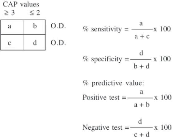
sen-sitivity and the specificity of the different antigens, 2 × 2
tables were carried out. In these tables, the number of
sera from patients diagnosed with Anisakis sensitisation
and CAP values ≥ 3 and ≤ 2 against the different antigens
were represented for each immunoglobulin (Ig’s, IgG, IgM, IgA, IgE). Further, different O.D. cutting points were se-lected in function of the positive and negative predictive values, as well as, the sensitivity and the specificity, in the case of each assayed immunoglobulins and antigens (AK, PAK, and PAS) (Fig. 3).
RESULTS
ELISA antibody determinations in the sera from pa-tients diagnosed with Anisakis sensitisation using A. sim-plex antigens purified by affinity chromatography - Thirty-eight sera obtained from the Servicio de Alergia del Hos-pital del Aire de Madrid were assayed. All the sera showed
positive CAP values to Anisakis by FEIA, which varied
from values of CAP = 1 (3%), CAP = 2 (26%), CAP = 3 (47%), CAP = 4 (10%), CAP = 5 (8%), to CAP = 6 (5%).
The 96 well microtiter plates were coated with 100 µl/
well (1 µg/ml) of the crude extract (CE) antigens of A.
sim-plex or A. suum (AK and AS antigens), as well as, the
antigens purified using the columns of anti-A. simplex or
anti-A. suum IgG (PAK and PAS antigens, respectively)
and the antigen eluted from the latest column (EAS anti-gen). The sera were diluted at 1/400, and, after their incu-bation, the Ig’s, IgM, IgG and IgA were measured. The mean O.D. were calculated after the subtraction of the BSA and negative control values.
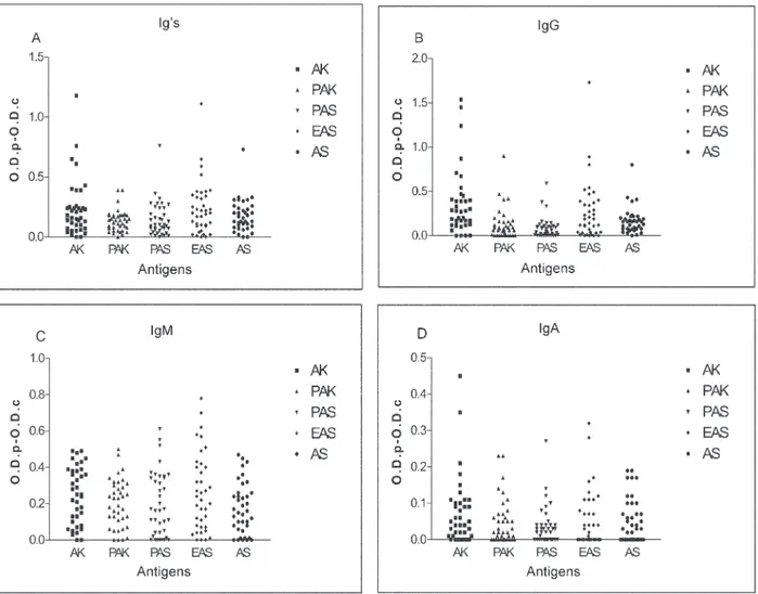
With all the assayed sera from the patients diagnosed
with Anisakis sensitisation, the highest values were
ob-tained when the ELISA test were performed against the A.
simplex CE (AK antigen). The IgG was the most abundant immunoglobulin, followed by the IgM and the Ig’s, whilst the lowest values were observed in the case of the IgA (Fig. 1).
When the sera were tested against the A. suum crude
extract (AS antigen) 45% of them showed values equal or superior than 0.2 in the case of the specific Ig’s were de-tected. Contrarily, these values were modified when the IgG antibodies were tested. In this case, 24% of the sera
showed anti-A. suum IgG levels equal or superior than
0.2. On the other hand, IgM values were detected in 39% of the sera. In the case of the IgA, the levels were lower, because only 13% of the sera showed O.D. values equal or higher than 0.15 (Fig.1).
When the sera were tested against the purified anti-genic fractions (PAK and PAS antigens), in both cases, the highest values were observed when the IgM immuno-globulins were tested, followed by the Ig’s and the IgG. Finally, the lowest values belonged to the IgA.
All the detected O.D. values using the purified prepa-rations (PAK and PAS antigens) were lower than the
ob-served using the A. simplex CE (AK antigen) with the
highest diminution in the case of the IgG (0.36 - 0.08/AK-PAS antigens).
When the sera were tested against the “eluted of
As-caris” (EAS antigen) the mean values of the IgG increased (O.D. = 0.26) compared to the observed using the purified antigens (Fig. 1).
ELISA antibody determinations in the sera from pa-tients diagnosed with different helminth parasitic infec-tions using A. simplex antigens purified by affinity chro-matography - The evaluation by ELISA of the A. simplex larval antigens purified by affinity chromatography was
completed using sera from patients diagnosed with L. loa,
O. volvulus, A. lumbricoides, T. trichiura, T. canis, S. mansoni, S. intercalatum, E. granulosus,and F. hepatica.
These sera were tested against the CE antigens of A.
simplex (AK antigen) and A. suum (AS antigen), as well
as the antigen eluted from the column of anti-A simplex
rabbit IgG (PAK antigen), the A. simplex antigen purifed
by the column of anti-A. suum rabbit IgG (PAS antigen),
and the eluted from the mentioned column (EAS antigen). The specific Ig’s, IgG, IgM, and IgA were measured.
Only the sera from the patients diagnosed with S.
mansoni or O. volvulus parasitic infections were negative
against the A. simplex antigen (AK antigen) and its
puri-fied fractions (PAK, PAS, and EAS antigen). However, all
the sera showed cross-reactions with the A. suum one
(AS antigen) when the Ig’s, IgG, IgM and IgA antibodies were measured (Table I).
When the PAK antigen were tested, in all the patients,
except O. volvulus, A. lumbricoides, and S. mansoni cases,
the IgG levels enhanced with respect to the A. simplex
antigen (AK antigen). On the contrary, when the sera were tested against the PAS antigen, the IgG levels suffered a
reduction higher than 50% for the T. trichiura, S.
in-tercalatum, and E. granulosus patients and were
nega-tive for the O. volvulus, A. lumbricoides, and S. mansoni
patients. Finally, when the EAS antigen was tested, the IgG levels enhanced more than twice with respect to the
PAS antigen in the case of the T. trichiura, S. intercalatum,
E. granulosus, and F. hepaticapatients.The IgM levels unchanged with respect to the PAS antigen, as well as, the total immunoglobulins (Table I).
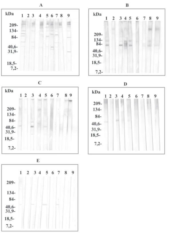
Western blot antibody determinations in the sera from patients diagnosed with different helminth parasitic in-fections using A. simplex antigens purified by affinity chromatography - When the sera were tested against the A. simplex antigen, all the sera except the O. volvulus case, reacted with proteins of high molecular masses
(> 84 kDa). The sera from the L. loa, A. lumbricoides, and
S. mansoni patients reacted with the 60 kDa protein. These same sera also reacted with a 40 kDa protein, as well as,
296 296 296 296
296 Purified antigen to identify anisakidosis • M Rodero et al.
patients, while the 32 kDa protein only was
immuno-recognised for the F. hepatica serum. When the sera were
tested against the PAK antigen, the immunorecognition against the 60 kDa protein enhanced in intensity in the
case of the L. loa and A. lumbricoides sera. This reaction
disappeared with the S. mansoni serum and appeared
us-ing the T. canis, E. granulosus, and F. hepatica sera. In
addition, in this last patient, the immunorecognition against the 32 kDa protein disappeared. Against the PAS antigen, only was immunorecogniton of the 60 kDa
pro-tein in the case of the L. loa, A. lumbricoides, and T. canis
sera (Fig. 2).
Determination of the sensitivity and the specificity of
the A. simplex crude extract and the PAK and PAS
puri-fied antigens with sera from patients diagnosed with A. simplex sensitisation - When the A. simplex crude extract was used, variations of the sensitivity and the specificity were detected according to the selected immunoglobulin.
The cut point of O.D.≥ 0.15 for the IgG was selected as a
first reference because this immunoglobulin showed the greatest sensitivity (82.7%). When this parameter was
applied, 70% of the patients diagnosed with A. simplex
sensitisationwere positive. When the second
determina-tion was performed by using the PAK antigen and the cut
point of O.D. ≥ 0.07 for the IgG, a decrease of 20% in the
positive group was produced. Finally, the cut point of
O.D. ≥ 0.05 for the IgA was selected against the PAS
anti-gen. Only seven of the 38 tested sera were positive (Table II).
When these three parameters were applied to the sera from the patients diagnosed with several parasitic infec-tions, only the sera from the patients diagnosed with
parasitisms of E. granulosus and F. hepatica were
posi-tive.
DISCUSSION
In this work, the specificity and the sensitivity of the A. simplex antigen purified by affinity chromatography previously described (Rodero et al. 2001, 2002), were evalu-ated by ELISA, using sera from patients previously diag-nosed with several parasitic infections.
297 297 297 297 297 Mem Inst Oswaldo Cruz, Rio de Janeiro, Vol. 100(3), M ay 2005
IgA) were tested by ELISA, against the A. simplex
anti-gen, as well as, its purified fractions, only the sera from
the patients diagnosed with S. mansoni or O. volvulus
infections were negative. These results were maintained
using the serum from an O. volvulus patient when
as-sayed by Western blot as a diagnostic method. Any im-munoreactive band did not appear when the total
immu-noglobulins were tested. However, the S. mansoni patient
serum showed several bands of high molecular masses
against the A. simplex antigen (AK antigen) and others
immunoreactive bands of around 60 and 40 kDa. This difference between the antibody determination using both techniques (ELISA and Western blot), could be due to the different techniques and metodologies utilised in order to determine the different
immunoglobu-TABLE I
Ig’
s, IgG
, IgM, and IgA
levels of the patients diagnosed of dif
ferent helminthic infections
against
A: CE of
Anisakis simplex
(AK antigen), B: P
AK antigen, C: P
AS antigen, D: EAS
antigen, and E: CE of
Ascaris suum (AS antigen) A B C D E Patient Ig ’s Ig G IgM Ig A Ig ’s Ig G IgM Ig A Ig ’s Ig G IgM Ig A Ig ’s Ig G IgM Ig A Ig ’s Ig G IgM Ig A Loa loa 0,25 0,29 0,69 0,01 0,41 0,61 0,77 0,07 0,32 0,16 0,59 0,00 0,32 0,42 0,56 0,00 0,89 1,58 1,35 0,14 Onchocerca volvulus 0,05 0,00 0,05 0,00 0,07 0,07 0,08 0,00 0,03 0,02 0,05 0,00 0,05 0,13 0,06 0,00 1,20 0,57 0,07 0,08 Ascaris lumbricoides 0,26 0,00 0,62 0,04 0,31 0,00 0,66 0,00 0,28 0,00 0,54 0,00 0,35 0,19 0,59 0,00 0,34 0,22 0,72 0,13 T richuris trichiura 0,89 1,33 0,28 0,12 0,68 1,86 0,38 0,12 0,36 0,82 0,31 0,00 0,80 1,66 0,39 0,08 1,54 2,43 0,79 0,42 T oxocara canis 0,16 0,00 0,25 0,00 0,55 0,12 0,39 0,00 0,16 0,01 0,25 0,00 0,30 0,12 0,34 0,00 0,75 0,59 0,70 0,00 Schistosoma intercalatum 0,42 0,57 0,11 0,04 0,54 0,96 0,08 0,00 0,23 0,35 0,05 0,00 0,37 0,67 0,06 0,00 1,82 2,03 0,22 0,37 Schistosoma mansoni 0,07 0,04 0,04 0,00 0,02 0,00 0,04 0,00 0,08 0,00 0,06 0,00 0,06 0,00 0,07 0,00 0,16 0,00 0,02 0,05 Echinococcus granulosus 0,78 0,36 0,11 0,45 0,55 0,53 0,10 0,52 0,54 0,21 0,06 0,58 0,77 0,51 0,06 0,54 2,24 1,49 0,08 0,51 Fasciola hepatica 1,49 0,51 0,59 0,38 1,39 0,83 0,52 0,30 1,35 0,36 0,56 0,26 2,10 1,12 0,69 0,31 2,59 1,75 0,60
0,42 Positive human sera (¸) against Anisakis simplex AK, PAK,TABLE II and PAS antigens for the different cut points selected
Cut point
AK PAK PAS
Patient/CAP IgG ≥0,15 IgG ≥ 0,07 IgA ≥ 0,05 2936/2
3056/3 ¸ ¸
3423/3 ¸
3461/3 ¸ ¸
3475/3 ¸ ¸
3500/3
3524/3 ¸ ¸ ¸
3530/2 ¸
3544/3 ¸
3604/3
3619/3 ¸ ¸ ¸
3638/4 3679/5
3720/1 ¸ ¸
3755/2 ¸
3782/3 ¸ ¸ ¸
3801/6 ¸ ¸
3807/2 ¸
3837/2 ¸ ¸ ¸
3857/3 ¸ ¸
3866/5 ¸
3878/3 ¸ ¸
3883/2
3884/3 ¸ ¸
3903/3 ¸
3906/4 ¸ ¸
3913/2
3914/3 ¸
3938/3 ¸
3949/3 ¸
3975/2 ¸
3990/6 ¸ ¸ ¸
4009/4 ¸ ¸ ¸
4016/3 ¸ ¸ ¸
4118/4
4153/2 ¸ ¸
4159/5 ¸ ¸
4189/2 ¸
298 298 298 298
298 Purified antigen to identify anisakidosis • M Rodero et al.
lin class levels, like the serum dilution, as well as, the antigenic concentration.
Noya et al. (1995) detected IgG, IgM, IgA, and IgE specific antibodies by ELISA when tested the sera of 30 S. mansoni infected children, while, by Western blot, ap-peared reactive bands of high molecular masses, as well as, 45, 36, and 30 kDa for IgG and 77 kDa for IgM
antibod-ies, indicating that the 36 kDa protein was usefull for
im-munodiagnostic. In the case of S. intercalatum the
re-sults obtained, in our experimental conditions, were com-pletely different. High levels of Ig’s and IgG antibodies, and basal levels for IgM and IgA were detected by ELISA. These diferences among immunoglobulins could be due to the different parasitic locations in function of the
299 299 299 299 299 Mem Inst Oswaldo Cruz, Rio de Janeiro, Vol. 100(3), M ay 2005
tion time and the type of immunological response that the worms can provoke in the host.
In the serum from the T. trichiura patient the highest
antibody levels detected by ELISA against the crude
ex-tract (CE) A. simplex and its purified fractions were
ob-served when the Ig’s and IgG antibodies were measured. Turner et al. (2002) mentioned that people who live in endemic areas have high levels of IgG1 and IgG4 antibod-ies when tested against their homologous antigen. This increase of antibodies could be related with the high IgG
levels detected by us when the A. simplex antigen and its
purified fractions were tested by ELISA, due to both
para-sites (A. simplex and T. trichiura) are nematodes and the
high probability of existence of common proteins.
How-ever, the antigenic cross-reactivity between Ascaris and
Anisakis antigens is known. Sakanari et al. (1988) sug-gested the existence of taxonomically related epitopes between both nematodes. Further, Iglesias et al. (1996)
observed high cross-reactions among A. simplex and other
ascarids as Hystherotylacium aduncum, T. canis and A.
summ, indicating that neither A. simplex
excretory-secre-tory nor crude extract antigen are eminently good for the serodiagnosis.
Lillywhite et al. (1991) showed the presence of
cross-reactions between T. trichiura and A. lumbricoides and T.
canis when IgG, IgE, and IgM antibodies were measured. In the same manner, we detected in that, the serum from the T. trichiura patient, the presence of unspecific
anti-bodies was higher against the A. suum than the A.
sim-plex antigen.
In the serum of the patient diagnosed with E.
gra-nulosus infection the highest antibody levels corre-sponded to Ig’s, IgA, and IgG. In this disease, the anti-body responses during the infection are measured against the CE antigen or parcially purified hydatidic cyst fluid. Some workers have mentioned that the carbohydrated structures play an important role in the survival of some parasites (Butterworth et al. 1988, Dunne 1990, Ouaissi et
al. 1991). In the E. granulosus case, 23% of the IgG
re-sponses against surface protoescolex antigens are leaded by the carbohydrated epitopes (Hernández & Nieto 1994).
When the Western blot were carried out using the A.
simplex antigen, in the serum from the E. granulosus pa-tient, the presence of immunoreactive bands of high mo-lecular masses were observed when the Ig’s were tested while, in the case of the PAK antigen, a band of around 60 kDa appeared.
The first purification step consisted in binding the
larval A. simplex antigen to homologous antibodies from
rabbits experimentally immunized with this antigen and eluted from the column in order to eliminate the major
cross-reactive molecules. The protein patterns of the A.
simplex crude extract (AK antigen) by SDS-PAGE showed proteins of 205, 120, 66-45, 40, 31-21, and 14 kDa. After its
purification across the rabbit anti-A. simplex IgG column
(PAK antigen), the same proteins were obseved but in a different proportion and, in the case of the human IgG column, the protein of 40 kDa was in a high concentration (Rodero et al. 2002).
When the anti-F. hepatica serum was tested, the
high-est antibody levels corresponded to Ig’s, IgM, IgG, and
finally IgA, against the CE A. simplex antigen and its
pu-rified fractions. However, by Western blot, only appeared bands of high molecular masses, as well as, other around
30 kDa, when the A. simplex CE antigen was tested. In
patients diagnosed with F. hepatica there are elevated
IgM and IgG antibody levels, being the IgG1 and the IgG4 the most prevalent isotypes (Chen & Mott 1990, O’Neill et al. 1998), while the IgA levels are not altered (Sampaio Silva et al. 1985).
In the sera from the patients diagnosed with A.
lumbricoides and T. canis infections there were neither
IgG nor IgA antibodies against any A. simplex antigen.
However, IgM antibodies were detected which were re-sponsible of the Ig’s level augmentation. Matsumura et
al. (1984) observed that anti-Toxocara circulating
anti-bodies can be detected in puppies six months old, as well as, in adult dogs, due to an increase of IgM levels in-duced by small amounts of excretory-secretory materials from the larvae maintained in the tissues.
Iglesias et al. (1996) confirmed by immunoblotting the high degree of cross-reactivity between the somatic
anti-gens of A. simplex and other ascaridoids, such as, A. suum,
T. canis, and H. aduncum, although several A. simplex components, in the 11-18 kDa range, were only
recog-nized by sera from mice infected with A. simplex.
After the immunobloting using the T. canis and A.
lumbricoides sera, we observed immunorecognition of
proteins of high molecular mass when the CE A. simplex
antigen was tested. In 1990, Maizels and Page, observed
that the T. canis larva released high quantities of
glyco-proteins during the in vitro culture, belonging to the ex-cretory-secretory products and Mc Williams et al. (1987)
demonstrated a type I allergy cross-reaction between A.
suum and T. canis in which the allergens responsible for
the cross-reactivity were predominantly of high molecu-lar masses.
When the PAS antigen was tested, immunoreactive bands around 40 and 30 kDa proteins were present. When the protein pattern of the PAS antigen was studied after Fig. 3: calculation of sensitivity, specificity, and predictive value
(positive and negative) for the different immunoglobulins (Ig’s, IgG, IgM, and IgA) against crude extract of Anisakis simplex and purified antigens PAK, and PAS. The different cut points selected corresponded of values of O.D. ≥ 0.05
CAP values ≥ 3 ≤ 2
a b O.D.
c d O.D.
a % sensitivity = x 100
a + c
d % specificity = x 100
b + d
% predictive value: a Positive test = x 100
a + b
d Negative test = x 100
300 300 300 300
300 Purified antigen to identify anisakidosis • M Rodero et al.
its purification using the anti-A. suum column, the
un-binding proteins (specific proteins) were of 120, 66-45, 40, 31-21, and 14 kDa. Higher concentrations of specific pro-teins were seen compared to the unpurified samples (Rodero et al. 2002).
Finally, in both sera (T. canis and A. lumbricoides) a
band around 60 kDa appeared against the EAS antigen
responsible of the cross-reaction with the A. simplex
an-tigen. Nunes et al. (1997) detected at least one band with molecular weight around 55-66 kDa that seems to be
re-sponsible for the cross-reactivity between T. canis and A.
suum, which disappears when previous absorption of the
serum samples with A. suum antigens was performed.
Previously, the discriminating capability of both anti-gens (PAK and PAS) were assayed in sera from rabbits
immunized with larval A. simplex or adult A. suum CE or
inoculated with embryonated T. canis eggs and we
ob-served that this capability for discriminate between A.
sim-plex and A. suum was improved in the case of the PAS
antigen (Rodero et al. 2002).
To calculate the sensitivity and the specificity param-eters, it is neccesary to select the “gold standard” which can evaluate the accuracy of the diagnostic test on the basis that the evaluated disease is truly present of absent in the selected patients. In our case, the CAP value was selected as a “gold standard”, although is not a very good diagnostic method but is routinely used to select the Anisakis sensitised patients.
The sensitivity and the specificity determinations were
performed testing sera from patients diagnosed with A.
simplex sensitisation, against the CE (AK antigen), as
well as, the purified PAK and PAS A. simplex antigens, in
order to observe the possible variations produced during the purification process. These evaluations were carried out against all the tested immunoglobulins (Ig’s, IgG, IgM, IgA, IgE), to determine the working conditions (immuno-globulin and antigen) for the human anisakidosis diagno-sis.
When the A. simplex crude extract was used,
varia-tions of the sensitivity and the specificty were detected according to the immunoglobulin selected. For the same cut point of O.D. = 0.15, the sensitivity varied from values of 83%, for the IgG, to only 20.7%, in the case of the IgA. This fact showed the difficulty of the immunodiagnosis of anisakidosis when only a specific immunoglobulin type is studied.
In our experimental conditions, we showed the useful
of testing the sera in a first step against the A. simplex
crude extract and then carrying out a second determina-tion against the PAK and the PAS purified antigens.
This does not imply an additional cost of time. For
this, the cut point of O.D. ≥ 0.15 for the IgG was selected
as a first reference. This immunoglobulin showed the greater sensitivity (82.7%). In the first step of the diag-nostic investigations the sensitive tests are the most use-ful (Fletcher et al. 1998). When this parameter was ap-plied, 70% of the sera were positive, in spite of all the sera
except one that had a CAP value ≥ 2. The CAP System has
a poor specificity and shows a high rate of false positive
results. This fact was also observed by Lorenzo et al. (2000) when evaluated several immunological techniques
in order to make the diagnosis of Anisakis allergy,
ob-serving 50% of specificity with the CAP system assay. The second determination was performed by using
the PAK antigen and the cut point of O.D. ≥ 0.07 for the
IgG. This value was selected because in the middle of the diagnostic investigation, showed medium values of speci-ficity and sensitivity (58.6 and 54.5%, respectively). When all the assayed sera were evaluated, a decrease of 20% in the positive group was observed. Only two sera,
previ-ously negative against the A. simplex crude extract, were
maintained positive. These results are in accordance with
the high predictive value (80%) observed in the A.
sim-plex crude extract, although a probability of 20% (six sera)
of the existence of false positive results is always present. In our case the decrease observed was greater (from 28
positive sera for A. simplex crude extract to 20 for the
PAK antigen).
Finally, the cut point of O.D. ≥ 0.05 for the IgA was
selected against the PAS antigen. This immunoglobulin showed the highest specificity (81.8%) and predictive value (88.9%).
Likewise, for the detection of IgA, a little amount of serum (1 µl) is required in contrast to the IgE, in which case 200 µl of serum are necessary.
When these parameters were applied, only seven of the 38 tested sera were positive. All these sera were also
positive against both the A. simplex CE and the PAK
pu-rified antigens.
When the cut points were selected, we applied these parameters to the sera from the patients diagnosed of sev-eral parasitic infections and we observed that, against the
AK antigen (O.D.≥ 0.15 for the IgG), the sera from the
patients diagnosed with parasitisms by L. loa, T. trichiura,
S. intercalatum, E. granulosus, and F. hepatica were posi-tive. This fact indicated the presence of common
anti-genic proteins among the Anisakis antigens and other
parasites. Likewise, one of the fundamental aspects of the immunodiagnosis is the selection of adequate anti-gens to carry out the diagnostic tests.
When the sera were tested against the PAK antigen
(OD ≥ 0.07 for the IgG), the same sera were positive again
but, when the third determination was carried out using
the PAS antigen (OD ≥ 0.05 for the IgA), only were
posi-tive the sera from the patients diagnosed with parasitisms of E. granulosus and F. hepatica. Previously, we had ob-served this same fact, when the capability of the different antigens to discriminate among the sera from the patients diagnosed with several parasitic infections were deter-mined, when the lowest values were detected using the
anti-Echinococcus or anti-Fasciola sera. It is necessary
to mention that, in the purification process of the A.
sim-plex antigen, an hyperimmnune serum from rabbits
immunised with A. suum was used and no sera from
ani-mals immunised with the other parasites that are infecting the different patients utilised in this assay. However, a remarkable augmentation of the sensitivity and
specific-ity in the fractions of A.simplex antigen purified, was
301 301 301 301 301 Mem Inst Oswaldo Cruz, Rio de Janeiro, Vol. 100(3), M ay 2005
REFERENCES
Águila C, Cuéllar C, Fenoy S, Guillén JL 1987. Comparative study of assay detecting circulating immune-complexes and specific antibodies in patients infected with Toxocara canis. J Helminthol 61: 196-202.
Bradford M 1976. A rapid a sensitive method for the quantitation of microgram quantities of protein utilizing the principle of protein-dye-binding. Ann Biochem 72: 248.
Butterworth A, Dunne D, Fulford A, Capron M, Koch D, Ouma J, Sturrock R 1988. Immunity in human schistosomiasis mansoni: cross-reactive IgM and IgG2 anticarbohydrate antibodies block the expression of immunity. Biochemistry 70: 1053-1063.
Cuéllar C, Fenoy S, del Águila C, Guillén JL 1990. Evaluation of chemotherapy in experimental toxocarosis by determi-nation of specific immune complexes. J Helminthol 64: 279-289.
Chen MG, Mott KE 1990. Progress in morbidity due to Fas-ciola hepatica infection. Trop Dis Bull 87: 1-37.
Dune D 1990. Schistosome carbohydrates. Parasitol Today 6: 45-48.
Fletcher RH, Fletcher SW, Wagner EH 1998. Epidemiología Clinica. Aspectos Fundamentales, Masson-Williams & Wilkins, Barcelona
García-Palacios L, González ML, Esteban MI, Mirabent E, Perteguer MJ, Cuéllar C 1996. Enzyme-linked immu-nosorbent assay, immunoblot analysis and RAST fluoroimmunoassay analysis of serum responses against crude larval antigens of Anisakis simplex in a Spanish ran-dom population. J Helminthol 70: 281-289.
Guillén JL, Cuellar C, del Águila C 1986. Fotodependencia del desarrollo embrionario de Toxocara canis (Werner, 1782). Stiles, 1905. Rev Ibér Parasitol 46: 67-74.
Hames BD 1986. An introduction to polyacrilamide gel electrphoresis. In BD Hames, D Rickwood (eds), Gel Elec-trophoresis in Proteins, IRL Press, Oxford.
Hernández A, Nieto A 1994. Induction of protective immunity against murine secondary hydatidosis. Parasite Immunol 16: 537-544.
Iglesias R, Leiro J, Ubeira FM, Santamarina MT, Navarrete I, Sanmartín ML 1996. Antigenic cross-reactivity in mice be-tween third-stage larvae of Anisakis simplex and other nema-todes. Parasitol Res 82: 378-381.
Ishikura H, Kikuchi K, Nagasawa K, Ooiwa T, Takamiya H, Sato N, Sugane K 1993. Anisakidae and Anisakiosis. In T Sun, Progress in Clinical Parasitology, Springer-Verlag, New York.
Kennedy MW, Tierney J, Ye P, McMoanagle FA, McIntosh A, McLaughlin D, Smith JW 1988. The secreted and somatic antigens of the third stage larva of Anisakis simplex, and antigenic relationship with Ascaris suum, Ascaris lumbricoides, and Toxocara canis. Mol Biochem Parasitol 31: 35-46.
Laemmli UK 1970. Cleavage of structural proteins during the assembly of the head of bacteriophage T4. Nature 227: 680-685.
Lillywhite JE, Bundy DA, Didier JM, Cooper ES, Bianco AE 1991. Humoral immune responses in human Infection with
the whipworm Trichuris trichura. Parasite Immunol 13: 491-507.
Lorenzo S, Romaris F, Iglesias R, Audícana MT, Alonso J, Leiro J, Ubeira FM 2000. O-glycans as a source of cross-reactiv-ity in determinations of human serum antibodies to Anisakis simplex antigens. Clin Exp Allergy 30: 551-559.
Maizels RM, Page AP 1990. Surface glycoproteins from Toxo-cara canis larvae parasites. Act Trop 47: 355-364. Mc Williams AS, Stewart GA, Turner KJ 1987. An
immuno-logical investigation of the allergens from Ascaris suum pe-rienteric fluid. Cross-reactivity, molecular weight distribu-tion and correladistribu-tion with phosphorilcholine-containing com-ponents. Int Arch Allergy Appl Immunol 82: 125-132. Matsumura K, Kazuta Y, Endo R, Tanaka K 1984. Detection of
circulating toxocaral antigens in dogs by sandwich enzyme-immunoassay. Immunology 51: 609-613.
Noya O, Fermin Z, Alarcon de Noya B, Losada S, Clomenares C, Hermoso T 1995. Humoral immune response of children with chronic schistosomiasis. Isotype recognition of adult worm antigens. Parasite Immunol 17: 319-328.
Nunes CM, Tundisi RN, García JF, Heinemann MB, Ogassawara S, Richtzenhain LJ 1997. Cross-reactions between Toxo-cara canis and Ascaris suum in the diagnosis of visceral larva migrans by western blotting technique. Rev Inst Med Trop São Paulo 39: 253-256.
O’Neill SM, Parkinson M, Strauss W, Angles R, Dalton JP 1998. Immunodiagnosis of Fasciola hepatica infection (fasciolosis) in a human population in the Bolivian Altipl-ano using purified cathepsin L cysteine proteinase. Am J Trop Med Hyg 58: 417-423.
Ouaissi MA, Taibi A, Loyens M, Martín U, Afchain D, Maidane C, Cardioti C, Cornete J, Martelleur A, Velge P 1991. Try-panosoma cruzi: a carbohydrate epitope defined by a mono-clonal antibody as a posible marker of the cute phase of human Chaga’s disease. Am J Trop Med Hyg 45: 214-225. Perteguer MJ, Cuéllar C 1998. Isotype-specific immune
re-sponses in murine experimental anisakiasis. J Vet Med 45: 603-610.
Rodero M, Jimenez A, Chivato T, Laguna R, Cuellar C 2001. Purification of Anisakis simplex antigen by affinity chro-matography. Parasitol Res 87: 736-740.
Rodero M, Jiménez A, Cuellar C 2002. Evaluation by ELISA of Anisakis simplex larval antigen purified by affinity chroma-tography. Mem Inst Oswaldo Cruz 97: 247-252.
Sakanari JA, Loinaz M, Deardorff TL, Raybourne RB, McKerrow JH, Frierson JG 1988. Intestinal anisakiasis. A case diagnosed by morphological and immunologic meth-ods. Am J Clin Pathol 90:107-113.
Sakanari JA, McKerrow JH 1989. Anisakiasis. Clin Microbiol Rev 2: 278-284.
Sampaio Silva ML, Vindimian M, Wattré P, Capron A 1985. Etude des anticorps IgE dans la distomatose humaine á Fas-ciola hepatica. Pathol Biol 33: 746-750.
Turner J, Faulkner H, Kamgno J, Else K, Boussinesq M, Brad-ley JE 2002. A comparison of cellular and humoral immune responses to trichuroid derived antigens in human trichu-riasis. Parasite Immunol 24: 83-93.