Tumorigenesis through BASCs Amplification
Carla Micucci*, Silvia Orciari, Alfonso Catalano
Department of Clinical and Molecular Sciences, Polytechnic University of Marche, School of Medicine, Ancona, Italy
Abstract
Oncogenic K-Ras represents the most common molecular change in human lung adenocarcinomas, the major histologic subtype of non–small cell lung cancer (NSCLC). The presence of K-Ras mutation is associated with a poor prognosis, but no effective treatment strategies are available for K-Ras -mutant NSCLC. Epidemiological studies report higher lung cancer mortality rates in patients with type 2 diabetes. Here, we use a mouse model of K-Ras-mediated lung cancer on a background of chronic hyperglycemia to determine whether elevated circulating glycemic levels could influence oncogenic K-Ras-mediated tumor development. Inducible oncogenic K-Ras mouse model was treated with subtoxic doses of streptozotocin (STZ) to induce chronic hyperglycemia. We observed increased tumor mass and higher grade of malignancy in STZ treated diabetic mice analyzed at 4, 12 and 24 weeks, suggesting that oncogenic K-Ras increased lung tumorigenesis in hyperglycemic condition. This promoting effect is achieved by expansion of tumor-initiating lung bronchio-alveolar stem cells (BASCs) in bronchio-alveolar duct junction, indicating a role of hyperglycemia in the activity of K-Ras-transformed putative lung stem cells. Notably, after oncogene K-Ras activation, BASCs show upregulation of the glucose transporter (Glut1/Slc2a1), considered as an important player of the active control of tumor cell metabolism by oncogenic K-Ras. Our novel findings suggest that anti-hyperglycemic drugs, such as metformin, may act as therapeutic agent to restrict lung neoplasia promotion and progression.
Citation:Micucci C, Orciari S, Catalano A (2014) Hyperglycemia Promotes K-Ras-Induced Lung Tumorigenesis through BASCs Amplification. PLoS ONE 9(8): e105550. doi:10.1371/journal.pone.0105550
Editor:Srikumar P. Chellappan, H. Lee Moffitt Cancer Center & Research Institute, United States of America
ReceivedMarch 31, 2014;AcceptedJuly 23, 2014;PublishedAugust 21, 2014
Copyright:ß2014 Micucci et al. This is an open-access article distributed under the terms of the Creative Commons Attribution License, which permits unrestricted use, distribution, and reproduction in any medium, provided the original author and source are credited.
Data Availability:The authors confirm that all data underlying the findings are fully available without restriction. All relevant data are within the paper and its Supporting Information files.
Funding:AC was supported by Italian Ministry of Research (Progetti di ricerca di interesse nazionale, PRIN, grant number 20098FRBAK_002) and Italian Association for Cancer Research (AIRC, grant number 6348-MFAG Regional Program). The funders had no role in study design, data collection and analysis, decision to publish, or preparation of the manuscript.
Competing Interests:The authors have declared that no competing interests exist.
* Email: [email protected]
Introduction
Lung cancer is the leading cause of cancer mortality in the United States and worldwide: the 5-year relative survival rate has not been significantly improved during the last 30 years and remained limited to about 15%. Surgery remains the only curative option for most of the patients with localized disease for whom systemic treatment usually fails to reach a complete and durable response, mostly in the advanced level of this disease [1]. Four histologic types comprise the majority of lung cancers that include Small Cell Lung Cancer (SCLC-20%) and three Non-Small Cell Lung Cancer (NSCLC-80%) types: adenocarcinomas (AC), squamous-cell carcinoma (SCC) and large cell carcinoma. Among those, AC is the most frequent subtype and oncogenic K-Ras is expressed in about 30% of AC [2]. A growing body of evidence indicates that K-Ras mutation is important in the initiation of lung adenocarcinoma development. Indeed, K-Ras mutation has been identified in atypical adenomatous hyperplasia (AAH) lesions, which are thought to precede the development of lung adenocar-cinoma [3,4]. However, beside the genetic factors, other factors are known to influence the high incidence and mortality of lung cancer. Among them, environmental factors, particularly diabetes, may play a role in development and progression of lung cancer. In a recent study, conducted by the independent academic
coordi-nating center of the Emerging Risk Factors Collaboration (ERFC), the hazard ratio for death in lung cancer was found moderately higher among participants with diabetes compared to those without diabetes [5].
Diabetes is a multisystemic disorder. It is unclear whether diabetes has a direct effect due to hyperglycemia that refers to an elevated level of glucose in the bloodstream or an indirect one due to insulin resistance, hyperinsulinemia or other shared risk factors (e.g. obesity) [4,6]. Some mechanisms have been hypothesized to understand why the impaired fasting glucose concentration is a risk factor for many cancers. An excess level of glucose can lead to accumulation of glycation end-products in the cells that causes the degenerative changes in cell metabolism leading to the carcino-genic mutation [7–9]. Moreover high glucose concentration can better sustain the intensive proliferation of typical cancer cells [10,11].
4-Reagents
Antibodies purchased for these studies include anti-Glut2 (NBP2-22218) (Novus Biologicals), anti-insulin (LS-B2526) (Life-Span BioSciences), anti-Prosurfactant Protein C (AB3786) (Milli-pore), CC10 (T-18): sc-9772 (Santa Cruz Biotechnology), anti-Glut1 (07-1401) (Millipore). Other purchased reagents include biotinylated anti-rabbit IgG (BA-1000), biotinylated anti-goat IgG (BA-5000) and Vectastain ABC Kit (Vector Laboratories), Texas Red Conjugated goat Anti-Rabbit IgG (31506) (Thermo Scientif-ic).
Animals
The genetic model used was the K-Ras (+/LSLG12Vgeo); RERTn (ert/ert) mice described by Guerraet al[13]. The mouse model was a gift from M. Barbacid, director of the Spanish National Cancer Research Center (CNIO). Mice were maintained on a 12 h light/dark cycle. The studies of transgenic mice were reviewed and approved by the committee ‘‘Comitato Etico per la Sperimentazione Animale (C.E.S.A.)’’ of Polytechnic University of Marche, Ancona, Italy.
Experimental groups and protocols
For induction of K-RasV12expression, mice of, at least, 4 weeks of age were injected intraperitoneally with 4-hydroxy-tamoxifen (4-OHT) as previously reported [13]. Mice were divided into four groups with at least three mice per group: wild type (Untreated-WT), K-Ras (+/LSLG12Vgeo); RERTn (ert/ert) treated with 4-OHT (Untreated-K-RasV12), K-Ras (+/LSLG12Vgeo); RERTn (ert/ert) treated with 4-OHT and with streptozotocin (STZ-K-RasV12), and wild type treated with streptozotocin (STZ-WT). To activate the K-Ras oncogene in untreated-K-RasV12 group, K-Ras (+/LSLG12Vgeo); RERTn (ert/ert) mice were treated with 4-OHT (0,1 mg 3 times a week for 2 weeks) [13]; to obtain the STZ-K-RasV12mice, after the 4-OHT treatment, we started with the intraperitoneal administration of 40 mg/kg of streptozotocin dissolved in sodium citrate buffer (pH 4.5) for 5 consecutive days as reported in Figure S1 [15]. The control groups consist in wild type mice treated with streptozotocin, to create the STZ-WT, and wild type mice without any treatment. Blood glucose was measured before treatment with STZ and then after 4, 8, 12 and 18 weeks in all mice groups, after 3 hours fast (Figure S1). Tail-vein glucose was measured between 10 A.M. and 12:00 P.M., and mice were considered diabetic when blood glucose level were.200 mg/dl in two consecutive measurements using the GLUCOCARD G+ meter (A. Menarini Diagnostics s.r.l, Firenze, Italy)
Tissue collection and immunohistochemical analysis Mice were sacrificed at different time points. After 4 weeks from streptozotocin treatment (and it means 5 weeks after 4-OHT
primary antibody as follows: for Glut2, Insulin, SP-C and CC10, sections were incubated at 4uC overnight; for Glut1, sections were incubated with primary antibody for 45 min at room temperature. After primary antibody incubation, sections were washed with PBS and then incubated with biotinylated anti-rabbit or biotinylated anti-goat IgG (1:200) for 30 min, then washed and incubated with ABC-horseradish peroxidase. Antibody binding was visualized with diaminobenzidine and counterstained with hematoxylin. Finally, sections were dehydrated through graded alcohol, cleared in xylene, and coverslipped.
b-Gal staining for tissues and cells
Cells and tissues were fixed with Glutaraldehyde solution for 7 minutes and for 60 minutes respectively and washed 3–4 times with PBS for 5 minutes each and stain with Staining solution: Na2HPO4 80 mM, NaH2PO4 20 mM, MgCl2 1,3 mM, K3Fe
(CN)63 mM, K6Fe (CN)63 mM, X-Gal 1 mg/ml for 24 hours.
Both cells and tissues are then paraffin-embedded.
BASC cells in high-glucose condition
Cells derived from K-Ras (+/LSLG12Vgeo); RERTn (ert/ert) mice were plated in suspension to select BASC cells. After first generation spheres, cells were dissociated mechanically and enzymatically with trypsin and plated in adherent condition at 12000 cells per well in a 24 multi-well. The medium used was DMEM supplemented with BPE 1%, EGF (20 ng/ml), B27 and FBS 1%. The culture was divided into four conditions, resembling thein vivo situations. An half of the cells were cultured in the medium described above and a part was treated with 0,6mM of 4-OHT; the second half was plated in medium supplemented with 30 mM of glucose (high glucose) and a part was treated with 0,6mM of 4-OHT as well. The cell count was performed after 5 days of culture, in three wells per condition, using the Scepter Cell Counter (EMD Millipore).
Statistical analysis
The data were expressed as mean 6SEM (standard error of mean). Statistical differences between groups were analyzed using Student’s t-test. The difference was considered statistically significant atpvalue,0.05. To obtain comparable results, data of at least three mice from each group were used for statistical analysis.
Results
Phenotype of hyperglycemic and euglycemic K-RasV12 mice
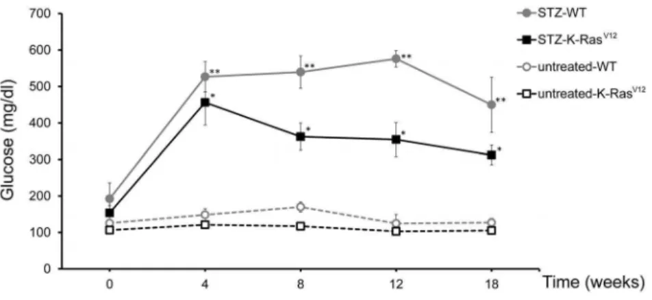
STZ induced diabetes is a well-documented model of experi-mental diabetes [15,16]. Administration of multiple low doses of STZ (40 mg/kg65 day, i.p.) produces significant hyperglycemia
with control groups untreated-WT and untreated-K-RasV12, respectively (Figure 1). The animals maintained high glucose level up to 18 weeks. No statistical differences in the diabetic state were observed between untreated-WT and untreated-K-RasV12
mice (Figure 1). In a separate experiment, STZ treated and untreated mice were killed at 12 weeks for histological and immunohistochemical analysis of pancreas. For WT and K-RasV12 mice, the architecture of islets after STZ treatment was
Figure 1. Average blood glucose levels in WT and K-RasV12mice before and after STZ treatment.Tail-vein glucose was measured after
3 hours of fast. Mean6standard error of mean; *p,0.05versusuntreated-K-RasV12; **p
,0.05versusuntreated-WT (Student’st-test). doi:10.1371/journal.pone.0105550.g001
Figure 2. Histology and immunostaining of pancreas in treated and untreated mice with STZ. A.(i, iv) H-E of a pancreatic islet in untreated and STZ-WT mice; (ii, v) and (iii,vi) reduction of Glut2 and insulin expression in STZ-WT mice compared to control mice.B.(i, iv) H–E of a pancreatic islet in untreated and STZ-K-RasV12mice; (ii, v) and (iii,vi) reduction of Glut2 and insulin expression in STZ-K-RasV12mice compared to untreated K-RasV12control mice. Magnification 2006. Calibration bar: 50mm.
largely disrupted (Figure 2A-iv, 2B-iv), weaker Glut2 positive-cells were observed (Figure 2A-v, 2B-v) and the intensity of insulin staining was reduced (Figure 2A-vi, 2B-vi). On the other hand, a normal shape of pancreatic islet and a normal expression
of both Glut2 and insulin were found in untreated control WT and K-RasV12 mice, represented in Figure 2A-i, ii, iii and Figure 2B-i, ii, iii, respectively [17].
Figure 3. Early and late time representation of tumor formation and progression, respectively. A. Early time, after 4 weeks from STZ treatment, no tumor formation in untreated-K-RasV12mice (i) and some tumors in K-RasV12mice treated with STZ (ii).B. Late time, after 24 weeks from STZ treatment, well defined masses in untreated-K-RasV12mice (i) and several multifocal tumor distribution in K-RasV12treated with STZ (ii).C. Assessment of tumors at low and high grade in untreated-K-RasV12and STZ- K-RasV12.Left: percent of low (white bar) and high (black bar) tumors in STZ- K-RasV12and untreated-K-RasV12mice (mean6SEM; *,p,0.01 by X2test).Right: representative H-E of low and high lung tumor grade. H-E, magnification 1006. Calibration bar: 100mm.
Chronic hyperglycemia increases lung tumorigenesis in K-RasV12 mice
To assess the role of hyperglycemia in oncogenic K-Ras-initiated lung tumorigenesis, mice were killed and examined for lung tumors. No visible lesions were found during the period of observation in untreated-WT and STZ-WT mice (data not shown). Immunohistochemical examination for the earliest detectable lesions (after 4 weeks from STZ treatment) revealed the presence of few small lesions in the STZ-K-RasV12mice, while no adenoma were observed in untreated-K-RasV12mice (Figur-e 3Ai-ii). At 24 w(Figur-e(Figur-eks (lat(Figur-e tim(Figur-e), w(Figur-e notic(Figur-ed som(Figur-e mass(Figur-es in untreated-K-RasV12 mice, with a well-defined distribution, in accordance with the mouse model [13,18] (Figure 3B-i). In contrast, several masses with multifocal distribution were present in K-RasV12mice treated with STZ (Figure 3B-ii). Furthermore, we observed frequently higher grade adenocarcinomas in STZ-K-RasV12 mice than untreated-K-RasV12 group (3460.3% vs 2061.2% respectively; mean 6 SEM; p,0.01, Figure 3C). Consistently, quantitative analysis of visible tumors revealed, at late time, a significant increase of lung lesions in STZ-K-RasV12 mice compared to untreated- K-RasV12ones (1661.5 vs 6.561.2; p,0.05;Figure 4A). The tumor size of STZ-K-RasV12mice was similar to the untreated K-RasV12(Figure 4B). The total amount of cancer cells spread, all over the lung, indicated as tumor burden index, is two fold higher in STZ-K-RasV12mice than untreated K-RasV12ones (Figure 4C). All together, these data indicate that chronic hyperglycemia promotes tumor initiation and progression during K-RasV12-driven tumorigenesis.
Hyperglycemia promotes K-Ras-mediated bronchio-alveolar stem cell expansion
The fact that the STZ- K-RasV12 mice develop more lung tumors, yet at early time of our observation, suggests that hyperglycemia could influence the initial step of K-RasV12 -induced lung tumorigenesis. The clonal expansion of bronchio-alveolar stem cells (BASCs), putative regional stem cells that reside at the terminal bronchioles is thought to be a key event in early step of lung tumor development, at least in this mouse model [13]. Therefore, we assessed whether the presence of high glucose affects the expansion of BASCs expressing oncogenic K-Ras, after 4-OHT injection. To this end, we identified and quantified BASCs in paraffin sections of mouse lungs, by immunohistochemical staining in sequential sections for surfactant protein c (SP-C) and the Clara cell-specific protein (CC10). Within the terminal
bronchioles, in STZ-K-RasV12 and untreated-K-RasV12 mice, columnar epithelial cells showed a typical CC10 staining (Figure 5A-i, 5A-iii). At the bronchio-alveolar duct junction, adjacent the alveolar space, occasional epithelial cells were, also, positive for SP-C staining (Figure 5A-ii, 5A-iv). SP-C positive cells appeared to be strongly increased in hyperglycemic respect to euglycemic K-RasV12 mice. CC10 and SP-C immunostainings were, also, carried out on lung samples from STZ-WT and untreated-WT mice, showing similar CC10 positive cell distribu-tion, but few sporadic or no cells positive for SP-C (data not shown). Quantitive analysis of lung tissue sections, at early and late time, revealed an increase number of BASCs in STZ-K-RasV12 mice compared to other groups (Figure 5B). Thus the hypergly-cemic status enhances BASCs amplification particularly when K-RasV12 is activated. Consistently with BASCs expansion as early event in lung tumorigenesisin vivo, we observed a positive effect of hyperglycemia on oncogenic K-Ras induced proliferation of BASC cultures. Indeed, BASC-like cells derived from K-Ras (+/ LSLG12Vgeo); RERTn (ert/ert) mice, exposed to high glucose media and treated with 4-OHT, underwent a low, but reproduc-ible, increase of about 20% in cell number, compared to control cultures (Figure S2).
K-Ras-mutated BASCs are sensitive to hyperglycemia due to Glut1 expression
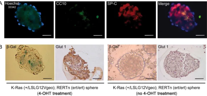
Glucose is the major source of energy for cells and glucose transport 1 (Glut1) is the most common glucose transporter. Glut1 has been found to be abberrantly expressed in K-Ras expressing cells [19–22], suggesting that mutated BASCs exhibit enhanced expansion and promote tumor formation in high glucose conditions owing to enhanced glucose uptake and glycolysis. Accordingly, we found that expression of Glut1 protein was markedly higher in terminal buds of STZ-K-RasV12compared to untreated-K-RasV12 mice (Figure 6A) or STZ-WT (data not shown). As expected the Glut1 protein was found in tumor masses from STZ-K-RasV12(Figure 6B). To further test the specificity of the up-regulation of Glut1, BASC-like cells were isolated using the colony formation assay from K-Ras (+/LSLG12Vgeo); RERTn (ert/ert) mice. After 4-OHT treatmentin vitro, we showed BASC-like cells as double-positive sub-population within spheres, using immunofluorescence labeling with CC10 and SP-C antibodies (Figure 7A). In those spheres we verified the K-Ras oncogene activation byb-Gal staining, taking advantage of the reporter gene in the mouse model (Figure 7B) and we noticed a Glut1 up
regulation if compared to the control spheres which were not treated with 4-OHT and not positive for b-Gal staining (Figure 7B). Therefore, oncogene K-Ras activation induces Glut1 expression in BASCs cells, promoting glucose affinity, and hyperglycemia increases BASCs cell autonomous growth in Glut1 positive BASCs.
Discussion
Numerous epidemiologic studies suggest that diabetes may significantly increase mortality in patients with cancers, including lung cancer [5]. However, metformin, the most-widely used drug for type-2 diabetes, has been shown to have anti-neoplastic activity bothin vitroandin vivoin different studies [23]. Recent animal studies show that treating aggressive lung cancer with metformin along with radiation and chemotherapy may slow down tumor
growth and recurrence [24]. Although these data clearly suggest a direct role of the diabetic milieu in tumor development, these studies do not dissect a pathophysiologic mechanism underlying this phenomenon. A critical question is whether the impact of diabetes on cancer development is due to shared risk factors (obesity, poor diet, physical inactivity and aging) or whether diabetes itself and the specific metabolic derangements, typical of diabetes (e.g., hyperglycemia, insulin resistance, hyperinsulinemia), have a tumor-promoting activity for some types of cancer. Here, we investigated in a mouse model whether hyperglycemia, specifically, affects the growth and progression of lung cancer.
To this end, we used STZ to induce hyperglycemia in a mouse model of K-Ras-driven lung adenocarcinoma. As previously reported, STZ treatment resulted in apoptosis of pancreatic b -cells and consequent inflamatory response contributed and
Figure 5. Analysis of BASCs expansion. A.Immunohistochemical analysis of CC10 and SP-C in terminal bronchioles of STZ-K-RasV12(i, ii) and untreated-K-RasV12(iii, iv) mice. Magnification 2006. Calibration bar: 50mm.B.Bar graph indicates percentage of terminal bronchioles with 1 or more BASCs at early and late time (STZ-K-RasV12vsSTZ-WT: *p
Figure 6. Immunofluorescent staining of tissue sections to detect the Glut1 expression (red). A.Terminal bronchioles of both STZ-K-RasV12and untreated-K-RasV12mice;B.Tumoral masses of STZ-K-RasV12mice. Magnification 1006. Calibration bar: 100mm.
doi:10.1371/journal.pone.0105550.g006
Figure 7. Oncogenic K-Ras induces Glut1 expression in BASCs. A.Immunofluorescent analysis of CC10 (green) and SP-C (red) dual positive BASCs of K-Ras (+/LSLG12Vgeo); RERTn (ert/ert) mice after 4-OHT treatmentin vitro.B.Spheres from K-Ras (+/LSLG12Vgeo); RERTn (ert/ert) mice treated with 4-OHTin vitroshowb-Gal staining and Glut1 expression compared with no Glut1 staining in control spheres from K-Ras (+/LSLG12Vgeo); RERTn (ert/ert) mice not treated with 4-OHT. Magnification 2006. Calibration bar: 50mm.
that elevated circulating glucose levels in K-Ras-driven lung tumorigenesis play a role as tumor promoter and progressor. Indeed, K-Ras-driven tumors exposed to hyperglycemiain vivo grew faster than euglycemic hosts (at early time we, yet, observed tumor mass in STZ-treated mice,Figure 3A–B) and showed a more malignant growth behavior (Figure 3C). Moreover, our current study provides compelling evidence that hyperglycemia, after activation of oncogenic K-Ras, exerts its pro-tumorigenic effects, at least in part, by maintaining a sub-population of cancer cells, namely tumor-initiating lung bronchio-alveolar stem cells (BASCs). Though it is still unclear whether BASCs represent regional lung stem cells, strong circumstantial evidence indicates that they are involved in tumor initiation in the mouse lung. BASCs undergo expansion and transformation in response to K-Ras activation [12]. Genetic and/or pharmacological disruption of multiple key oncogenic pathway genes involved in K-Ras-mediated tumorigenesis, lead to inhibition of BASC expansion and K-Ras-mediated tumor formationin vivo[26]. Human lung adenocarcinomas, which frequently harbor K-Ras mutation, often develop at the bronchio-alveolar duct junction and display either bronchial or alveolar airway differentiation, or both [27,28], suggesting that some of these tumors may have originated from BASC-like cells.
On the other hand, K-Ras oncogene is known to induce aerobic glycolysis [29]. Recently, it has been shown that K-Ras-mutated cells show rapid upregulation of specific glycolytic metabolic enzymes (e.g. the glucose transporter Glut1/Slc2a1) and their pathways prior to any discernible biological impact (e.g., morphological or proliferative changes), a finding consistent with the active control of tumor cell metabolism by oncogenic K-Ras [30]. Our data provide the evidence that the role of hyperglycemia in lung mutated BASCs is cell autonomous. The K-Ras-derived lung tumors show Glut1 staining in tumor cells, with little to no staining in tumor associated-stroma or morphological normal lung epithelium. More importantly, at the bronchio-alveolar duct junction we observed a strong expansion of oncogenic BASCs expressing the glucose transporter Glut1. Thus, while many metabolic changes produced by the tumor microenvironment play prominent roles in the invasive and metastatic properties of lung tumor cells, our data demonstrate that oncogenic K-Ras specifically enhances glycolytic flux in BASCs to support the autonomous growth of these tumor-initiating cells. Our data do not exclude a contributory role of other factors that influence diverse aspects of diabetes (e.g., hyperinsulinemia or chronic inflammation). However, our studies provide new insight into a largely unappreciated role for hyperglycemia in the regulation of tumor-initiating BASCs behavior. In this regard, it is interesting to note that Glut1 expression has been observed to be elevated in oncogenic K-Ras-positive human lung adenocarcinomas [31], suggesting that our results may also be translated in human malignancy.The tumor-promoting activity of hyperglycemia on
epigenetic mechanism, high glucose condition significantly induces histone acetylation, NF-kB activation, and proinflammatory cytokine (e.g., IL-6 and TNF-alpha) release [36]. Although future studies are needed to uncover the specific chromatin events and molecular mechanisms induced by hyperglycemia that influences lung cancer progression, these data further support our hypothesis that the tumor-promoting activity of hyperglycemia can be associated with several aspects of oncogenesis.
Taken together, our data provide compelling evidence that diabetes accelerates lung cancer progression. We hypothesize that due to oncogenic K-Ras activation and increased Glut1 expres-sion, the BASC-like cells are more susceptible to mitogenic and survival signals induced by hyperglicemic milieu. Lung carcino-genesis in diabetic patients represents a two-hit phenomenon, with an oncogene (e.g., K-Ras or others) acting as a tumor initiator and hyperglycemia, at least in part, acting as a tumor promoter. The present study has an important clinical relevance and significant public health implications. Early detection and therapeutic correction of hyperglycemia may help to reduce lung cancer morbidity and mortality, particularly in those expressing onco-genic K-Ras. Furthermore, our data provide the rationale for some diabetes medications as chemopreventive agents in those patients with a risk to develop lung cancer (e.g. smokers, etc.).
Supporting Information
Figure S1 Schematic representation of experimental protocol: 4-OHT treatment, STZ treatment, glucose measurements and sacrifices at different time points are shown.
(TIF)
Figure S2 Effect of high glucose on BASCs proliferation derived from K-Ras (+/LSLG12Vgeo); RERTn (ert/ert) mice.Cell number of BASCs after five days of culture in basal (no Glucose) or in high glucose medium, in the presence or absence of 4-OHT (mean6SEM, n = 3). Cell number was determined by Scepter Cell Counter.
(TIF)
Acknowledgments
We thank M. Barbacid (CNIO, Madrid) for the generous gift of K-Ras (+/ LSLG12Vgeo); RERTn (ert/ert) mouse model and Elena Belloni (IEO, Milan) for suggestions and discussion support. We are, also, grateful to Daniele Giannini for technical support.
Author Contributions
References
1. Siegel R, Naishadham D, Jemal A (2013) Cancer statistics, 2013. CA Cancer J Clin; 63: 11–30.
2. Travis WD, Brambilla E, Muller-Hermelink HK, Harris CC (2004) World Health Organization Classification of Tumours. Pathology and Genetics of Tumours of the Lung, Pleura, Thymus and Heart.IARC Press. pp 9–122. Available: http://www.iarc.fr/en/publications/pdfs-online/pat-gen/bb10/ bb10-chap1.pdf
3. Aviel-Ronen S, Blackhall FH, Shepherd FA, Tsao MS (2006) K-ras mutations in non-small-cell lung carcinoma: a review.Clin Lung Cancer8: 30–38. 4. Mori M, Tezuka F, Chiba R, Funae Y, Watanabe M, et al. (1996) Atypical
adenomatous hyperplasia and adenocarcinoma of the human lung: their heterology in form and analogy in immunohistochemical characteristics.Cancer 77: 665–674.
5. Seshasai SR, Kaptoge S, Thompson A, Di Angelantonio E, Gao P, et al. (2011) Diabetes mellitus, fasting glucose, and risk of cause-specific death. N Engl J Med364(9): 829–41.
6. Pia˛tkiewicz P, Czech A (2011) Glucose metabolism disorders and the risk of cancer.Arch Immunol Ther Exp59(3):215–30.
7. Stopper H, Schinzel R, Sebekova K, Heidland A (2003) Genotoxicity of advanced glycation end products in mammalian cells.Cancer Lett190:151–156. 8. Yamagishi S, Nakamura K, Inoue H, Kikuchi S, Takeuchi M (2005) Possible participation of advanced glycation end products in the pathogenesis of colorectal cancer in diabetic patients.Med Hypotheses64:1208–1210. 9. Yamagishi S, Matsui T, Nakamura K (2008) Possible involvement of
tobacco-derived advanced glycation end products (AGEs) in an increased risk for developing cancers and cardiovascular disease in former smokers. Med Hypotheses71:259–261.
10. Macheda ML, Rogers S, Best JD (2005) Molecular and cellular regulation of glucose transporter (GLUT) proteins in cancer.J Cell Physiol202:654–662. 11. Hitosugi T, Kang S, Vander Heiden MG, Chung TW, Elf S, et al. (2009)
Tyrosine phosphorylation inhibits PKM2 to promote the Warburg effect and tumor growth.Sci Signal2(97): ra73.
12. Kim CF, Jackson EL, Woolfenden AE, Lawrence S, Babar I, et al. (2005) Identification of bronchioalveolar stem cells in normal lung and lung cancer. Cell121: 823–835.
13. Guerra C, Mijimolle N, Dhawahir A, Dubus P, Barradas M, et al. (2003) Tumor induction by an endogenous K-ras oncogene is highly dependent on cellular context.Cancer Cell4(2):111–20.
14. Hogan B, Beddington R, Costantini F, Lacy E (1994) Manipulating the mouse embryo: a laboratory manual, second edition. Cold Spring Harbor, NY;Cold Spring Harbor Laboratory Press.
15. Tian HL, Wei LS, Xu ZX, Zhao RT, Jin DL, et al. (2010) Correlations between blood glucose level and diabetes signs in streptozotocin-induced diabetic mice. Global Journal of Pharmacology4(3): 111–116.
16. Arora S, Ojha SK, Vohora D (2009) Characterization of Streptozotocin induced diabetes mellitus in Swiss albino mice.Global Journal of Pharmacology3(2): 81– 84.
17. Chia JS, McRae JL, Thomas HE, Fynch S, Elkerbout L, et al. (2013) The protective effects of CD39 overexpression in multiple low-dose streptozotocin-induced diabetes in mice.Diabetes62(6):2026–35.
18. Jackson EL, Willis N, Mercer K, Bronson RT, Crowley D, et al. (2001) Analysis of lung tumor initiation and progression using conditional expression of oncogenic K-ras.Genes & Dev15: 3243–3248.
19. Ganapathy V, Thangaraju M, Prasad PD (2009) Nutrient transporters in cancer: relevance to Warburg hypothesis and beyond.Pharmacol Ther121(1):29–40. 20. Szablewski L (2013) Expression of glucose transporters in cancers. Biochim
Biophys Acta1835(2):164–9.
21. de Geus-Oei LF, van Krieken JH, Aliredjo RP, Krabbe PF, Frielink C, et al. (2006) Biological correlates of FDG uptake in non-small cell lung cancer.Lung Cancer55(1):79–87.
22. Sasaki H, Shitara M, Yokota K, Hikosaka Y, Moriyama S, et al. (2012) Overexpression of GLUT1 correlates with Kras mutations in lung carcinomas. Mol Med Rep5(3):599–602.
23. Pollak MN (2012) Investigating metformin for cancer prevention and treatment: the end of the beginning.Cancer Discov2(9):778–90.
24. Storozhuk Y, Hopmans SN, Sanli T, Barron C, Tsiani E, et al. (2013) Metformin inhibits growth and enhances radiation response of non-small cell lung cancer (NSCLC) through ATM and AMPK. Br J Cancer 28; 108(10):2021–32.
25. Liadis N, Murakami K, Eweida M, Elford AR, Sheu L, et al. (2005) Caspase-3-dependent beta-cell apoptosis in the initiation of autoimmune diabetes mellitus. Mol Cell Biol25(9):3620–9.
26. Yang Y, Iwanaga K, Raso MG, Wislez M, Hanna AE, et al. (2008) Phosphatidylinositol 3-kinase mediates bronchioalveolar stem cell expansion in mouse models of oncogenic K-ras-induced lung cancer.PLoS One21;3(5). 27. Morrisey EE, Hogan BL (2010) Preparing for the first breath: genetic and
cellular mechanisms in lung development.Dev Cell18: 8–23.
28. Sutherland KD, Berns A (2010) Cell of origin of lung cancer.Mol Oncol4: 397– 403.
29. Weinberg F, Hamanaka R, Wheaton WW, Weinberg S, Joseph J, et al. (2010) Mitochondrial metabolism and ROS generation are essential for Kras-mediated tumorigenicity.Proc Natl Acad Sci U S A107(19):8788–93.
30. Ying H, Kimmelman AC, Lyssiotis CA, Hua S, Chu GC, et al. (2012) Oncogenic Kras maintains pancreatic tumors through regulation of anabolic glucose metabolism.Cell149(3):656–70.
31. Maki Y, Soh J, Ichimura K, Shien K, Furukawa M, et al. (2013) Impact of GLUT1 and Ki-67 expression on early-stage lung adenocarcinoma diagnosed according to a new international multidisciplinary classification.Oncol Rep 29(1):133–40.
32. Agrimi G, Brambilla L, Frascotti G, Pisano I, Porro D, et al. (2011) Deletion or overexpression of mitochondrial NAD+carriers in Saccharomyces cerevisiae alters cellular NAD and ATP contents and affects mitochondrial metabolism and the rate of glycolysis.Appl Environ Microbiol77(7):2239–46.
33. Semenza GL (2013) HIF-1 mediates metabolic responses to intratumoral hypoxia and oncogenic mutations.J Clin Invest123(9):3664–71.
34. Shyamasundar S, Jadhav SP, Bay BH, Tay SS, Kumar SD, et al. (2013) Analysis of epigenetic factors in mouse embryonic neural stem cells exposed to hyperglycemia.PLoS One8 (6).
35. Reddy MA, Tak Park J, Natarajan R (2013) Epigenetic modifications in the pathogenesis of diabetic nephropathy.Semin Nephrol33(4):341–53. 36. Kim HJ, Kim SH, Yun JM (2012) Fisetin inhibits hyperglycemia-induced