Sharing More than Friendship — Nasal Colonization with
Coagulase-Positive Staphylococci (CPS) and
Co-Habitation Aspects of Dogs and Their Owners
Birgit Walther1*, Julia Hermes2, Christiane Cuny3, Lothar H. Wieler1, Szilvia Vincze1, Yassmin Abou Elnaga1, Ivonne Stamm4, Peter A. Kopp4, Barbara Kohn5, Wolfgang Witte3, Andreas Jansen2, Franz J. Conraths6, Torsten Semmler1, Tim Eckmanns2, Antina Lu¨bke-Becker1
1Institute of Microbiology and Epizootics, Veterinary Faculty, Freie Universita¨t Berlin, Germany,2Department for Infectious Disease Epidemiology, Robert Koch Institute, Berlin, Germany,3National Reference Centre for Staphylococci, Robert Koch Institute, Wernigerode Branch, Germany,4Vet Med Labor GmbH, Division of IDEXX Laboratories, Ludwigsburg, Germany,5Clinic of Small Animals, Veterinary Faculty, Freie Universita¨t Berlin, Berlin, Germany,6Institute of Epidemiology, Friedrich-Loeffler-Institut, Federal Research Institute for Animal Health, Wusterhausen, Germany
Abstract
Background:Since the relationship between dogs and their owners has changed, and dogs moved from being working
dogs to family members in post-industrial countries, we hypothesized that zoonotic transmission of opportunistic pathogens like coagulase positive staphylococci (CPS) is likely between dogs and their owners.
Methodology/Principal Findings:CPS- nasal carriage, different aspects of human-to-dog relationship as well as potential interspecies transmission risk factors were investigated by offering nasal swabs and a questionnaire to dog owners (108) and their dogs (108) at a dog show in 2009.S. aureuswas found in swabs of 20 (18.5%) humans and two dogs (1.8%), and spatypes which correspond to well known humanS. aureuslineages dominated (e.g. CC45, CC30 and CC22). Multilocus sequence typing (MLST) of the two canine strains revealed ST72 and ST2065 (single locus variant of ST34). Fifteen dogs (13.9%) and six owners (5.6%) harbouredS. pseudintermedius, including onemecA-positive human isolate (MRSP). Pulsed field gel electrophoresis (PFGE) revealed that one dog/owner pair harboured indistinguishableS. pseudintermedius-isolates of ST33. Ten (48%) of the 21S. pseudintermedius-isolates showed resistance towards more than one antimicrobial class. 88.9% of the dog owners reported to allow at least one dog into the house, 68.5% allow the dog(s) to rest on the sofa, 39.8% allow their dogs to come onto the bed, 93.5% let them lick their hands and 52.8% let them lick their face. Bivariate analysis of putative risk factors revealed that dog owners who keep more than two dogs have a significantly higher chance of being colonized withS. pseudintermediusthan those who keep 1–2 dogs (p,0.05).
Conclusions/Recommendations:In conclusion, CPS transmission between dog owners and their dogs is possible. Further
investigation regarding interspecies transmission and the diverse adaptive pathways influencing the epidemiology of CPS (including MRSA and MRSP) in different hosts is needed.
Citation:Walther B, Hermes J, Cuny C, Wieler LH, Vincze S, et al. (2012) Sharing More than Friendship — Nasal Colonization with Coagulase-Positive Staphylococci (CPS) and Co-Habitation Aspects of Dogs and Their Owners. PLoS ONE 7(4): e35197. doi:10.1371/journal.pone.0035197
Editor:Herman Tse, The University of Hong Kong, Hong Kong
ReceivedJanuary 17, 2012;AcceptedMarch 12, 2012;PublishedApril 18, 2012
Copyright:ß2012 Walther et al. This is an open-access article distributed under the terms of the Creative Commons Attribution License, which permits unrestricted use, distribution, and reproduction in any medium, provided the original author and source are credited.
Funding:S. Vincze is financed by the BMBF-founded interdisciplinary expert network project on zoonotic Staphylococcus aureus (MedVet- Staph: IP8, 01KI1014F:). S. Vincze and Torsten Semmler were supported by the Federal Ministry of Education and Research (MedVet- Staph, Grant no. 01KI1014F) and (FBI-Zoo, Grant no. 01KI1012A). No additional external funding was received for this study. The funders had no role in study design, data collection and analysis, decision to publish, or preparation of the manuscript.
Competing Interests:Dr. Stamm and Dr. Kopp are employees of IDEXX Vet Med Labor GmbH (Ludwigsburg). While these authors are company employees, this does not alter their adherence to all the PLoS ONE policies on sharing data and materials. All other authors have declared that no competing interests exist.
* E-mail: walther.birgit@vetmed.fu-berlin.de
Introduction
In post-industrial countries, the socio-economic relationship between many dogs and their owners has changed dramatically during the last decades [1]. Instead of having working dogs (watch dogs, sheepdogs etc.) living in stables or kennels, people keep pets who live in the household almost as family members. In particular people who feel disconnected from society tend to substitute social contacts by pets, including supportive anthropomorphic traits (‘‘humanisation’’ of non-human beings) [2,3]. As a result of this development, transmission of microorganisms between humans and
their dogs may increase [4]. Especially microorganisms that reside on the skin or mucosal surfaces of dogs might easily be transmitted to pet owners either by direct contact or by sharing the same environment in the household, e.g. sofas, beds and bath tubs.
Commensals usually consist of a wide range of different bacterial species, including coagulase-positive staphylococci (CPS) like S. aureusandS. pseudintermedius. WhileS. aureusis a common colonizer in humans and widely distributed among mammalian hosts in general,
Opportunistic pathogens like CPS seem to be of special interest, since reports about the general transferability of these bacteria between pets and humans have increased during recent years, including methicillin resistant variants ofS. aureus(MRSA) and S.
pseudintermedius(MRSP) [9,10,11,12,13,14].
MRSA have evolved to one of the major nosocomial pathogens in healthcare systems, causing an increased mortality risk for patients and enormous costs for health insurance companies worldwide [15]. In the field of veterinary medicine, nosocomial outbreaks caused by MRSA have recently gained attention, e.g. in equine and small animal clinics [16,17]. Nasally colonized veterinary personnel seem to have an impact on rising infection rates in animal patients, just like health care workers in hospitals [14].
As far as CPS are concerned,S. pseudintermediusseems to be more common among dogs and cats than S. aureus [10]. Moreover, certain strains ofS. pseudintermediushave become a major threat in veterinary medicine due to their frequent methicillin- and multidrug resistant appearance [18,19].
The first serious human infections with S. pseudintermedius
(including MRSP) have recently been reported [20,21]. Owners of dogs suffering from dermatitis seem to be frequently colonized withS. pseudintermediusstrains (including MRSP) [13,22,23].
In general, data about the colonization of dogs and their owners with CPS in the community are scarce. Hanselmanet al.(2009) reported that S. aureus, S. pseudintermedius and S. schleiferi ssp.
coagulans (including methicillin-resistant variants) were harboured by dogs living in households. In addition, concurrent human and animal colonization by indistinguishable S. aureus and S. pseudin-termediusstrains have been observed [10]. Against this backdrop, the question has arisen how changes in the human-to-dog relationship influence inter-species CPS transmission.
Therefore, the aim of this study was to assess the nasal colonisation of dogs and their owners with CPS and to gain insights into potential risk factors for CPS transmission among them.
Materials and Methods
Questionnaires and ethical considerations
The study procedure and protocols were based on informed consent of the participants.
The sampling procedures in dogs had been approved by the Ethical Committee of the veterinary department of the Free University of Berlin (date: 03.03.2009).
Sampling of dog owners was approved by the Ethical Committee of the Charite´, Campus Virchow-Clinic (date: 10.10.2008, Berlin).
After the dog owners had been informed about the study, they were given a questionnaire to collect demographic data on owners (e.g. gender, age and profession), number and keeping conditions of the dogs and information on the quality of contacts they shared (e.g. frequency and type of dog handling, including physical contact). Questionnaires were completed by the owners just before or after the nasal swabs were taken.
Sampling procedure and bacterial growth
Nasal swabs (MASTASWABTM; MAST Diagnostica GmbH, Rheinfeld Germany), were simultaneously collected from volun-tarily participating pet owners (by physicians) and dogs (by veterinarians) as a convenience sample on a single day at a dog show event in Berlin in March 2009.
Until processing, all nasal samples were stored for approxi-mately 36 h at 4uC. Swabs were plated directly on columbia agar
containing 5% sheep blood and BBLTMCHROMagarTMMRSA II (Becton Dickinson GmbH, Heidelberg, Germany). All incubat-ed plates were investigatincubat-ed twice, first after 18 h and a second time after 36 h of incubation at 37uC. CPS suspected colonies were picked (at least two per plate) and subcultured.
Identification and typing of isolates
Phenotypic characterisation of CPS was performed as previ-ously reported [8,24]. Briefly, staphylococcal isolates were identified as CPS based on colony morphology, a positive catalase reaction, gram stain appearance and a positive tube coagulase reaction. All CPS were further screened formecA by PCR andnuc
for species verification in case ofS. aureus[25]. Species verification ofS. pseudintermediuswas performed by the MboI restriction analysis method ofptaas described before [26].
Automated antimicrobial susceptibility testing ofS. pseudinterme-dius was performed by Vet Med Labor GmbH (Ludwigsburg, Germany) using the bioMerieux VITEKH2 system according to the manufacturer’s instructions including penicillin, ampicillin-sulbactam, oxacillin, gentamicin, kanamycin, enrofloxacin, mar-bofloxacin, erythromycin, clindamycin, tetracycline, nitrofuranto-in, chloramphenicol and trimethoprim- sulfamethoxazole, follow-ing the CLSI guidelines.
AllS. aureusstrains underwentspa-typing and PCR detection of Panton-Valentine leukocidin (lukS-PV, lukF-PV) as previously reported [27,28]. MLST was performed on canineS. aureusstrains as described [29]. Possible associations between spa types and corresponding clonal complexes (CC) were determined according to the Ridom (http://spaserver.ridom.de/) and the MLST database (www.mlst.net).
MLST analysis onS. pseudintermediuswas performed according to a scheme published by Bannoehret al.(2007) includingpta,cpn60,
tuf, 16S rRNA andagrD [30]. Allele numbers and new STs were assigned according to the curator of the MLST database V. Perreten (vincent.perreten@vbi.unibe.ch).
PFGE analysis on S. pseudintermedius was carried out using endonuclease SmaI according to Ruscheret al.2010 [19]. PCR-based detection of the bicomponent leukotoxin Luk-I (lukS and
lukF) was carried out as described previously [31]. The presence of the exfoliative toxin ExpA (formerly named EXI) was determined by PCR with the primersexpA-F GCGCGTCCTTCTGATCCA-GAACT andexpA-R AACGTCCCCCTTTACCTACGTGAAT (according to AB489850.1, [32] at an annealing temperature of 58uC (25 cycles) including strain IMT21652 (JN604832) as a positive control. Detection of the gene encoding ExpB was performed as described previously [33].
Statistical analysis
Statistical analysis was conducted using STATA, version 11 (StataCorp). For bivariate analysis of risk factors for human CPS colonization, Odds Ratios (OR) were calculated and potential associations checked for statistical significance using the 2-tailed Fisher exact test. A value of p,0.05 was considered significant. Results were adjusted for age and sex.
Results
Description of study population
The mean age of the study participants was 48.5 years (range: 23–87 years, median 49 years), 75% were female and all but one were German residents. Analysing the zip codes (first three digits) of the places where the dog owners lived revealed a wide geographic distribution over Germany (data not shown). 50.9% of the participants declared to own only one dog, while 47.2% of them possessed two or more dogs (median: one dog, mean 2.2 dogs, range 1–11 dogs). Nine of the dog owners worked in a healthcare associated area (hospital, nursing home, medical practise) and three were veterinarians. On the day of sampling, only one dog that accompanied the owner was included in the study.
Behavioural aspects of human-pet cohabitation
The majority of the study subjects (96/108; 88.9%) reported to share their habitation with one or more dogs, while ten (9.3%) kept the dogs in a kennel or garden. Seventy-four dog owners (68.5%) allowed their dogs to rest on the sofa, 43 (39.8%) permitted them to come onto their beds, 101 (93.5%) let their dogs lick their hands, 57 (52.8%) allowed them to lick their face, and 35 (32.4%) reported washing their dogs in their own bath tubs (Table 1).
S. aureusnasal carriage in healthy dogs and their owners and strain characterisation
Nasal carriage ofS. aureuswas found in 20 (18.5%) dog owners and two dogs (1.8%). With regard toS. aureus-positive individuals, no simultaneous colonization of dog and dog owner was observed in this study. One of the three veterinarians and one of the nine participants with a known healthcare association were found to be
S. aureuscarriers. None of theS. aureus-isolates harboured the genes encoding for PVL or methicillin resistance.
Spa-typing of all 22 S. aureus isolates showed a broad heterogeneity and wide distribution, including two new spa
variants. According to the spa database, the twenty S. aureus
isolates of human origin showed the following results: Four strains were assigned to spa types which presumptively correspond to CC45, three strains to CC59 and three others to CC7. Two strains may correspond to CC30 and two more to CC22. Another sixspa
types t346, t495, t156, t1294, t8641 (new) and t8640 (new) remained unassigned (Table 2).
MLST-analysis of the two canineS. aureus-isolates revealed that one belonged to the genetic lineage ST72 (IMT18888), and the other to ST2065 (IMT18809), a single locus variant of ST34 (Table 2: bold letters).
S.pseudintermediusnasal carriage in healthy dogs and their owners and strain characterisation
Six owners (5.6%) and fifteen dogs (13.9%) were carriers ofS. pseudintermedius. In addition, one of the six human isolates (IMT18885) proved to bemecA-positive (MRSP). None of them were veterinarians or healthcare workers.
MLST analysis of all 21S. pseudintermediusisolates of human and canine origin revealed a heterogenic distribution of already known and 9 new sequence types (Fig. 1).
Table 1.Descriptive characteristics of dog owners.
Answers
positive negative no data
Dog owner characteristics
(n = 108) No. % No. % No. %
Sex 0
Female 81 75.0
Male 27 25.0
Keeps dog/dogs in the house 96 88.9 10 9.3 2 1.9
Dog breeding 22 20.4 80 74.1 6 5.6
Number of dogs in household 2 1.9
one dog 55 50.9
two dogs 25 23.1
three or more dogs (range: 3 to 11)
26 24.1
Allows to lick the hands (total)
101 93.5 5 4.6 2 1.9
Allows to lick the face (total) 57 52.8 37 34.3 14 13.0
Washes dog in bath tub (total)
35 32.4 53 49.1 20 18.5
Allows dog to rest on sofa (total)
74 68.5 28 25.9 6 5.6
Allows dog to rest on bed (total)
43 39.8 53 49.1 12 11.1
doi:10.1371/journal.pone.0035197.t001
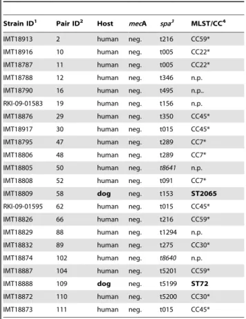
Table 2.Molecular characteristics ofS. aureusfrom the nasal cavity of dog owners and dogs.
Strain ID1 Pair ID2 Host mecA spa3 MLST/CC4
IMT18913 2 human neg. t216 CC59*
IMT18916 10 human neg. t005 CC22*
IMT18787 11 human neg. t005 CC22*
IMT18788 12 human neg. t346 n.p.
IMT18790 16 human neg. t495 n.p..
RKI-09-01583 19 human neg. t156 n.p.
IMT18876 29 human neg. t350 CC45*
IMT18917 30 human neg. t015 CC45*
IMT18795 47 human neg. t289 CC7*
IMT18806 48 human neg. t289 CC7*
IMT18805 50 human neg. t8641 n.p.
IMT18808 52 human neg. t091 CC7*
IMT18809 58 dog neg. t153 ST2065
RKI-09-01595 62 human neg. t015 CC45*
IMT18826 66 human neg. t216 CC59*
IMT18829 88 human neg. t1294 n.p.
IMT18832 89 human neg. t275 CC30*
IMT18874 102 human neg. t8640 n.p.
IMT18887 104 human neg. t5201 CC59*
IMT18888 109 dog neg. t5199 ST72
IMT18872 110 human neg. t5200 CC30*
IMT18873 111 human neg. t015 CC45*
1Strain collection number: IMT: Institute of Microbiology and Epizootics, RKI:
Robert Koch-Institute (Wernigerode branch).
2Individual number for each of the participating dog/dog owner pairs. 3italic:newspatypes.
4ST determined according to MLST result (bold),
Concurrent owner and dogS. pseudintermedius-colonization was demonstrated for the isolates IMT18797 (human) and IMT18798 (canine) by PFGE pattern analysis, revealing one pulsotype for both isolates, which belongs to ST33 (Fig. 1, pair 36).Two further
S. pseudintermedius-isolates had indistinguishable PFGE patterns but were obtained from unrelated canine and human individuals of pair 11 and 74, respectively (Fig. 1; IMT18789 and IMT18828). All 21 S. pseudintermedius isolates were positive for LukI, one yielded a positive signal for ExpA and two for ExpB (Fig. 1).
Each of the 21 S. pseudintermedius isolates showed at least penicillin resistance (100%), while 12 (57%) were resistant towards kanamycin, 10 (48%) to erythromycin, 8 (38%) to chloramphen-icol, 7 (33%) to tetracycline, 4 (19%) to clindamycin, 2 (9%) to gentamicin, one (4%) to enrofloxacin and one (4%) to marbo-floxacin. The isolates were susceptible to the remaining antimi-crobials (Fig. 1).
S. schleiferiissp.coagulansnasal carriage in healthy dogs and their owners
A single strain of S. schleiferii ssp. coagulans (IMT18886) was isolated from the nostrils of one dog. This isolate was not further analysed.
Statistical analysis of potential risk factors associated with S. pseudintermediuscarriage in humans
Dog owners who kept more than two dogs had a significantly higher chance of being colonized withS. pseudintermediusthan those who kept 1–2 dogs (p,0.05). No other suspected risk factor proved statistically significant, but elevated chances forS. pseudintermedius
colonization were observed for dog breeders (OR = 3.9), for owners who allowed dogs to lick their faces (OR = 3.5), to rest on the sofa (OR = 2), or to sleep in their bed (OR = 1.25). A summary of the risk factor analysis is provided in Table 3. The result remained unchanged when adjusted for age and sex.
Discussion
This study aimed at elucidating aspects of the relationship and behavioural patterns between dog owners and their pets with regard to nasal colonisation with CPS. In addition, comparative analysis of CPS-carriage among dogs and humans allowed insights into the proportions of CPS carriage for each group, with a special focus on CPS as zoonotic pathogens.
Substantial proportions of the participating dog owners not only shared their house (88.9%) with their pets, but also their sofa (68.5%) or their beds (39.8%). Many dogs had obviously gained a status nearly equal to that of a (human) family member, i.e. the animals were given privileges formerly exclusively meant for humans, which are behavioural aspects typical for anthropomor-phism.
Zoonotic transmission of CPS has been described [11,13,34] and it appears likely that the intensive daily contact between dogs and their owners may increase the likelihood of getting colonized by interspecies-transmission of CPS for both sides.
It is noteworthy that individual refusals to answer certain questions reached up to 11%, which may have biased the study results (Tables 1 and 3, columns ‘‘no data’’ and ‘‘valid response’’). Furthermore, almost half of the participating dog owners (47.2%) stated that they keep more than just the one dog that was available for sampling on the day of sampling. While the CPS-colonisation status of the other dog(s) remains unknown, it can be assumed that all dogs of one owner are likely to live in similar co-habitation conditions.
Longitudinal studies regarding S. aureus nasal carriage in humans report approximately 20% (range 12–30%) of individuals who are persistently colonized and 50% (range 16–69%) non-carriers depending on the study protocols and methods (e.g. use of enrichment broth, kind of study population) [35]. A recent study demonstrated 19.1% nasal colonisation in 278 healthy humans in Spain [36]. Eventually, the observed 18.5%S. aureus-colonization
Figure 1. Comparative PFGE analysis and strain characteristics ofS. pseudintermediusfrom human and canine origin.Dendrogram (percent similarity) showing DNA restriction pattern after digestion with SmaI for all 21 MSSP and MRSP isolates. PFGE analysis by use of bionumericsH(unweighted-pair group method using average linkages), dice coefficient, 1.2% tolerance and 0.5% optimization. Pair 36 comprised the isolates IMT18797 (canine) and IMT18798 (human), which show indistinguishable PFGE pattern. The canine isolate IMT18789 and IMT18828 (human) share an indistinguishable pulsotype as well, although they did not compose a dog/dog owner pair.1Strain collection number: IMT: Institute of Microbiology and Epizootics. RKI: Robert Koch-Institute (Wernigerode branch).2Individual number for each of the participating dog/dog owner pairs. 3
Determined by use of VITEKH2 (bioMerieux) according to the manufacturers instructions.4Allelic profile of the genespta,cpn60,tuf, 16S rRNA and agrD. Abbreviations: PEN, Penicillin G; GEN, Gentamicin; KAN, Kanamycin; ENR, Enrofloxacin; MAR, Marbofloxacin; ERY, Erythromycin; CLI, Clindamycin; TET, Tetracycline; CHL, Chloramphenicol; i: susceptibility testing result: intermediate. Luk-I: Leukotoxin I (lukS andlukF),expA: Exfoliative toxin A (primer: this study),expB: Exfoliative toxin B.
in the human participants of our study is marginally lower but in overall agreement with formerly published results and probably reflecting the abandonment of enrichment broth.
At present, data on the colonization of healthy dogs withS. aureusgained in a community environment is very limited. In this study, 1.9% nasalS. aureus- carriage was found among those dogs. When studies on S. aureus nasal carriage in dogs are compared, care should be taken with regard to individual study backgrounds, as the kind and selection of study participants, the use of enrichment broth, sampling in a clinical or in a non-clinical environment must be expected to influence the study results. However, Rubin et al. found 13 of 167 (7.8%) nasal S. aureus -colonization among clinically healthy dogs presented for vaccina-tion in a veterinary college [37], while Fazakerleyet al. reported 2.3% (1 of 43) nasal colonization in healthy dogs [38]. In addition, we reported aboutS. aureus-(nasal) carriage of 5.7% in dogs on admission to an animal hospital [14].
Spa-typing of all human and canine S. aureus revealed a high diversity of spa types as previously observed for community associated S. aureus [36]. The majority of spa types of human isolates (16/20) seemed to be associated with well known human lineages (e.g. CC45, CC30, CC22) and the remaining six had not been described in any context to animal associated lineages before. Therefore, a risk factor analysis for S. aureus-carriage in humans could not be conducted.
Interestingly, one of the two canine MSSA belonged to ST72, a genetic background also displayed by the USA700 type strain (CA-MRSA) and further MSSA and MRSA of human origin (http:// saureus.mlst.net). The second canine strain is a single nucleotide variant of ST34, an ST that had also previously been associated with MRSA of human origin [39]. In conclusion, both canineS. aureusstrains isolated in this study may have once originated from a human host. These strains could belong to extended host spectrum genotypes (EHSGs), which were previously reported for
MRSA strains of equine origin [40]. Consequently, dogs can be colonized with EHSG S. aureus strains (including MRSA) and represent a potential source of (re-) infection for humans [11].
Surprisingly, 5.5% of dog owners were found to be positive for S.pseudintermediusnasal carriage, including MRSP in one case. A recent study by Paul and co-workers found a nasal colonisation in 3.9% of 128 veterinarians attending a conference for animal dermatologists [41]. All strains were proven to be MRSP and their nasal carriage persisted for at least one month. While human infections withS. pseudintermediushave occasionally been reported in the past, an increased awareness of these CPS in human medicine ensued very recently because of the multi-drug resistant phenotype which is frequently associated with MRSP [19,41,42]. We found one dog/owner pair contemporarily colonized with one particularS. pseudintermediusstrain, as has previously been reported for MRSP in clinical cases [13,43]. Moreover, another person and a dog who had no relationship according to their pair ID’s were colonized with strains displaying an indistinguishable PFGE-pattern, which emphasizes the transferability ofS. pseudintermedius. WhileS. pseudintermediusis considered as a common colonizer in the first line [7], the antibiotic resistance phenotypes demonstrated that ten of the 21 isolates showed resistance towards four or more antimicrobials. Among these isolates was strain IMT18884, which showed resistance towards 7 agents (Fig. 1).
It is striking that all six dog owners who carriedS. pseudintermedius
kept their dogs in the house, and had three or more dogs (one person did not answer the respective question). Statistical analysis supports the result by revealing that keeping two or more dogs is significantly associated with S. pseudintermedius colonization in humans (p = 0,0006). Other suspected risk factors were not statistically significantly associated withS. pseudintermediuscarriage in our analysis. Interpretation of statistical results needs to take the small number of cases into account.
Table 3.Risk factors forS. pseudintermediuscolonization among dog owners sampled during a dog show event in Berlin in 2009 (n = 108) (bivariate analysis).
Exposure Valid response Nasal colonisation state of dog owners
Bivariate analysis (2-sided Fisher’s exact test)
S. pseudintermedius S. pseudintermedius
positive negative
no (%)
no (% of valid responses)
no (% of valid
responses) OR p-value 95% CI
Sex 108 (100%) f 4 (66.6%) 77 (75.5%)
m 2 (33.3%) 25 (24.5%) 1.54 0.6 0.1–11.3
Keeps dog/s in the house 106 (98.1%) 6 (100%) 90 (90.0%) -* 1 0.2
-Breeds dogs 102 (94.4%) 2 (50.0%) 20 (20.4%) 3.9 0.2 0.3–55.9
Keeps.2 dogs 106 (98.1%) 5 (100%) 21 (20.8%) -* 0.0006 4.7
-Dog/s allowed to lick the face 94 (87.0%) 5 (83.3%) 52 (59.1%) 3.5 0.4 0.4–168.2
Dog/s allowed to lick hands 106 (98.1%) 6 (100%) 95 (95.0%) -* 1 0.07
-Dog/s in bath tub 88 (81.5%) 1 (20.0%) 34 (41.0%) 0.4 0.6 0.0–3.9
Dog/s allowed on sofa 102 (94.4%) 5 (83.3%) 69 (71.9%) 2 1 0.2–95.9
Dog/s allowed in bed 96 (88.9%) 3 (50.0%) 40 (44.4%) 1.25 1 0.2–9.8
Abbreviations: f, female; m, male; *not calculated. OR: Odds ratio. CI: Confidence interval.
As a matter of fact, more dogs will result in more saliva as well as more dog scurf and hair harbouring commensals (including CPS) in the household environment shared by both, dogs and owners. In addition, domestic contamination due toS. pseudinter-medius has been reported for households of MRSP-infected pets before, and it was assumed that dust particles may play a role in this finding [34].
Furthermore, it was thought that certain MRSP lineages (e.g. ST71) may have a greater ability to adapt to the human host [41] than MSSP. However, it is important to note that the MRSP and MSSP strains from human origin detected in this study comprised six different STs (Fig. 1). Regarding the exfoliative toxins in MSSP reported on here, one human isolate yielded a positive PCR result for ExpA, while two canine strains were found positive for ExpB, indicating that these virulence factors may be more frequently associated with cases of superficial dermatitis in dogs [32,33].
The single MRSP (IMT18885) isolated during this study was of human origin, showed an ST41 background and was susceptible to most of the tested antimicrobials. In addition, IMT18885 had an oxacillin MIC of 2 mg/l and expressed a comparatively low MIC (4/2 mg/l) towards ampicillin-sulbactam (Fig. 1). Interest-ingly, this ST was originally described for a canine isolate (mecA negative) in the USA [30]. It seems likely that acquisition events of
mecA harbouring staphylococcal chromosomal cassettes will occur in accessible strains over time.
In conclusion, exposure of humans to pet dogs seems to be associated with the possibility of getting colonized by S. pseudintermedius, regardless whether these bacteria are
methicillin-resistant or not. Methicillin-methicillin-resistant CPS attract more scientific interest because of the clinical relevance of MRS, but according to our findings the inter-species transferability does not seem to be necessarily linked with methicillin-resistance at all. However, it remains unknown whether S. pseudintermedius colonization in humans is transient or permanent. Further investigations regard-ing inter-species transmission and the diverse adaptive pathways influencing the epidemiology of CPS (including MRSA and MRSP) in the community are needed. Especially the close relationship between pets and their owners underline the importance of interdisciplinary research including human and veterinary microbiologists as well as epidemiologists as demon-strated by our study results.
Acknowledgments
The authors thank C. Ruscher, K. Heidemanns, C. Probst and A. Hilbert for their help in collecting specimens and E. Anta˜o for critical reading of the manuscript. We particularly thank the German kennel club (Verband fu¨r das Deutsche Hundewesen; VDH) for the excellent cooperation.
Author Contributions
Conceived and designed the experiments: BW BK FJC TE LHW ALB. Performed the experiments: BW AJ FJC CC ALB SV YAE IS PAK. Analyzed the data: BW JH CC WW FJC TS. Contributed reagents/ materials/analysis tools: BW JH CC WW TS IS PAK. Wrote the paper: BW JH ALB FJC BK IS LHW.
References
1. Franklin A (1999) Animals and modern cultures: a sociology of human-animal relations in modernity. London: SAGE publications Ltd.
2. Blouin DD (2008) All in the Family? Understanding the Meaning of Dogs and Cats in the Lives of American Pet Owners [dissertation]. Bloomington: Indiana University, USA.
3. Epley N, Waytz A, Akalis S, Cacioppo JT (2008) When we need a human: motivational determinants of anthropomorphism. Social Cognition 26: 143–155.
4. Wieler LH, Ewers C, Guenther S, Walther B, Lu¨bke-Becker A (2011) Methicillin-resistant staphylococci (MRS) and extended-spectrum beta-lacta-mases (ESBL)-producing Enterobacteriaceae in companion animals: nosocomial infections as one reason for the rising prevalence of these potential zoonotic pathogens in clinical samples. Int J Med Microbiol 301: 635–641.
5. Cuny C, Friedrich A, Kozytska S, Layer F, Nu¨bel U, et al. (2010) Emergence of methicillin-resistant Staphylococcus aureus (MRSA) in different animal species. Int J Med Microbiol 300: 109–117.
6. Mahoudeau I, Delabranche X, Prevost G, Monteil H, Piemont Y (1997) Frequency of isolation of Staphylococcus intermedius from humans. J Clin Microbiol 35: 2153–2154.
7. Perreten V, Kadlec K, Schwarz S, Gronlund Andersson U, Finn M, et al. (2010) Clonal spread of methicillin-resistantStaphylococcus pseudintermediusin Europe and North America: an international multicentre study. J Antimicrob Chemother 65: 1145–1154.
8. Ruscher C, Lu¨bke-Becker A, Wleklinski CG, Soba A, Wieler LH, et al. (2009) Prevalence of Methicillin-resistant Staphylococcus pseudintermedius isolated from clinical samples of companion animals and equidaes. Vet Microbiol 136: 197–201.
9. Cohn LA, Middleton JR (2010) A veterinary perspective on methicillin-resistant staphylococci. J Vet Emerg Crit Care (San Antonio) 20: 31–45.
10. Hanselman BA, Kruth SA, Rousseau J, Weese JS (2009) Coagulase positive staphylococcal colonization of humans and their household pets. Can Vet J 50: 954–958.
11. Manian FA (2003) Asymptomatic nasal carriage of mupirocin-resistant, methicillin-resistantStaphylococcus aureus(MRSA) in a pet dog associated with MRSA infection in household contacts. Clin Infect Dis 36: e26–28. 12. Nienhoff U, Kadlec K, Chaberny IF, Verspohl J, Gerlach GF, et al. (2009)
Transmission of methicillin-resistantStaphylococcus aureusstrains between humans and dogs: two case reports. J Antimicrob Chemother 64: 660–662.
13. Vincze S, Paasch A, Walther B, Ruscher C, Lu¨bke-Becker A, et al. (2010) Multidrug- and methicillin resistantStaphylococcus pseudintermediusas a cause of canine pyoderma: a case report. Berl und Mu¨nch Tiera¨rztl Wochenschr 123: 335–358.
14. Walther B, Wieler LH, Friedrich AW, Kohn B, Brunnberg L, et al. (2009)
Staphylococcus aureusand MRSA colonization rates among personnel and dogs in a
small animal hospital: Effect on nosocomial infections. Berl Mu¨nch Tierarztl Wochenschr 122: 178–185.
15. Engemann JJ, Carmeli Y, Cosgrove SE, Fowler VG, Bronstein MZ, et al. (2003) Adverse clinical and economic outcomes attributable to methicillin resistance among patients withStaphylococcus aureussurgical site infection. Clin Infect Dis 36: 592–598.
16. Ishihara K, Shimokubo N, Sakagami A, Ueno H, Muramatsu Y, et al. (2010) Occurrence and molecular characteristics of methicillin-resistantStaphylococcus aureus and methicillin-resistant Staphylococcus pseudintermedius in an academic veterinary hospital. Appl Environ Microbiol 76: 5165–5174.
17. Weese JS, Rousseau J, Willey BM, Archambault M, McGeer A, et al. (2006) Methicillin-resistant Staphylococcus aureus in horses at a veterinary teaching hospital: frequency, characterization, and association with clinical disease. J Vet Intern Med 20: 182–186.
18. Black CC, Solyman SM, Eberlein LC, Bemis DA, Woron AM, et al. (2009) Identification of a predominant multilocus sequence type, pulsed-field gel electrophoresis cluster, and novel staphylococcal chromosomal cassette in clinical isolates of mecA-containing, methicillin-resistantStaphylococcus pseudinter-medius. Vet Microbiol 139: 333–338.
19. Ruscher C, Lu¨bke-Becker A, Semmler T, Wleklinski CG, Paasch A, et al. (2010) Widespread rapid emergence of a distinct methicillin- and multidrug-resistant
Staphylococcus pseudintermedius(MRSP) genetic lineage in Europe. Vet Microbiol 144: 340–346.
20. Chuang CY, Yang YL, Hsueh PR, Lee PI (2010) Catheter-related bacteremia caused byStaphylococcus pseudintermediusrefractory to antibiotic-lock therapy in a hemophilic child with dog exposure. J Clin Microbiol 48: 1497–1498. 21. Kempker R, Mangalat D, Kongphet-Tran T, Eaton M (2009) Beware of the pet
dog: a case ofStaphylococcus intermediusinfection. Am J Med Sci 338: 425–427. 22. Frank LA, Kania SA, Kirzeder EM, Eberlein LC, Bemis DA (2009) Risk of
colonization or gene transfer to owners of dogs with meticillin-resistant
Staphylococcus pseudintermedius. Vet Dermatol 20: 496–501.
23. Loeffler A, Pfeiffer DU, Lloyd DH, Smith H, Soares-Magalhaes R, et al. (2010) Meticillin-resistant Staphylococcus aureus carriage in UK veterinary staff and owners of infected pets: new risk groups. J Hosp Infect.
24. Becker K, von Eiff C (2011) Staphylococcus, Micrococcus and other catalase-positive cocci. In: Murray P, Baron E, Jorgensen J, Landry ML, Pfaller M, eds. Manual of Clinical Microbiology, 10th ed. Washington: American Society for Microbiology (ASM) Press. 308 p.
26. Bannoehr J, Franco A, Iurescia M, Battisti A, Fitzgerald JR (2009) Molecular diagnostic identification ofStaphylococcus pseudintermedius. J Clin Microbiol 47: 469–471.
27. Lina G, Piemont Y, Godail-Gamot F, Bes M, Peter MO, et al. (1999) Involvement of Panton-Valentine leukocidin-producingStaphylococcus aureusin primary skin infections and pneumonia. Clin Infect Dis 29: 1128–1132. 28. Harmsen D, Claus H, Witte W, Rothganger J, Turnwald D, et al. (2003) Typing
of methicillin-resistantStaphylococcus aureusin a university hospital setting by using novel software forsparepeat determination and database management. J Clin Microbiol 41: 5442–5428.
29. Enright MC, Day NP, Davies CE, Peacock SJ, Spratt BG (2000) Multilocus sequence typing for characterization of resistant and methicillin-susceptible clones ofStaphylococcus aureus. J Clin Microbiol 38: 1008–1015. 30. Bannoehr J, Ben Zakour NL, Waller AS, Guardabassi L, Thoday KL, et al.
(2007) Population genetic structure of theStaphylococcus intermediusgroup: insights into agr diversification and the emergence of methicillin-resistant strains. J Bacteriol 189: 8685–8692.
31. Futagawa-Saito K, Sugiyama T, Karube S, Sakurai N, Ba-Thein W, et al. (2004) Prevalence and characterization of leukotoxin-producingStaphylococcus intermedius
in Isolates from dogs and pigeons. J Clin Microbiol 42: 5324–5326. 32. Futagawa-Saito K, Makino S, Sunaga F, Kato Y, Sakurai-Komada N, et al.
(2009) Identification of first exfoliative toxin inStaphylococcus pseudintermedius. FEMS Microbiol Lett 301: 176–180.
33. Iyori K, Hisatsune J, Kawakami T, Shibata S, Murayama N, et al. (2010) Identification of a novelStaphylococcus pseudintermediusexfoliative toxin gene and its prevalence in isolates from canines with pyoderma and healthy dogs. FEMS Microbiol Lett 312: 169–175.
34. van Duijkeren E, Kamphuis M, van der Mije IC, Laarhoven LM, Duim B, et al. (2011) Transmission of methicillin-resistantStaphylococcus pseudintermediusbetween
infected dogs and cats and contact pets, humans and the environment in households and veterinary clinics. Vet Microbiol.
35. Wertheim HF, Melles DC, Vos MC, van Leeuwen W, van Belkum A, et al. (2005) The role of nasal carriage inStaphylococcus aureusinfections. Lancet Infect Dis 5: 751–762.
36. Lozano C, Gomez-Sanz E, Benito D, Aspiroz C, Zarazaga M, et al. (2011)
Staphylococcus aureus nasal carriage, virulence traits, antibiotic resistance mechanisms, and genetic lineages in healthy humans in Spain, with detection of CC398 and CC97 strains. Int J Med Microbiol 301: 500–505.
37. Rubin JE, Chirino-Trejo M (2010) Pharyngeal, rectal and nasal colonization of clinically healthy dogs withStaphylococcus aureus. Vet Microbiol 143: 440–441. 38. Fazakerley J, Nuttall T, Sales D, Schmidt V, Carter SD, et al. (2009)
Staphylococcal colonization of mucosal and lesional skin sites in atopic and healthy dogs. Vet Dermatol 20: 179–184.
39. Aires de Sousa M, Crisostomo MI, Sanches IS, Wu JS, Fuzhong J, et al. (2003) Frequent recovery of a single clonal type of multidrug-resistantStaphylococcus aureusfrom patients in two hospitals in Taiwan and China. J Clin Microbiol 41: 159–163.
40. Walther B, Monecke S, Ruscher C, Friedrich AW, Ehricht R, et al. (2009) Comparative molecular analysis substantiates a zoonotic potential of equine Methicillin- resistantStaphylococcus aureus(MRSA). J Clin Microbiol 47: 704–710. 41. Paul NC, Moodley A, Ghibaudo G, Guardabassi L (2011) Carriage of Methicillin-Resistant Staphylococcus pseudintermediusin Small Animal Veterinari-ans: Indirect Evidence of Zoonotic Transmission. Zoonoses and Public Health. 42. Van Hoovels L, Vankeerberghen A, Boel A, Van Vaerenbergh K, De Beenhouwer H (2006) First case ofStaphylococcus pseudintermediusinfection in a human. J Clin Microbiol 44: 4609–4612.