Case Report
378
BASSO MP, CHRISTIANO AB, GUERRER LV, GONÇALVES-FILHO FA, NETINHO JG. Non-Hodgkin lymphoma as a cause of acute intestinal obstruction/perforation in patients with adenocarcinoma of the sigmoidcolon: a case report. J Coloproctol, 2011;31(4):378-381.
AbstRACt: Report of a rare case of an 83-year-old patient with lymphoma of the terminal ileum causing obstructive/perforated acute abdomen synchronous with sigmoid colon adenocarcinoma and review of literature data about small bowel malignancies, particularly lymphomas. It seems to correspond to a rare disease (2% of all bowel cancers), more prevalent in elderly and immunocompromised patients, whose symptoms are vague and early diagnosis is dificult, often making it impossible to establish the correct therapy.
Keywords: gastrointestinal neoplasm; lymphoma; adenocarcinoma; intestinal obstruction; intestinal perforation.
Non-Hodgkin lymphoma as a cause of acute intestinal
obstruction/perforation in patients with adenocarcinoma of the
sigmoidcolon: a case report
MARCELO PANDOLFI BASSO¹, ADRIANA BORGONOVI CHRISTIANO¹, LETÍCIA VIEIRA GUERRER²,
FRANCISCO DE ASSIS GONÇALVES-FILHO³, JOÃO GOMES NETINHO4
¹Resident, Coloproctology, Hospital de Base da Faculdade de Medicina de São José do Rio Preto (FAMERP) – São José do Rio Preto (SP), Brazil. ²Resident, General Surgery, Hospital de Base da FAMERP – São José do Rio Preto (SP), Brazil. ³Assistant Physician, Coloproctology, Hospital de Base da FAMERP – São José do Rio Preto (SP), Brazil. 4Head of
Coloproctology, Hospital de Base da FAMERP – São José do Rio Preto (SP), Brazil.
Study carried out at the Hospital de Base da Faculdade de Medicina de São José do Rio Preto – São José do Rio Preto (SP), Brazil. Financing source: none.
Conlict of interest: nothing to declare.
Submitted: 07/14/2012 Approved on: 08/03/2010
INtRODUCtION
Acute abdomen refers to the group of signs and symptoms, especially pain, of an intra-abdominal dis-ease, for which the surgical treatment is most indicated1.
Obstructive acute abdomen is an urgent or emerg-ing clinical situation caused by interrupted
gastrointesti-nal low. This condition is one of the most frequent acute
abdominal diseases, associated with high morbidity and mortality rates, especially in case of late diagnosis or treatment start, and, in these circumstances, it may reach 20% mortality2.
The main causes of intestinal obstruction vary with the level of obstruction, with brides and adhesions, internal hernia and large stomach tumors as causes of high obstruction, and colorectal diseases, volvulus and
stenosis from intestinal inlammatory disease (terminal
ileum) as causes of low obstruction, both in order of
decreasing frequency2.
Perforated acute abdomen is the perforation of any
hollow viscera resulting from inlammatory, infectious
and/or neoplastic processes. In addition, it can have trau-matic causes, including iatrogenesis.
The main intra-abdominal places of gastrointesti-nal tract perforation are the stomach and duodenum, fol-lowed by colons and other small bowel regions3.
Perforation of small bowel segments is considered
Linfoma não-Hodgkin como causa de abdome agudo
obstrutivo/perfurativo em paciente com adenocarcinoma de sigmoide: relato de caso
Marcelo Pandoli Basso et al.
379 Journal of Coloproctology
October/December, 2011
Vol. 31 Nº 4
In patients with non-Hodgkin lymphoma, perfora-tion or obstrucperfora-tion has been described, especially in the terminal ileum. However, this situation is uncommon2,4.
CAsE REPORt
A.A.O., male, 83 years old, white, retired farm worker, formerly a smoker, was receiving multidisci-plinary outpatient care due to systemic arterial hyperten-sion, coronary artery disease, chronic kidney disease and chronic obstructive pulmonary disease. He had history of abdominal surgery to correct an aorta aneurism.
The patient was admitted to another outpatient care in June 2008, due to a subocclusive acute abdomen, with improved conditions after a non-surgical treatment dur-ing the hospitalization, as well as episodes of hematoche-zia that started in January 2009.
Without following the recommendations to in-vestigate intestinal bleeding, the patient returned to the Emergency Service of the Hospital de Base in May 2009, reporting, one day before admission, strong abdominal pain in the mesogastric region, associated with vomiting,
hyporexia, non-quantiied weight loss, intestinal consti -pation, as well as occasional episodes of hematochezia, which had started 6 months before. The patient had with him the result of a recent colonoscopy made at another service, which showed a vegetative injury in the sigmoid colon, with no biopsy result.
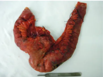
Three days after the symptoms appeared, the patient presented intense abdominal pain, abdominal distension and fecaloid vomiting (obstructive acute abdomen from probable neoplastic etiology), and was submitted to an emergency exploratory laparotomy. It showed neoplasm in rectum-sigmoid transition with macroscopic invasion of urinary bladder, without upstream colon distension, as well as presence of injury probably from the secondary graft in the terminal ileum, at approximately 15 cm from the ileocecal valve, associated with the obstruction, dis-tension of the loops of small intestine and blocked per-foration. The selected treatment was right-side entero-colectomy (Figures 1 to 3) and production of a Mickulicz ileotransversostomy, as well as exhaustive wash of the peritoneal cavity with physiological solution. The rectum-sigmoid transition injury was not addressed due to the pa-tient’s emergency situation, bladder size and invasion and as it was not the cause of the acute abdomen observed. The patient to the Intensive Care Unit in the immediate
postop-Figure 1. Resected terminal ileum segment, with apparent area of secondary graft.
Figure 2. Region of previously blocked perforation.
Linfoma não-Hodgkin como causa de abdome agudo
obstrutivo/perfurativo em paciente com adenocarcinoma de sigmoide: relato de caso
Marcelo Pandoli Basso et al.
380 Journal of Coloproctology
October/December, 2011
Vol. 31 Nº 4
erative period, progressed with clinical decompensation of comorbidities and died on the second postoperative day.
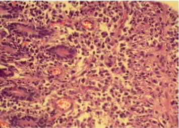
The anatomopathological exam of the vegetat-ing injury of the sigmoid observed in the colonoscopy showed a very differentiated and ulcerated tubular ad-enocarcinoma and the anatomopathological and im-munohistochemical exams (Figure 4) of the injury in the distal ileum showed diffuse large B-cell lympho-ma expressing immunophenotyping (CD20+).
DIsCUssION
The small bowel is rarely the place of neoplastic injuries, which represent only 1 to 5% of all gastrointes-tinal tract tumors4. The incidence of malign neoplasm
is even lower, around 1 to 2%5. The terminal ileum is
where malign injuries occur more frequently (50%), with the others equally occurring between the duode
-num and the jejune. In contrast, the frequency of benign
tumors seems to increase starting at the duodenum to-wards the ileum4,6.
Only 10% of the large intestine tumors are
symp-tomatic and the frequency of benign injuries is ten times the frequency of malign injuries4.
Lymphomas are some of the malign neoplasms of the small bowel (7–25%). Primary lymphomas corre-spond to less than 2% of all intestinal tumors, with
preva-lence of intestine iniltration by neoplastic cells from other
locations. The terminal ileum is the most common place of lymphoma occurrence, due to the higher concentration of lymphoid tissue associated with the intestine. The main
risk factor to be considered is the immunosuppression,
es-pecially the one caused by the human immunodeiciency
virus (HIV), chemotherapies and/or corticotherapy. In ad-dition, celiac disease presents increased risk for the devel-opment of primary lymphomas3,5.
The intestinal invasion by lymphomas may not
present speciic symptoms and remain silent for a long
time. Their initial manifestation can be obstruction or per-foration (25%). In general, they present vague symptoms, common to other histopathological types: unclear abdom-inal pain, nauseas, alteration to the intestabdom-inal rhythm, an-orexia and weight loss7.
Gastrointestinal tract lymphomas are most of non-Hodgkin type. The diffuse lymphoma of large B
cells corresponds to the most frequent histopathologi -cal and immunohistochemi-cal type8, more prevalent in
people in their 70s and older.
The preoperative diagnosis is dificult, consid -ering the insidious clinical manifestations that many times cause late search for specialized medical atten-tion. In addition, given the similar symptoms and signs to other common diseases in the digestive tract, the correct diagnosis can only be obtained after the histo-morphological and immunohistochemical analysis of the injury, many times after the patient is submitted to a surgical procedure.
In contrast, the distinction between primary
lym-phomas and the secondary iniltration of the gastro -intestinal tract can be made through a biopsy of the bone marrow or other organs under suspicion of be-ing affected by the disease. When this procedure is not possible, the consolidated and accurate criteria of Dawson9 can be used: non-palpable peripheral
lymph-adenopathy, normal thorax radiography, leukometry with differential showing no alterations, injury affect-ing particularly the gastrointestinal tract – only re-gional lymph nodes, and not the liver or spleen. The presence of these four criteria indicates the primary origin of lymphomas.
The case reported refers to an elderly patient with intermittent symptoms, suggesting a malign
in-testinal disease, conirmed through complementary
exams (colonoscopy with biopsies). Emergency lap-arotomy showed the disease already well advanced locally in the sigmoid colon, as well as injury sug-gesting distance metastasis (terminal ileum), with signs of intestinal obstruction and perforation. The
Linfoma não-Hodgkin como causa de abdome agudo
obstrutivo/perfurativo em paciente com adenocarcinoma de sigmoide: relato de caso
Marcelo Pandoli Basso et al.
381 Journal of Coloproctology
October/December, 2011
Vol. 31 Nº 4
REFERENCEs
1. Postier RG, Squires RA. Abdome agudo. In: Townsend Jr CM,
Beauchamp RD, Evers BM, Mattox KL, editors. Sabinston - Tratado de cirurgia. 19a ed. Rio de Janeiro: Elsevier; 2009. p. 1108-25.
2. Sallum EA. Abdome agudo obstrutivo. In: Gama-Rodrigues JJ, Machado MCC, Rasslan S, editors. Clínica cirúrgica. Barueri (SP): Manole; 2008. p. 1071-8.
3. Silva AL, Barbosa CA. Obstrução intestinal aguda. In: Lopes AC, editor. Tratado de clínica médica. São Paulo: Roca; 2006. p. 1092-109.
4. Steinman E. Perfuração de víscera oca. In: Gama-Rodrigues JJ, Machado MCC, Rasslan S (Eds). Clínica cirúrgica. Barueri (SP): Manole; 2008. p. 1079-94.
5. Evers BM. Intestino delgado. In: Townsend Jr CM, Beauchamp RD, Evers BM, Mattox KL, editors. Sabinston - Tratado de cirurgia. 19a ed. Rio de Janeiro: Elsevier; 2009. p. 1201-51.
6. Torricelli FCM, Lopes RI, Dias AR, Marchini GS, Bonafe WW, Lopes JM, et al. Linfoma ileal primário como uma causa de intussuscepção ileocecal recorrente. J Coloproctol 2008;28(2):246-50.
7. Rangel MF, Silva MVM, Fernandes MJC, Ferreira MAS, Nóbrega LPS, Souza MG. Tumores malignos do intestino delgado. Rev Col Bras Cir 2000;27(6):385-8.
8. Silva FE, Scofano V, Ascoly MH, Arakaki Jr N, Reis OCA, Silva Sá MAG. Fístula êntero-vesical como complicação de linfoma intestinal. J Coloproctol 2003;23(3):200-4.
9. Dawson IMP, Comes JS, Morson BC. Primary malignant lymphoid tumours of the intestinal tract. Report of 37
cases with study of factors inluencing prognosis. Br J Surg
1983;49:80-9.
10. Jara RLS, Santos CHM, Alves LP, Miiji LNO. Tumor de reto e cárdia sincrônicos. Relato de caso e revisão da literatura. J Coloproctol 2007;27(1):80-2.
11. Santos CHM, Silva CN, Miiji LNO. Adenocarcinoma de cólon e carcinoma espinocelular de canal anal concomitante. Relato de caso e revisão da literatura. J Coloproctol 2005;25(2):162-4.
Correspondence to:
João Gomes Netinho
Rua San Francisco, 481, Condomínio Débora Cristina CEP 15093-030 – São José do Rio Preto (SP), Brazil E-mail: [email protected]
REsUmO: Relato de caso raro de um paciente de 83 anos, com linfoma de íleo terminal causador de abdome agudo obstrutivo/perfu-rativo sincrônico à adenocarcinoma de cólon sigmoide e revisão dos dados disponíveis na literatura acerca das neoplasias de intestino delgado, em especial os linfomas. Constata-se que corresponde a uma afecção rara (2% de todas as neoplasias intestinais), mais pre-dominante em pacientes idosos e imunodeprimidos, cuja sintomatologia é vaga e o diagnóstico precoce difícil, fato que impossibilita muitas vezes a instituição da terapêutica correta.
Palavras-chave: neoplasia gastrointestinal; linfoma; adenocarcinoma; obstrução intestinal; perfuração intestinal. anatomopathological analysis of the resected
intesti-nal segment showed a termiintesti-nal ileum lymphoma syn-chronous with adenocarcinoma of the sigmoid colon. With no proper time for further investigations of the patient’s conditions and with the criteria of Dawson described above, we concluded that the lymphoma in the reported case was a primary lymphoma of the
gastrointestinal tract, since it fulilled all criteria es -tablished, and was synchronous with the neoplasms presented by the patient.
Synchronization between intestinal
neo-plasms is frequent, especially in the case of sig -moid colon adenocarcinoma, and it should always be investigated10,11. The incidence of a second
synchro-nous neoplasm with sigmoid colon adenocarcinoma, regardless of the histological type and the location in
the gastrointestinal tract, ranges between 1 and 6.8% in general population10.
CONCLUsION
Small bowel neoplasms are rare, and the benign
type is more frequent. Gastrointestinal tract lympho -mas are among the malign injuries, primary or
sec-ondary to a neoplastic iniltration. The most frequent
place of incidence is the terminal ileum, considering its histological peculiarities.
Signs and symptoms are vague and unspeciic,
which makes early diagnosis and accurate treatment
more dificult. However, these diseases should always