135
Brito MCB et al. Jejunal diverticulitis: a case report
Radiol Bras. 2011 Mar/Abr;44(2):135–136
Jejunal diverticulitis: a case report
*
Diverticulite jejunal: relato de caso
Mayra Carneiro Barbosa de Brito1, Beatriz Lavras Costallat1, Daniel Lahan Martins2, Adilson Prando3
Jejunal diverticulitis is a rare disease that primarily affects the elderly. Clinical signs and symptoms are nonspecific, a fact that complicates and delays the diagnosis. Computed tomography is very valuable in defining the diagnosis. The present report describes a case of jejunal diverticulosis complicated with acute perforated obstructive diverticulitis, and presents a brief discussion on jejunal diverticulitis and its complications.
Keywords: Jejunal diverticulitis; Computed tomography; Complications.
Diverticulite jejunal é doença rara que acomete principalmente os idosos. Os sintomas e sinais clínicos são inespecí-ficos, fato que dificulta e retarda o diagnóstico. A tomografia computadorizada é muito valiosa na definição do diag-nóstico. Neste relato será apresentado um caso de diverticulose jejunal complicada com diverticulite aguda perfurada bloqueada, assim como uma discussão breve sobre diverticulite jejunal e suas complicações.
Unitermos: Diverticulite jejunal; Tomografia computadorizada; Complicações.
Abstract
Resumo
* Study developed at Centro Radiológico Campinas – Hospi-tal Vera Cruz, Campinas, SP, Brazil.
1. MDs, Trainees in Radiology at Centro Radiológico Campi-nas – Hospital Vera Cruz, CampiCampi-nas, SP, Brazil.
2. Titular Member of Colégio Brasileiro de Radiologia e Diag-nóstico por Imagem (CBR), MD, Radiologist at Centro Radioló-gico Campinas – Hospital Vera Cruz, Physician Assistant, Depart-ment of Radiology, Universidade Estadual de Campinas (Uni-camp), Campinas, SP, Brazil.
3. Titular Member of Colégio Brasileiro de Radiologia e Diag-nóstico por Imagem (CBR), Head of the Department of Radiology, Centro Radiológico Campinas – Hospital Vera Cruz, Campinas, SP, Brazil.
Mailing Address: Dra. Beatriz Lavras Costallat. Rua Ezequiel Magalhães, 26, Jardim das Paineiras. Campinas, SP, Brazil, 13092-522. E-mail: [email protected]
Received June 20, 2010. Accepted after revision November 19, 2010.
Brito MCB, Costallat BL, Martins DL, Prando A. Jejunal diverticulitis: a case report. Radiol Bras. 2011 Mar/Abr;44(2):135–136.
0100-3984 © Colégio Brasileiro de Radiologia e Diagnóstico por Imagem
In the present report, the authors de-scribe a case of jejunal diverticulitis with intestinal obstruction, highlighting the most typical imaging findings and possible complications of this condition.
CASE REPORT
A male, 70-year-old patient was admit-ted to the emergency service with a three-day history of diffuse abdominal pain, with interrupted fecal and gaseous elimination. At clinical examination, the patient pre-sented fever, prostration, with diffuse ab-dominal pain at palpation. Complete blood count demonstrated leukocytosis. Previous surgical history: appendectomy and tran-surethral prostatectomy for benign pros-tatic hyperplasia. The patient reported to be diabetic and smoker.
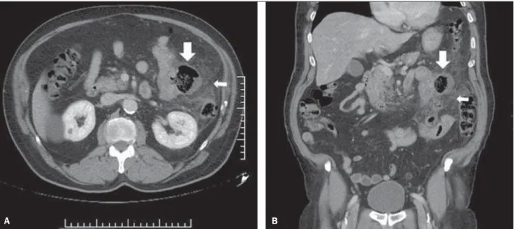
Whole abdomen CT scan was requested by the emergency medical team and was performed in 16-channel multidetector-row CT equipment in two phases: pre- and post-intravenous contrast (venous phase at 90 seconds). The following tomographic find-ings were observed: parietal thickening of the proximal jejunal loop and saccular im-age with a gaseous content corresponding to jejunal diverticulum associated with extensive densification of the adjacent mesenteric fat (Figure 1).
The patient was submitted to explor-atory laparotomy for resection of the
diver-CASE REPORT
ticulum and of the affected loop (Figure 2), that confirmed the diagnosis of jejunal di-verticulitis.
DISCUSSION
Jejunal diverticula are constituted of a thin mucosal wall herniated through a mus-cular layer(4). They affect only the mucosal, submucosal and serous layers, hence the denomination pseudodiverticula, and occur adjacent to the mesenteric border(1,4). Its eti-ology still remains unclear, but it is known that changes in peristalsis, intraluminal pres-sure and intestinal dyskinesia are included in this entity pathogenesis. Jejunal diver-ticula tend to be larger, with a higher num-ber of them occurring in the proximal je-junum, with its frequency progressively decreasing towards the distal jejunum, ex-cept for the terminal ileum region where multiple diverticula may be found(1–5). As-sociation with colonic diverticular disease occurs in 35–75% of cases(5–9). Clinical symptoms and signs are vague and nonspe-cific and, considering the rarity of this con-dition, the diagnosis is always delayed and difficult to be achieved(1–5,9). In many cases, CT is essential for defining the diagnosis. Diverticulosis complications are rare, with incidence of 6–13%(4). Acute diverticulitis, pseudo-intestinal obstruction and chronic diverticulitis with development of diverticu-lum are the most frequent complications(6).
INTRODUCTION
136
Brito MCB et al. Jejunal diverticulitis: a case report
Radiol Bras. 2011 Mar/Abr;44(2):135–136
Tomographic findings of jejunal diver-ticulitis are similar to the ones of colonic diverticulitis: inflammatory mass contain-ing gas and/or fecal residues, wall thicken-ing in the affected segment (increased con-trast uptake), with distension and edema of adjacent tissues (densification of the fascia and mesenteric fat)(4,6,7). In the present case, the imaging findings are compatible with the ones described in the literature and
Figure 1. A: Contrast-enhanced axial CT image demonstrating a diverticulum at the mesenteric border of jejunal loop (downwards arrow) and extensive den-sification of the adjacent fat (leftwards arrow), signs of acute diverticulitis. B: Reconstruction of contrast-enhanced, coronal, whole abdomen CT image. Proxi-mal jejunum diverticulum with signs of acute diverticulitis. Saccular image with gaseous content in the jejunal loop (downwards arrow) associated with exten-sive densification of the adjacent fat (leftwards arrow).
Figure 2. Images of surgical specimen: jejunal diverticulum complicated with diverticulitis, demonstrat-ing inflammatory process and obstructed perforation.
considered as typical signs of disease by many authors(5,7,10).
Among the possible complications of acute jejunal diverticulitis the most severe one is perforation followed by peritonitis, with a risk for mortality > 40%, and pos-sible development of abscesses, bleeding, adhesions and fistulas(1,4,6).
Because of its rarity and the nonspe-cificity of symptoms, acute jejunal
diver-ticulitis is rarely considered in the clinical differential diagnosis with other more prevalent conditions. In this context, CT becomes fundamental for determining its diagnosis, complications, and for ruling out other causes of acute abdomen.
REFERENCES
1. Peters R, Grust A, Gerharz CD, et al. Perforated jejunal diverticulitis as a rare cause of acute ab-domen. Eur Radiol. 1999;9:1426–8.
2. Furtado E, Machado BFF. Divertículo intralumi-nal do duodeno: relato de caso. Radiol Bras. 2003;36:389–90.
3. Setubal R, Souza RP, Endo E, et al. Intussuscep-ção jejunojejunal em adulto – relato de um caso e revisão da literatura. Radiol Bras. 1996;29: 331–4.
4. Coulier B, Maldague P, Bourgeois A, et al. Diver-ticulitis of the small bowel: CT diagnosis. Abdom Imaging. 2007;32:228–33.
5. Graña L, Pedraja I, Mendez R, et al. Jejuno-ileal diverticulitis with localized perforation: CT and US findings. Eur J Radiol. 2009;71:318–23. 6. Spiegel RM, Schultz RW, Casarella WJ, et al.
Massive hemorrhage from jejunal diverticula. Radiology. 1982;143:367–71.
7. Gliustra PE, Killoran PJ, Root JA, et al. Jejunal diverticulitis. Radiology. 1977;125:609–11. 8. Ramos AM, Piantá CD, Alves JM, et al.
Diverti-culite de intestino delgado em paciente idoso. Revista AMRIGS. 2005;49:41–3.
9. Nacif MS, Rocha VMB, Mello RAF, et al. Análise retrospectiva do trânsito do delgado em um ser-viço de radiologia de hospital geral. Radiol Bras. 2004;37:179–83.
10. Veen M, Hornstra BJ, Clemens CHM, et al. Small bowel diverticulitis as a cause of acute abdomen. Eur J Gastroenterol Hepatol. 2009;21:123–5.