Karyotypic and fluorescent
in-situ hybridization study of the centromere of
chromosome 7 in secondary myeloid neoplasms
1
Cytogenetics Laboratory, Serviço de Hematologia do Hospital das Clínicas, Faculdade de Medicina, Universidade de São Paulo – USP, São Paulo, SP, Brazil 2
Pathology Service, Faculdade de Medicina, Universidade de São Paulo – USP, São Paulo, SP, Brazil
Roberta Sandra da Silva Tanizawa1
Cristina Aiko Kumeda1
Raymundo Soares de Azevedo Neto2
Aline de Medeiros Leal1
Patrícia de Barros Ferreira1
Elvira Deolinda Rodrigues Pereira Velloso1
Background: Secondary myeloid neoplasms comprise a group of secondary diseases following exposure to myelotoxic agents or due to congenital diseases. The improvement of anticancer agents and immunosuppressive drugs seem to be associated with an increased incidence of secondary myeloid neoplasms. Karyotyping of bone marrow is essential for diagnosis and prognosis. Previous use of alkylating agents and radiation are associated with clonal abnormalities such as recurrent unbalanced -5/5q-, -7/7q- and complex karyotypes, whereas topoisomerase-II inhibitors lead to changes such as the balanced 11q23 rearrangement, t(8;21), t(15;17) and inv(16).
Objective: To study the clinical and cytogenetic data of patients with secondary myeloid neoplasms who took antineoplastic and/or immunosuppressive drugs or progressed from aplastic anemia.
Methods: The clinical and cytogenetic characteristics of 42 patients diagnosed with secondary myeloid neoplasms in one institution were retrospectively evaluated. Of these, 25, 11 and 6 patients had had oncological diseases, aplastic anemia and other diseases, respectively. Conventional cytogenetic and FISH analyses were performed for monosomy 7.
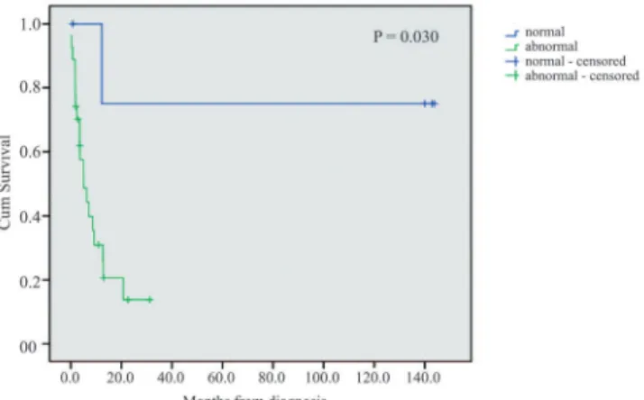
Results: The cytogenetic study was conclusive in 32 cases with 84.4% of clonal abnormalities. Monosomy 7 and complex karyotypes were present in 44.4% and 37%, respectively. A high prevalence of unbalanced abnormalities (96.3%) was observed. Monosomy 7 was more prevalent in patients with myelodysplastic syndromes/myeloid neoplasms after aplastic anemia (66.6%). The median survival after diagnosis of myeloid neoplasms was only 5.7 months. Normal cytogenetics was associated to better survival (p-value = 0.03). There was a slightly worse trend of survival for patients with complex karyotypes (p-value = 0.057). Abnormal karyotype was an independent risk factor for poor survival (p-value = 0.012).
Conclusion: This study enhances the importance of cytogenetic analysis of patients at the time of diagnosis of secondary myeloid neoplasms.
Keywords: Cytogenetic analysis; Myelodysplastic syndromes; Leukemia, myeloid
Introduction
The myelodysplastic syndromes (MDS), acute myeloid leukemias (AML) and secondary myeloproliferative syndromes (MPS) constitute a group of myeloid neoplasias (MNs) which result from exposure to risk factors such as chemotherapy and radiotherapy, acquired aplastic anemia associated with immunosuppression and congenital diseases.(1-5) A cytogenetic study of the bone marrow, fundamental for the
characterization of the secondary MNs, provides essential information for the diagnosis and prognosis of the disease. Between 80 and 95% of patients with MDS/MN present abnormal and frequently complex karyotypes (an average of 5.3 abnormalities per case). Chromosome 7 monosomy is the commonest alteration in patients with secondary MN after aplastic anemia (AA).(6) It is estimated that 10-20% of myeloid neoplasia cases are
secondary to the use of chemotherapeutic agents and/or radiotherapy.(5) The progress of
therapeutic regimens used to treat primary neoplasias, as well as the new conditions of autologous hematopoietic stem cell transplantations (HSCT), have led to an increase in the rate of therapy-related myeloid neoplasia (MN-t) cases.(7) Some patterns of
chromosomal abnormalities are recurrent after exposure to cytotoxic agents. The previous use of alkylating agents/radiation can favor the development of MN-t, which is usually associated with cytopenia and dysplasia and often involves unbalanced clonal genetic alterations including monosomies or deletions of chromosomes 5 and/or 7, and complex karyotypes with numerous marker chromosomes that may progress to point alterations in the P53 and AML1 genes. Previous use of topoisomerase II inhibitors (etoposide, anthracyclines) is usually associated with the development of AML, frequently with
Conflict-of-interest disclosure: The authors declare no competing financial interest
Submitted: 3/13/2011 Accepted: 9/5/2011
Corresponding author:
Roberta Sandra da Silva Tanizawa Laboratório de Citogenética Av. Dr. Enéas de Carvalho Aguiar, 155 1º andar, sala 30 – Cerqueira César 05403-000 – São Paulo, SP, Brazil Phone: 55 11 3061-5544 R-312 [email protected]
balanced chromosomal alterations involving 11q23 rearrangements, t(8;21), t(15;17), t(8;16) or inv(16).(8,9) The
high frequency of complex karyotypes in MN-t indicates a fast-emerging resistance to conventional treatments used in de novo leukemia cases.(10,11) Typically, the clinical course
progresses with high mortality and morbidity rates. Allogeneic HSCT is the only therapeutic modality with the potential to cure patients with MN-t.(7,12) Cytogenetic
aberrations which are present pre-transplantation are associated with low disease-free survival.(13) A study
conducted by the European Group for Blood and Marrow Transplantation (EBMT) on patients with MN-t who underwent allogeneic HSCT between 1981 and 2006 found, using multivariate analysis, that the progress of the disease was more favorable in patients with normal karyotypes.(7)
The development of secondary MDS/MN associated with treatment by immunosuppressants has been documented in AA. However, this subgroup also includes patients with autoimmune diseases and patients submitted to solid organ transplants.(2,14,15) Immunosuppressants such as cyclosporine
A (CyA), anti-lymphocyte or anti-thymocyte globulin (ALG/ATG), cyclophosphamide (CTX), azathioprine and the use of granulocyte colony-stimulating factor (G-CSF) appear to be involved in the etiology of MN.(16)
The cytogenetic abnormalities most frequently found in MDS/MN after AA involve chromosomes 6, 7 and 8.(17) In
some cases these alterations may progress with transient cytogenetic abnormalities, such as chromosome 8 trisomy, which is responsive to immunosuppression treatment, and trisomy of chromosome 6. However, monosomy 7, the most frequent cytogenetic abnormality, occurs in patients with minimal clinical response, in patients who relapsed with severe pancytopenia(18) or in those who took G-CSF for a
long time.(14) Some authors admit the possibility of a
pre-existent clone with chromosome 7 monosomy being present at the diagnosis of AA.(18)
Cytogenetic abnormalities involving chromosomes 5 and 7 were found in patients with rheumatic diseases that progressed to secondary MN after being treated with cumulative doses of alkylating agents for long periods.(15)
Myeloid neoplasms are rare in patients who received solid organ grafts, such as in heart, liver and kidney transplantations. However, the occurrence of these neoplasms seems to be related to maintenance immunosuppression that is basically composed of prednisone, cyclosporine and azathioprine. Thus, abnormalities involving chromosome 7 have been described in patients who received solid organ transplants, particularly those who were treated with azathioprine, a potent immunosuppressant that leads to chromosome instability.(2)
The aim of the present work was to study the clinical and cytogenetic data of patients who were exposed to antineoplastic and/or immunosuppressive drugs and those that progressed from aplastic anemia to secondary myeloid neoplasia.
Methods
A retrospective study was carried out involving 42 patients with secondary MN diagnosed between September 1987 and December 2008 at the Hospital das Clínicas da Faculdade de Medicina da Universidade de São Paulo. Conventional cytogenetics was performed using bone marrow samples and in one case using peripheral blood, with supplemented RPMI 1640 culture medium without any mitogenic agent incubated for 24 and 48 hours in a CO2 incubator at 37°C, followed by G-banding.(19) The fluorescence
in situ hybridization (FISH) technique was used to investigate possible chromosome 7 monosomies in cases in which the karyotype showed no abnormalities or in which no metaphases could be obtained. The CEP 7® Chromosome Enumeration DNA FISH Probe (Vysis, Inc) was employed according to the manufacturer's recommendations. The cut-off point for the probe had previously been established in the laboratory following international guidelines.(20) For this
purpose, an inverted β function statistical test was used (Microsoft Excel), after analyzing 200 interphase nuclei from five negative control samples. For each batch of probes, a normal control sample was tested in parallel. Cases presenting more than 10% of interphase nuclei with a single signal for the chromosome 7 centromere, analyzed by two independent observers (RSST and CAK), were considered positive for monosomy 7. The molecular cytogenetic analysis was performed in samples previously fixed in Carnoy solution (methanol/acetic acid). The karyotype and FISH results were described according to the international nomenclature (ISCN 2009).(21) The karyotypes were classified as normal or abnormal
(chromosome 7 monosomy, complex karyotypes with 3 or more clonal alterations and other patterns with < 3 clonal alterations). Whenever possible, 20 metaphases were analyzed. To be considered a clone, the same structural alteration or the gain of the same chromosome in two metaphases or the loss of the same chromosome in three metaphases had to be observed.(21)
Statistical analysis was performed using the Kaplan-Meier product limit estimator (survival curves), log rank test to compare groups and Fisher's exact test for associations between variables. In addition to the cytogenetic parameters, other clinical, hematological, bone marrow aspirate and histological factors were submitted to the binary logistic regression test to evaluate overall survival (OS). The data were analyzed using version 16.0 of the Statistical Package for the Social Sciences (SPSS) Computer program.
Results
Clinical characteristics of the patients
neoplasias (n= 19), aplastic anemia (n= 11), solid tumors (n= 6), autoimmune diseases (n= 4) and solid organ transplantations (n=2). The treatments of the previous
The median age of patients with previous aplasia was 24 years (range: 15-61), six (54%) were male and the latency period until developing secondary MN was 97 months. Of the other 31 patients, median age was 60 years (range: 4-88), 16 (51.6%) were male and the median latency period was 76 months.
Cytogenetic characteristics
observed by conventional karyotyping in 25/32 (78.1%) cases.
FISH investigations (Table 2) for chromosome 7 monosomy were carried out in 11 patients; two (18.2%) were positive (Patients 1 and 25).
Altogether, the clonal abnormalities detected by conventional karyotyping and FISH accounted for 27/32 (84.4%) of the cases. Monosomy 7 was observed in 10/27 (37%), complex karyotypes in 12/27 (44.4%) and other abnormalities in 5/27 (18.5%) including deletion of the long arm of chromosome 7, chromosome 8 tetrasomy, t(3;21) and structural alterations involving chromosomes 1 and 21 (Figure 1). The frequency of unbalanced abnormalities was 26/27 (96.3%); a single case presented a balanced abnormality. Monosomy 7 was more prevalent in patients with secondary MN after AA (66.6%), whereas in the other cases the incidence of complex karyotypes was higher (52.2%). The description of the karyotypes according to the ISCN 2009 and the classification of the abnormalities are shown in Table 2.
Figure 2 exemplifies a complex karyotype, Figure 3 shows a karyotype with a balanced translocation and Figure 4 is a FISH image showing a nucleus with monosomy 7.
Figure 1 – Incidence of chromosome abnormalities (%) in secondary myeloid neoplasms (n = 27)
Figure 2 – Therapy-related complex karyotype (Patient 14) showing del(5q), trisomy 8 and del(13q). The therapy for follicular lymphoma (previous disease) included cyclophosphamide and fludarabine phosphate
Figure 3 – Therapy-related anomaly (Patient 9) showing t(3;21) (q22;q26). The therapy for plasma cell myeloma (previous disease) included melphalan
Figure 4 – FISH study using centromeric probe for chromosome 7. Therapy-related abnormality (Patient 25) one interphase cell (only one green signal) demonstrating monosomy 7. The therapy for rhabdomyosarcoma (previous disease) included cyclophosphamide, etoposide, doxorubicin and carboplatin.
Cytogenetics and prognosis
The median overall survival of the 42 patients was 6 months; a little shorter (5.7 months) when the date of HSCT, a procedure used for eight patients, was delayed.
and with complex karyotypes (p-value = 0.123). Binary logistic regression analysis for overall survival-related factors showed that the presence of an abnormal karyotype was an independent factor for reduced survival (p-value = 0.012).
Discussion
The results obtained from our sample are concordant with those of other authors, reinforcing the importance of cytogenetics, both in the diagnosis and prognosis of secondary MN.
The high rate of clonal abnormalities (84.4%) observed here is similar to other studies that reported karyotype abnormalities in 80-95% of patients with secondary MN.(6)
Also, the predominance of unbalanced abnormalities (96.3%), the high frequency of complex karyotypes (44.4%) and chromosome 7 monosomy (37%) are consistent with the literature.(6,7,9) In particular, monosomy 7 was described in
patients following AA; this cytogenetic abnormality has been reported to be recurrent in secondary MN of patients with medullar failure treated with immunosuppressants and G-CSF.(8,17,22) Curiously, the only case of balanced abnormality
with t(3;21) was a patient (Patient 9) who had been treated for multiple myeloma using an alkylating agent (melphalan).
Balanced abnormalities involving the CBFB (16q22),
RARA (17q21) and AML1 (21q22) genes have been reported more frequently after the use of topoisomerase inhibitors. The presence of the t(3;21)(q26;q22) translocation that leads to the fusion of the EVI1(3q26) and AML1 or RUNX1
(21q22) genes has been frequently reported in MN-t and in the blast crisis stage of CML.(23) In the present sample,
no rearrangements of the 11q23 region were observed; the importance of investigating the integrity of the MLL gene by FISH must be remembered, given the fact that the MLL
rearrangement may remain cryptic by conventional cytogenetic methods.(24)
Cytogenetic abnormalities are known to be independent prognostic factors in de novo MDS and are incorporated in the most commonly used prognostic scores such as the
International Prognostic Scoring System (IPSS) and WHO Prognostic Scoring System (WPSS).(25,26) Yet in secondary
MN, the prognostic factors are less well studied, with variables such as age, primary disease, platelet count, hemoglobin concentration, serum protein and C-reactive protein levels, cytogenetic analysis, medullar fibrosis and CD34 cell expression in bone marrow histology, all being associated with survival.(27-30) The prognosis of MN-t is highly
reserved, with a median survival time estimated at 6 months; the cytogenetic study plays an important role, not only for prognosis, but in the follow-up after HSCT.(7,12) In this study,
abnormal karyotype proved to be an independent factor for poorer survival (p-value = 0.03), while complex karyotype alterations showed a slight tendency towards a reduced survival (p-value = 0.057) compared to the other cytogenetic abnormalities.
Secondary MNs are becoming more common; this is the price of longer survival of patients with neoplasias. The treatment of chronic lymphoproliferative diseases with purine analogs associated with alkylating agents, as well as high-intensity therapeutic regimens, followed by autologous transplantation are current examples of this scenario. Moreover, in the field of non-neoplastic hematological diseases, a clear increase in the incidence of MDS is observed in patients with AA. Despite the unfavorable prognosis of secondary MNs, karyotype analysis allows the stratification of these patients. New drugs, such as hypomethylating agents, are the subject of research as a "bridge" until a suitable donor for allogeneic HSCT can be found.
References
1. Luna-Fineman S, ShannonKM, Lange BJ. Childhood monosomy 7: Epidemiology, biology, and mechanistic implications. Blood. 1995;85(8):1985-99.
2. Huebner G, Karthaus M, Pethig K, Freund M, Ganser A. Myelodysplastic syndrome and acute myelogenous leukemia secondary to heart transplantation. Transplantation. 2000;70(4): 688-90.
3. Naschitz JE. Rheumatic syndromes: clues to occult neoplasia. Curr Opin Rheumatol. 2001;13(1):62-6.
4. Jesus G, Barcelos A, Neves C, Crespo J. Manifestações reumáticas e neoplasias. Acta Reum Port. 2006;31(4):305-21.
5. Vardiman JW, Arber DA, Brunning RD, Larson RA, Matiutes E, Baumann I, et al. Therapy-related myeloid neoplasms. In: Swerdlow SH, Campo E, Harris NL, Jafe ES, Pileri SA, Stein H, et al., editors. WHO Classification of Tumours of Haematopoietic and Lymphoid Tissues. Lyon: IARC Press; 2008. p.127-9.
6. Heim S. Cytogenetics findings in primary and secondary MDS. Leuk Res.1992;16(1):43-6.
7. Kröger N, Brand R, van Biezen A, Zander A, Dierlamm J, Niederwieser D, Devergie A, Ruutu T, Cornish J, Ljungman P, Gratwohl A, Cordonnier C, Beelen D, Deconinck E, Symeonidis A, de Witte T; Myelodysplastic Syndromes Subcommittee of The Chronic Leukaemia Working Party of European Group for Blood and Marrow Transplantation (EBMT). Risk factors for therapy-related myelodysplastic syndrome and acute myeloid leukemia treated with allogeneic stem cell transplantation. Haematologica. 2009; 94(4):542-9. Comment in: Haematologica. 2009;94(4):454-9. Figure 5 – Kaplan-Meier survival curves according to the cytogenetic
8. Pedersen-Bjergaard J, Andersen MT, Andersen MK. Genetic pathways in the pathogenesis of therapy-related myelodysplasia and acute myeloid leukemia. Hematology Am Soc Hematol Educ Program. 2007:392-7.
9. Hake CR, Graubert TA, Fenske TS. Does autologous transplantation directly increase the risk of secondary leukemia in lymphoma patients? Bone Marrow Transplant. 2007;39(2):59-70. 10. Larson RA. Etiology and management of therapy-related myeloid
leukemia. Hematology Am Soc Hematol Educ Program. 2007: 453-9.
11. Godley LA, Larson RA. Therapy-related myeloid leukemia. Semin Oncol. 2008;35(4):418-29.
12. Larson RA. Therapy-related myeloid neoplasms. Haematologica. 2009;94(4):454-9. Comment on: Haematologica. 2009;94(4): 542-9.
13. Chang C, Storer BE, Scott BL, Bryant EM, Shulman HM, Flowers ME, et al. Hematopoietic cell transplantation in patients with myelodysplastic syndrome or acute myeloid leukemia arising from myelodysplastic syndromes: similar outcomes in patients with de novo disease and disease following prior therapy or antecedent hematologic disorders. Blood. 2007;110(4):1379-87.
14. Young NS, Calado RT, Scheinberg P. Current concepts in the pathophysiology and treatment of aplastic anemia. Blood. 2006; 108(8):2509-19.
15. McCarthy CJ, Sheldon S, Ross CW, McCune WJ. Cytogenetic abnormalities and therapy-related myelodysplastic syndromes in rheumatic disease. Arthritis Rheum. 1998;41(8):1493-6.
16. Young NS. Pathophysiologic mechanisms in acquired aplastic anemia. Hematology Am Soc Hematol Educ Program. 2006: 72-7.
17. Maciejewski JP, Selleri C. Evolution of clonal cytogenetics abnormalities in aplastic anemia. Leuk Lymphoma. 2004;45(3): 433-40.
18. Bagby GC, Lipton JM, Sloand EM, Schiffer CA. Marrow failure. Hematology Am Soc Hematol Educ Program. 2004:318-36.
19. Yunis JJ, Sawyer JR, Ball DW. The characterization of high-resolution G-banded chromosomes of man. Chromosoma. 1978; 67(4):293-307.
20. Wolff DJ, Bagg A, Cooley LD, Dewald GW, Hirsch BA, Jacky PB, Rao KW, Rao PN; Association for Molecular Pathology Clinical
Practice Committee; American College of Medical Genetics Laboratory Quality Assurance Committee. Guidance for fluorescence in situ hybridization testing in hematologic disorders. J Mol Diagn. 2007;9(2):134-43.
21. Shaffer LG, Slovak ML, Campbel LJ, editors. ISCN: an International System for Chromosome Nomenclature. Basel: Karger; 2009. 22. Rubin CM, Arthur DC, Woods WG, Lange BJ, Nowell PC, Rowley
JD, et al. Therapy-related myelodysplastic syndrome and acute myeloid leukemia in children: correlation between chromosomal abnormalities and prior therapy. Blood. 1991;78 (11):2982-8. 23. Nimer SD. Myelodysplastic syndromes. Blood. 2008;111(10):
4841-51.
24. Leone G, Pagano L, Ben-Yehuda D, Voso MT. Therapy-related leukemia and myelodysplasia: susceptibility and incidence. Haematologica. 2007;92(10):1389-98.
25. Greenberg P, Cox C, Le Beau MM, Fenaux P, Morel P, Sanz G, et al. International scoring system for evaluating prognosis in myelodysplastic syndromes. Blood. 1997;89(6):2079-88. Comment in: Blood. 2001;98(6):1985; Blood. 1997;90(7): 2843-6.
26. Malcovati L, Della Porta MG, Pascutto C, Invernizzi R, Boni M, Travaglino E, et al. Prognostic factor and life expectancy in myelodysplastic syndromes classified according to WHO criteria: a basis for clinical decision making. J Clin Oncol. 2005;23 (30):7594-603.
27. Pedersen-Bjergaard J, Philip P, Larsen SO, Jensen G, Byrsting K. Chromosome aberrations and prognostic factors in therapy-related myelodysplasia and acute nonlymphocytic leukemia. Blood. 1990;76(6):1083-91.
28. Kantarjian HM, Keating MJ, Walters RS, Smith TL, Cork A, McCredie KB, et al. Therapy-related leukemia and myelodysplastic syndrome: clinical, cytogenetics, and prognostic features. J Clin Oncol. 1986;4(12):1748-57.
29. Takeyama K, Seto M, Uike N, Hamajima N, Ino T, Mikuni C, et al Therapy-related leukemia and myelodysplastic syndrome: a large-scale japanese study of clinical and cytogenetic features as well as prognostic factors. Inter J Hematol. 2000;71(2):144-52. 30. Orazi A, Cattoretti G, Soligo D, Luksch R, Lambertenghi-Deliliers
G. Therapy-related myelodysplastic syndromes: FAB classification, bone marrow histology, and immunohistology in the prognostic assessment. Leukemia. 1993;7(6):838-47.