Received from Instituto do Coração at Hospital das Clínicas, Medical School at Universi-dade de São Paulo, Brazil.
1. PhD; Medical School, Universidade de São Paulo (FM-USP); Professor, Medical School, Centro Universitário Lucíadas (UNILUS); Medical Supervisor, Anesthesiology Service at the Radiology Institute at Hospital das Clínicas, FM-USP
2. TE-AMIB; PhD, FM-USP; Medical Supervisor at Surgical ICU, Anesthesia Division at Central Institute, Hospital das Clínicas, FM-USP; Medical Coordinator, Surgical Patients Critical Care Unit, Anesthesiology Service, Hospital do Servidor Público Estadual de São Paulo
3. TE-AMIB; PhD, FM-USP; Anesthesiologist, Hospital das Clínicas, FM-USP, Ribeirão Preto
4. PhD, FM-USP; Medical Doctor Assistant, Instituto do Coração at Hospital das Clinicas, FM-USP
5. TE-AMIB; Associate Professor, Anesthesiology, FM-USP; Director, Anesthesia Division at Central Institute, Hospital das Clínicas, FM-USP
Submitted on March 29, 2011. Approved on August 3, 2011. Correspondence to:
Luiz Marcelo Sá Malbouisson, MD Av. Enéas Carvalho de Aguiar, 155
Prédio dos Ambulatórios 8º andar Bloco 3 Divisão de Anestesia Cerqueira César
05403900 – São Paulo, SP, Brazil E-mail: [email protected] SCIENTIFIC ARTICLES
Extracorporeal Circulation Interference on Emergence
from Anesthesia in Patients Submitted to Myocardial
Revascularization
Ricardo Antonio Guimarães Barbosa, TSA
1, Luiz Marcelo Sá Malbouisson, TSA
2,
Luciana Moraes dos Santos, TSA
3, Marilde de Albuquerque Piccioni
4, Maria José Carvalho Carmona, TSA
5Summary: Barbosa RAG, Malbouisson LMS, Santos LM, Piccioni MA, Carmona MJC – Extracorporeal Circulation Interference on Emergence
from Anesthesia in Patients Submitted to Myocardial Revascularization.
Background and objectives: Extracorporeal circulation (ECC) may change drug pharmacokinetics as well as brain function. The objectives of
this study are to compare emergence time and postoperative sedation intensity assessed by the bispectral index (BIS) and the Ramsay sedation scale in patients undergoing myocardial revascularization (MR) with or without ECC.
Method: Ten patients undergoing MR with ECC (ECC group) and 10 with no ECC (no-ECC group) were administered with sufentanyl, propofol
2.0 µg.mL-1 and pancuronium target controlled infusion. After surgery, propofol infusion was reduced to 1 µg.mL-1 and suspended when extubation was indicated. Patients BIS, Ramsay scale and time to wake up were assessed.
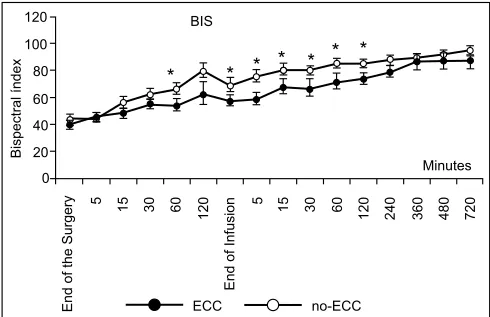
Results: The ECC group showed lower BIS values beginning at 60 minutes after surgery (no-ECC = 66 ± 13 and ECC = 53 ± 14, p = 0.01) until
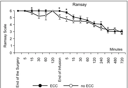
120 minutes after infusion (no-ECC = 85 ± 8 and ECC = 73 ± 12, p = 0.02). Sedation level measured by the Ramsay scale was higher in the ECC group at 30 minutes after the end of the surgery (no-ECC = 5 ± 1 and ECC = 6 ± 0, p = 0.021), at the end of infusion (no-ECC = 5 ± 1 and ECC = 6 ± 1, p = 0.012) and 5 minutes after the end of infusion (no-ECC = 4 ± 1 and ECC = 5 ± 0.42, p = 0.039). Emergence from anesthesia time was higher in the ECC group (no-ECC = 217 ± 81 and ECC = 319 ± 118, p = 0.038).
Conclusions: There was a higher intensity of sedation after the end of surgery and a longer wake up time in ECC group, suggesting changes in
the pharmacokinetics of propofol or effects of ECC on central nervous system.
Keywords: Deep Sedation; Extracorporeal Circulation; Pharmacokinetics; Propofol.
©2012 Elsevier Editora Ltda. All rights reserved.
INTRODUCTION
Extracorporeal circulation (ECC) may change plasma concen-trations of drugs used during anesthesia for cardiac surgery, as well as cause effects on the central nervous system lead-ing patients to a higher sedation level, which changes patients’ emergence from anesthesia time 1.
ad-BARBOSA, MALBOUISSON, SANTOS ET AL.
290 Revista Brasileira de Anestesiologia
Vol. 62, No 3, May-June, 2012 ditionally assessing the correlation between bispectral index
(BIS) monitoring and clinical assessment (sedation scale by Ramsay et al. 13).
METHODS
Study protocol was approved by the institutional Medical Eth-ics Committee. After clarifications regarding the study’s gen-eral objectives, patients signed the informed consent form.
Were studied 20 patients with chronic coronary insuffi-ciency, candidates to myocardial revascularization elective surgery, with left ventricle ejection fraction superior to 50%. Patients were randomically allocated in two non-randomized groups according to surgical practice: Myocardial Revascu-larization with Extracorporeal Circulation Group (ECC Group) (n = 10) and Myocardial Revascularization without Extracor-poreal Circulation Group (no-ECC Group) (n = 10).
Pre-anesthetic medication consisted of oral midazolam 0.1 to 0.2 mg.kg-1 dose, 30 minutes before surgery, reaching the maximum dose of 15 mg. Patients were monitored in the surgery room with electrocardiography, pulse oximetry, inva-sive blood pressure and central venous catheter.
Anesthesia induction was conducted with propofol through specific infusion pump (Diprifusor®, AstraZeneca, USA) with a 30-second infusion initial time and 2.0 µg.mL-1 as the tar-get concentration during the entire surgery, and sufenta-nyl initially infused at the 0.5 µg.kg-1 dose, followed by a 0.5 µg.kg-1.hour-1continuous infusion through infusion pump (Anne, Abbott®, USA) specially programmed with the patient’s weight and the specific drug. Continuous infusion was sus-tained during the entire surgery. Muscle relaxation was ob-tained with 0.1to 0.2 mg.kg-1 pancuronium bromide dose. Manual ventilation was used under mask and tracheal intuba-tion with proper diameter tube, followed by mechanical con-trolled ventilation (Cicero Dragger®, Germany) with 8 mL.kg-1 current volume, 10 respiratory incursions per minute for the respiratory frequency, I:E ratio = 1:2 and FiO2 = 0.6 (oxygen, compressed air and PEEP = 5 cm H2O).
After anticoagulation administration with sodium heparin 500 U.kg-1, ECC started using roller pump or centrifugal pump, with membrane oxygenator and initial perfusion of 1,600 mL Ringer Lactato solution. Perfusion flow was 60 to 80 mL.kg-1. min-1, using moderate hypothermia at 32oC to 34oC and se-rial gasometry control. During ECC, hypnosis was maintained with target controlled propofol continuous infusion aiming to keep a 2 µg.mL-1 plasma concentration.
At the end of the surgery, propofol target concentration was changed to 1.0 µg.mL-1. It was maintained constant dur-ing transportation to the Intensive Care Unit (ICU) until the moment tracheal extubation was indicated. Extubation was indicated when patients were considered to be normothermal, hemodynamically stable, conscious and showing response to verbal commands.
Postoperative sedation intensity was assessed by bispec-tral index (BIS) and Ramsay sedation scale. Ramsay scale is used to assess patient sedation level. It has been described
by Michael Ramsay as part of a study about Alphaxalone/Al-phadolone (Althensin) anesthetic effect published in 1974 13. It comprehends values to be attributed from 0 to 6, observing responses given by the patient after stimulus:
Level 1: anxious, restless;
Level 2: cooperative, orientated, tranquil; Level 3: sleepy, responding to commands;
Level 4: sleeping, brisk response to glabelar or vigorous sound stimulus;
Level 5: sleeping, sluggish response to glabelar or vigor-ous sound stimulus;
Level 6: sleeping, no response to stimulus.
Assessment of postoperative sedation intensity was per-formed using the BIS and Ramsay sedation scale in different moments:
1) At the end of the surgery;
2) 5 minutes after the end of the surgery; 3) 15 minutes after the end of the surgery; 4) 30 minutes after the end of the surgery; 5) 60 minutes after the end of the surgery; 6) 120 minutes after the end of the surgery; 7) at the end of the Propofol infusion.
When patients were hemodynamically stable and normo-thermal, propofol infusion was interrupted so that the patients could be extubated as fast as possible. Assessment of the BIS and Ramsay sedation scale continued at the moments described as follows in the two studied groups (ECC Group and no-ECC group):
1) 5 minutes after the end of the Propofol infusion; 2) 15 minutes after the end of the Propofol infusion; 3) 30 minutes after the end of the Propofol infusion; 4) 60 minutes after the end of the Propofol infusion; 5) 120 minutes after the end of the Propofol infusion; 6) 240 minutes after the end of the Propofol infusion; 7) 360 minutes after the end of the Propofol infusion; 8) 480 minutes after the end of the Propofol infusion; 9) 720 minutes after the end of the Propofol infusion.
Patient wake up time was also assessed, considered to be the time between the end of the propofol infusion and the mo-ment the patient started to respond to verbal commands.
Groups were compared considering weight, height, body mass index, surgery time, patient intubation time and extra-corporeal circulation time in the ECC group.
Statistical analysis
no-ECC group, considering a standard deviation of 75 minutes in both groups. For this estimation, a test power of 80% and p value = 0.05 were considered, as well as a required sample of at least nine patients in each group. Normal distribution of data was assessed by the Shapiro-Wilk test, and kurtosis and asymmetry tests. Data regarding age, weight, height, body mass index, surgery time, tracheal intubation time and com-plete wake up time were assessed by the Student t test for non-paired samples. The behavior of BIS values throughout the time in the groups with and without ECC was analyzed using a two-way variance analysis for repeated measures, fol-lowed by the Student-Newman-Keuls test to detect differenc-es between the groups at several time-points. The behavior of the Ramsay scale values between the groups were compared at several time-points of interest using the Wilcoxon test, as a result of Ramsay scale’s values non-normal distribution. The
Spearman test was used to assess the correlation between BIS and Ramsay sedation scale values. A p value < 0.05 was considered to be significant. All analyses were conducted using the statistical program STATA 11 (STATACorpTM, Tx, USA) and Sigmastat 3.5 (Systat Software Inc.TM, Ca, USA).
RESULTS
The two groups were compared considering weight, height, age and body mass index (BMI). Regarding the intubation time and emergence from anesthesia, the ECC group showed to have greater values for these variables, with a significant difference (Table I). The surgery time was greater in the no-ECC group, though showing no significant difference. All patients reported complete amnesia during the surgical pro-cedure. Population BIS mean values were pointed out versus time as illustrated in Figure 1.
Regarding BIS, significant differences were identified be-tween the ECC and no-ECC groups at the following time-points: 60 minutes after the end of the surgery, at the end of the infusion, and 5, 15, 30, 60 and 120 minutes after the end of the infusion, in which the ECC group showed BIS values smaller than the no-ECC group values (Figure 1).
Higher postoperative sedation intensity was observed in the ECC group patients assessed by the Ramsay sedation scale, with a significant difference at the following time-points: 30 minutes after the end of the surgery, at the end of the propofol infusion, and 5 minutes after the end of the propofol infusion (Figure 2).
In all cases, when an association between Ramsay seda-tion scale and BIS values was identified, such associaseda-tion had been considered to be inversed, i.e., the greater the BIS value was, the smaller was the Ramsay values (Figure 3)
Table I – Patient Demographic Data in MR and TV Groups (mean
± SD)
ECC Group No-ECC Group p
N 10 10
Gender M = 9 F = 1 M = 8 F = 2
Age (years) 62.20 ± 8.32 68.50 ± 6.57 0.0766
Weight (kg) 75.23 ± 10.55 74.87 ± 8.17 0.9329
Height (cm) 163.00 ± 0.04 166.00 ± 0.10 0.554
BMI 28.14 ± 4.63 27.58 ± 2.70 0.7466
ECC Time 79 ± 23.37
Surgery Time
(min) 277.50 ± 53.66 287.50 ± 77.04
0.7527
TI Time (min) 689.00 ± 123.89 568.50 ± 119.77 0.0402
Emergence
(min) 319.30 ± 118.99 217.00 ± 81.38
0.038
ECC: extracorporeal circulation; BMI: body mass index; TI: tracheal intubation.
End of the Surgery
End of Infusion
5 5
15 30 60 15 30 60
120 120 240 360 480 720
Minutes
ECC no-ECC 120
100
80
60
40
20
0
Bispectral índex
*
*
*
*
*
*
*
BIS
Figure 1 – BIS Mean Profile (± SD) according to the group and
as-sessment timepoint.
BIS Behavior throughout the study– Black circles (ECC group) and white circles (no-ECC group).*: p < 0.05. Data presented as mean and standard error.
Table II –BIS and Ramsay Correlation at Some Assessment
Timepoints in both Groups
Assessment Timepoint
ECC Group No-ECC Group
I II I II
End of Infusion - - -0.83 0.003
5 min after Infusion -0.44 0.209 -0.81 0.005 15 min after Infusion -0.53 0.118 -0.36 0.313 30 min after Infusion -0.69 0.026 -0.75 0.012 60 min after Infusion -0.66 0.039 -0.83 0.003 120 min after Infusion -0.41 0.244 -0.24 0.507 ECC: extracorporeal circulation.
I: Spearman Correlation Coefficient between BIS and Ramsay at some asses-sment timepoints.
BARBOSA, MALBOUISSON, SANTOS ET AL.
292 Revista Brasileira de Anestesiologia
Vol. 62, No 3, May-June, 2012 DISCUSSION
The results obtained showed that the group of patients under-going myocardial a revascularization with ECC differed their behavior regarding the emergence from anesthesia and post-operative sedation intensity after the interruption of the target controlled infusion.
The greater postoperative sedation intensity observed in the ECC group may be justified by hypothermia experienced by the patients in this group and by the central nervous system de-pression caused by ECC. Hypothermia also causes a reduction in the hepatic blood flow with consequent reduction in propofol metabolism. All these factors may increase patient wake up
time (calculation obtained between the end of the propofol infu-sion and the moment when the patient first responded to verbal commands) in this group, justifying the greater postoperative tracheal intubation time (Table I). The greatest sedation level observed in the ECC group shows a possible greater central nervous system depression caused by the ECC.
BIS is related to the anesthesia hypnotic component, with-out considering patient response movements or hemodynamic response to painful stimulus, providing the deepness level of the anesthesia 14,15. In the present study, monitoring results demonstrated an immediate BIS reduction after the anesthesia induction in the two investigated groups, a fact that could be ex-plained by the rapid action start and the rapid distribution of the hypnotic agent to the central nervous system. For the patients with coronary disease assessed in this study, the propofol in-fused dose was adequate for hypnosis.
The anesthesiologist may define propofol target concentra-tions that range from 2 to 6 µg.mL-1 for general anesthesia or 0.5 to 1.5 µg.mL-1 for sedation. Pharmacokinetics models of target controlled infusions rapidly reach the desired propofol target concentrations 16,17. However, caution must be taken when administering propofol infusion until the desired effect is obtained due to the subjects’ variability regarding propofol pharmacokinetics and pharmacodynamics 18.
Patient response to propofol during surgery is highly variable and infusion rate and administration dose will be determined according to patients’ individual needs. Factors that influence the propofol dose are: age, weight, pre-existing diseases, sur-gery type and concomitant medical treatments.
The short duration of propofol action of approximately 5 to 8 minutes may be explained by the drug elevated clearance and quick distribution. Propofol concentration at the action site also quickly increases due to the rapid balance reached between plasma and brain concentration (< 3 minutes). These propofol pharmacokinetics features lead to a fast manifestation of the hypnotic effect and loss of consciousness. Notwithstanding the compartmental model chosen, propofol distribution at the ac-tion site is considered to be nearly instantaneous, and the drug free fraction controls the pharmacological effect intensity. Pre-ceding studies reported up to a three fold increase in the drug free fraction during cardiac surgery with ECC 6. An increase in the free drug fraction of approximately 300% may contribute to ensure a rapid elevation in propofol concentration at the action site, with relevant effect in the pharmacokinetics of this agent.
Some studies have also showed a greater hypnotic effect of propofol as a result of the ECC 19. For this reason, ECC brain effects may interfere in the hypnosis level.
Results obtained in previous studies confirm that sufentanyl in the low concentrations used does not interfere in the propofol effect measured by BIS 20.
Patient clinical assessment during emergence from anesthe-sia at the ICU is extremely important during the postoperative time of a cardiac surgery, as it enables patient early extubation. This assessment performed using the sedation scale and also the BIS enables a better follow-up of the patients.
According to the present study, sedation intensity as mea-sured by BIS was greater in the ECC group when compared to the no-ECC group, suggesting changes in the propofol phar-macokinetics or in the ECC secondary effects on the level of sedation.
End of the Surgery
End of Infusion
5 5
15 30 60 15 30 60
120 120 240 360 480 720
Minutes
ECC no ECC 6
5
4
3
2
1
0
Ramsay Scale
*
*
*
Ransay
Ransay
6
5
4
3
2
1
0
0 20 40 60 80 100 120
Bispectral index
ECC no ECC
Figure 2 – Concentration Mean Profile (± SD) during ECC.
Behavior of the sedation deepness according to Ramsay scale throu-ghout the study– Black circles (ECC group) and white circles (no-ECC group).*: p < 0.05. Ramsay sedation scale values in groups described in the figure. Data presented as mean and standard error.
Figura 3 –BIS X Ramsay Correlation (ECC Group and no-ECC
Group).
REFERÊNCIAS/REFERENCES
1. Barbosa RAG, Santos SRCJ, White PF et al. – Effects of cardiopulmo-nary bypass on propofol pharmacokinetics and bispectral index during coronary surgery. Clinics, 2009;64:215-221.
2. Mets B – The pharmacokinetics of anesthetic drugs and adjuvants dur-ing cardiopulmonary bypass. Acta Anaesthesiol Scand, 2000;44:261-273.
3. Gouke CR, Keaveny JP, Kay B, Healy TE, Ryan M – The effect of cardiopulmonary bypass on the pharmacokinetics of drugs. Clin Phar-macokinet, 1982;7:234-251.
4. Buylaert WA, Herregods LL, Mortier EP, Bogaert MG – Cardiopulmo-nary bypass and the pharmacokinetics of drugs. Clin Pharmacokinet, 1989;17:10-26.
5. Wood M – Plasma drug binding: implications for anesthesiologists. Anesth Analg, 1979;65:786-804.
6. Wood M – Plasma drug binding: implications for anesthesiologists. Anesth Analg, 1986;65:786-804.
7. Hiraoka H, Yamamoto K, Morita T, Goto F, Horiuchi R – Changes in drug plasma concentrations of an extensively bound and highly extracted drug, propofol, in reponse to altered plasma binding. Clin Pharmacol Ther, 2004;75:324-330.
8. Bailey JM, Mora, CT, Shafer SL – Pharmacokinetics of propofol in adult patients undergoing coronary revascularization. Anesthesiology, 1996;84:1288-1297.
9. White PF – Intravenous anesthesia ana analgesia: what is the role of target-controlled infusion (TCI). J Clin Anesth, 1996;8:26-28. 10. Massey NJ, Sherry KM, Oldroyd S, Peaccock JE – Pharmacokinetics
of infusion of propofol during cardiac surgery. Br J Anaesth, 1990; 65:475-479.
11. Lee HS, Khoo YM, Chua BC, Tan SS, Chew SL – Pharmacokinetics of propofol infusion in Asian patients undergoing coronary artery bypass grafting. Ther Drug Monit,1995;17:336-341.
12. Hammaren E, Yli-Hankala A, Rosenberg PH, Hynynem M – Cardiopul-monary bypass-induced changes in plasma concentrations of propofol and in auditory evoked potentials. Br J Anaesth, 1996;77:360-364. 13. Ramsay MA, Savege TM, Simpson BR, Goodwin R – Controlled
seda-tion with alphaxalone-alphadolone. Br Med J, 1974;2:656-659. 14. Fisher DM – Development and clinical application of
electroencephalo-graphic Bispectrum monitoring. Anesthesiology, 2000;93:1336-1344. 15. Sigl JC, Chamoun NG – An introduction to bispectral analysis for the
electroencephalogram. J Clin Monit, 1994;10:392-404.
16. White PF – Intravenous anesthesia and analgesia: what is the role of target-controlled infusion (TCI). J Clin Anesth, 1996;8:26-28.
17. Kenny GNC, Sutcliffe N – European perspective. In: White PF. Text-book of intravenous anesthesia. Baltimore: Williams & Wilkins. 1997; pp.527-537.
18. Glass PSA, Markhan K, Ginsberg B, Hawkins ED – Propofol concen-trations required for surgery. Anesthesiology, 1989;71:A273. 19. Yoshitani H, Takeuchi M, Sakamoto K, Akasaka T, Yoshida K,
Yo-shikawa J – Effect of one or more co-morbid conditions on diagnostic accuracy of coronary flow velocity reserve for detecting significant left anterior descending coronary stenosis. Heart, 2005;91:1294-1298. 20. Lysakowski C, Dumont L, Pellegrini M, Clergue F, Tassony E – Effects