Original Article
Prevalence of Coronary Artery Disease and Preoperative Assessment
in Patients with Valvopathy
Roney Orismar Sampaio
1, Vívian Masutti Jonke
1, João L. Falcão
2, Sandra Falcão
1, Guilherme S. Spina
1, Flávio
Tarasoutchi
1, Max Grinberg
1Instituto do Coração, Departamento de Valvopatias1 e Departamento de Hemodinâmica2, São Paulo, SP - Brazil
Summary
Background: Coronary angiography has been indicated in the preoperative phase for patients with valvopathy over 35 years of age. However, the actual prevalence of obstructive coronary artery disease (CAD) in this population has been little studied.
Objective: To assess the prevalence of and the risk factors for CAD in candidates for valve surgery in Brazil.
Methods: Coronary angiography was performed in 3,736 patients who were candidates for valve surgery; prevalence of and risk factors for CAD associated with valvopathy were assessed.
Results: CAD was associated with valvopathy in 121 patients (prevalence of 3.42%). In 79 patients (68.1%), CAD was diagnosed by means of preoperative coronary angiography. Of these 79 patients, 50 (63.3%) had isolated aortic valvopathy or aortic valvopathy associated with mitral valvopathy. Smoking habit was observed in 54 patients (68.3%), hypertension in four (43%), family history in 24 (30.3%), diabetes mellitus in 15 (18.9%), and obesity in eight (10.1%). Of the 121 patients, 95.7% were over 50 years of age. Only five (4.3% of the patients with CAD) were below 50 years of age, and all of them had at least one risk factor for CAD.
Conclusions: CAD prevalence was low in the patients studied. Aortic valvopathy was the most frequent valvopathy associated with CAD, and most patients were over 50 years of age. The ideal age for routine preoperative coronary angiography in patients with valvopathy should be reassessed. (Arq Bras Cardiol 2008;91(3):183-186)
Key words: Coronariography angiography; preoperative care; heart valve diseases; coronary arteriosclerosis.
Mailing Adress: Roney Orismar Sampaio •
Av.Dr Eneas de Carvalho Aguiar, 44, Cerqueira Cesar, 05.403-000,São Paulo, SP - Brazil.
E-mail: [email protected]
Manuscript received June 12 2007; revised manuscript received January 07 2008; accepted February 26 2008.
Introduction
Preoperative coronary angiography has been routinely indicated for patients older than 35 years who have valvopathy and have to undergo surgery1. However, the actual prevalence
of coronary artery disease (CAD) in these patients has been little studied2,3.
The association between CAD and valvopathy generally worsens symptoms, accounts for a worse prognosis and increases surgery risk, especially in patients with aortic stenosis4.
CAD prevalence in valvopathies is inconsistent and may be influenced by the local degree of development, social conditions and eating habits of the population assessed. Therefore, the indication for preoperative coronary angiography could be changed according to the actual prevalence of obstructive CAD. In the United States, coronary
angiography is indicated before 35 years of age, even for those patients without a clinical history of or risk factors for CAD. In other countries, such as Spain, coronary angiography has been performed in male patients at 60 and female patients at 65 in view of the low prevalence of CAD, which is lower than 5%, in individuals under 60 years of age3.
To assess the prevalence of major CAD in patients with valvopathy, we studied all the preoperative coronary angiographies over a period of ten years in our institution.
Methods
From January 1990 to December 2000, 3,736 patients with valvopathy underwent surgery in our institution. We assessed the medical records of all patients submitted to valve surgery and myocardial revascularization. When necessary, some incomplete data, such as the presence of risk factors, were clarified by phone at a later stage. Patients with known ischemic mitral insufficiency were excluded. Major CAD was defined as an obstruction of at least 70% of one of the coronary arteries. An obstruction equal to or greater than 50% of the left coronary trunk was also considered significant. The study was submitted to the institution’s Ethics Committee and was duly approved.
Original Article
Arq Bras Cardiol 2008;91(3):183-186
Sampaio et al. Coronariography in valve disease
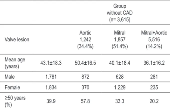
Table 1 – Data on �atients wit�o�t obstr�cti�e coronary artery disease (CAD)
Group without CAD
(n= 3,615)
Valve lesion
Aortic 1,242 (34.4%)
Mitral 1,857 (51.4%)
Mitral+Aortic 5,516 (14.2%) Mean age
(years) 43.1±18.3 50.4±16.5 40.1±18.4 36.1±16.2 Male 1.781 872 628 281 Female 1.834 370 1.229 235
≥50 years
(%) 39.9 57.8 33.3 20.2
Patients with major CAD were divided into two groups: previous CAD, comprised of patients with known coronary artery disease; and new CAD, comprised of those patients whose diagnosis of coronary artery disease was possible only after the preoperative coronary angiography was performed.
The following parameters were assessed: 1) gender and age;
2) type of valvopathy;
3) risk factors for CAD including: smoking habit5 (patients
were asked whether they smoked and the number of cigarretes smoked per day, and whether they were former smokers), obesity (defined as a body mass index above 30 kg/m2),
dyslipidemia (total cholesterol serum level above 200 mg/dL or low density lipoprotein level above 130 mg/dL), family history of CAD6 (defined as the presence of sudden death or
acute myocardial infarction before 55 and 65 respectively for male and female forebears), hypertension (defined as blood pressure higher than 140x90 mmHg) and patients diagnosed with diabetes mellitus (defined at the time the study began and maintained during the study; defined as fast glycemia above 140 mg/dL).
The statistical analysis was carried out using the SAS software and a value of p<0.05 was considered statistically significant. Quantitative variables for means and standard deviations were used. Paired and unpaired Student’s t tests were employed when appropriate. For qualitative data (for example, presence of hypertension or not, presence of diagnosis of diabetes mellitus or not), when they were compared for the hypotesis of equivalence of proportions, the Chi-square test was used; when limited, Fischer’s exact test was used.
Results
The association between major CAD and valvopathy occurred in 121 patients of the 3,736 patients who underwent surgery, thus resulting in a prevalence of 3.24%. Of these 3,736 patients, 1,905 (51%) had mitral valvopathy, 1,196 (32%) had aortic valvopathy and 635 (17%) had mitral-aortic valvopathy.
Among the 3,736 cases, there were 3,615 patients with non-obstructive CAD (table1). The association between CAD and valvopathy was more frequent in patients with aortic disease, either isolated or combined with mitral disease (3.94%), than in patients with isolated mitral valvopathy (2.26%).
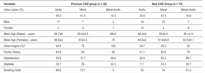
We retrieved complete data of 109 out of the 121 patients with CAD (figure1). For 30 patients there was information on previous CAD (“Previous CAD”). However, for 79 patients (72.5%) the diagnosis of CAD was possible only thanks to the routine preoperative coronary angiography (“New CAD”). The data on the 30 patients with “Previous CAD” and on the 79 patients with “New CAD”, including the number of risk factors, are detailed in table 2.
Of the 79 patients with New CAD, 55 were male (69.62%), with a mean age of 64.74±9.23 years of age. There were 50 patients with either isolated aortic or mitral-aortic valvopathy, and 29 with isolated mitral valvopathy. There was no significant difference between the means of age, diagnosis of diabetes mellitus or hypertension when the Previous CAD and New CAD
groups were compared (p = ns) (tab.2). Additionally, symptoms of chest angina in the Previous CAD group were more frequent than in the New CAD group (p<0.001).
In the New CAD group the following risk factors were observed: smoking habit in 54 patients, diagnosis of diabetes mellitus in 15, and obesity (body mass index ≥30 kg/m2) in 8
patients. Hypertension was observed in 34 and family history in 24 patients. Only five patients (4.6% of all patients with CAD and 0.13% of all the population with valvopathy) were below 50 years of age. All of these patients (one mitral, two aortic and two mitral-aortic) had at least one risk factor for CAD. Three of these were female and two were male; all were smokers; two had a family history; two had hypertension, one had diabetes and one had dyslipidemia.
Discussion
CAD prevalence has not been extensively studied, particularly in asymptomatic patients2,7. Enriquez-Sarano et
al.2 assessed patients with valvopathy from the United States
and found that approximately 35% of them had associated CAD, with no significant change in a ten-year follow-up. Recent data has shown that CAD accounts for more than half of all cardiovascular events in men and women over 75 years of age in the United States8, and that the lifetime risk of
developing CAD after 40 years of age is 49% for male and 32% for female subjects. Hypertension was a risk factor especially for black women8.
Our data showed a prevalence that was ten times lower (3.42%). Other studies showed a variable frequency depending on the population assessed. San Jose et al.3
demonstrated a prevalence of 20.3% in a Spanish study with 234 patients with a mean age of 64±10 years. Timmermans et al.9 identified a lower prevalence of CAD (approximately
14%) in Belgian subjects with aortic insufficiency. Studies in developing countries showed a prevalence between 5% and 15%10. These differences relate to older age, risk factors and
eating habits which differ among the populations studied. Our study showed a very low incidence of obstructive coronary
Original Article
Arq Bras Cardiol 2008;91(3):183-186
Sampaio et al.
Coronariography in valve disease
Table 2 –Data on �atients wit� Pre�io�s CAD and new CAD
Variables Pre�io�s CAD gro�� (n = 30) New CAD Gro�� (n = 79)
Valve Lesion (%) Aortic Mitral Mitral+Aortic Aortic Mitral Mitral+Aortic
48.3 41.4 10.3 35.9 47.4 16.6
Male 11 7 2 15 33 7
Female 3 5 1 13 4 6
Mean Age (Males) – years 66.7±8 65.9±5.8 66±2 68.3±9 62±6.4 58.1±14 Mean Age (Females) - years 66.6±4 67±4.6 78 65.6±2 61.9±6.8 63.5±9.1
Chest Angina (%)* 92.8 75 100 30.7 55.2 50
Family History 63.6 60 50 41.1 52.6 70
Hypertension 53.8 72.7 66.6 26.9 55.2 66.7
Diabetes 30.7 30 33.3 7.1 33.3 30.7
Smoking Habit 66.6 72.7 0 52 74 61.5
* P <0�001 (P�����u� CA� G��u� ����u� N�w CA� G��u�)�
Figure 1 -Breakdown of groups of patients who underwent surgery because of valvopathy, with or without obstructive coronary artery disease (CAD)
disease, that is, where revascularization was required. Rheumatic fever is still a common disease in Brazil, and the mitral valve is the valve that is most frequently affected. The absolute sample of patients with mitral valvopathy was high, including its association with CAD. However, when a proportional assessment was made, we observed a higher frequency of association between aortic stenosis and CAD, in the region of 10%, which is similar to the findings of other studies11,12. The older age group and the predominance
of male subjects may explain these findings. Additionally a high number of risk factors could also account for this association, especially dyslipidemia, about which our data was inconclusive. Data on cholesterol levels prior to valve surgery were available for only 35% of our patients, and 70% of these had high total cholesterol levels.
Preoperative coronary angiography is commonly performed in patients with valvopathy who are over 35 years of age in
the United States1, especially when they have risk factors
for CAD. Noninvasive evaluation techniques such as electrocardiography, chest X-ray, Doppler echocardiography, scintigraphy and, more recently, angiotomography, when combined with clinical examination, can clarify, in most cases, the etiology, the degree of valve lesion, and whether there is ventricular dysfunction. The hemodynamic study played a diagnostic role in only 10% of the patients with valvopathy 11-15. Additionally, there are issues relating to the high cost and
risks which are inherent to invasive tests, such as vascular lesion resulting from the insertion of the catheter, anaphylactic shock and/or kidney dysfunction due to the iodinated contrast media and even an epysode of cerebrovascular accident due to calcium embolism or thrombus13.
The performance of catheterization for the sole purpose of assessing CAD can be restricted further. Differences in CAD prevalence in different populations should be taken
Original Article
Arq Bras Cardiol 2008;91(3):183-186
Sampaio et al. Coronariography in valve disease
into account when we challenge coronary angiography as a preoperative exam, especially in younger patients with valvopathy. Noninvasive tests, such as treadmill exercise test and myocardial scintigraphy have been used in selected patients with the purpose of avoiding coronary angiography14,15. More recently, coronary
angiotomography16 has demonstrated higher coronary
calcium content in populations with a mean age that is higher than the mean age of the patients with valvopathy assessed in this study (43.1±18.3). However, there was disagreement in the data relative to the presence or not of obstructive CAD. The presence of risk factors seems to be a good indicator for the performance of preoperative coronary angiography17,18. Our data have shown that chest
angina was the most frequently found datum in the Previous CAD group, which reflects the importance of clinical history for these patients19,20.
The absence of risk factors is an important marker to exclude obstructive CAD in patients below 50 years of age. In fact, we found no patients below age 50 with no risk factor for CAD who actually had CAD associated with valvopathy. We consider a limitation the fact that this is a retrospective study. Thus, data on dyslipidemia and details about the progression may have been ommitted. However, these limitations do not invalidate the study, as they
emphasize the need to reassess the most appropriate age for the performance of preoperative coronary angiography.
Conclusion
In our institution, we identified a low prevalence of obstructive coronary disease in patients with valvopathy with indication for surgery. Aortic valvopathy was the most frequent valvopathy associated with CAD and most patients were over 50 years of age. The ideal age for the performance of routine preoperative coronary angiography in patients with valvopathy should be reassessed.
Potential Conflict of Interest
No potential conflict of interest relevant to this article was reported.
Sources of Funding
There were no external funding sources for this study.
Study Association
This study is not associated with any graduation program.
References
1. Bonow RO, Carabello BA, Chaterjee K, de Leon AC Jr, Faxon DP, Freed MD, et al. ACC/AHA 2006 Guidelines for the management of patients with valvular heart disease: a report of the American College of Cardiology/American Heart Association Task Force on Practice Guidelines (writing Committee to Revise the 1998 guidelines for the management of patients with valvular heart disease) developed in collaboration with the Society of Cardiovascular Anesthesiologists endorsed by the Society Cardiovascular Angiography and Interventions and the Society of Thoracic Surgeons. J Am Coll Cardiol. 2006; 48 (3): e1-148.
2. Enriquez-Sarano M, Klodas E, Garratt KN, Bailey KR, Tajik AJ, Holmes DR Jr. Secular trends in coronary atherosclerosis – analysis in patients with valvular regurgitation. N Engl J Med. 1996; 335: 316-22.
3. San Jose JCM, Galán LF, Cerrón IG, Carpentier MT, Garcia JB, Martin JA, et al. Coronariografia preoperatoria en pacientes valvulares: criterios de indicación en una determinada poblácion. Rev Esp Cardiol. 1997; 50: 467-73. 4. Lytle BW. Impact of coronay artery disease on valvular heart disease. Cardiol
Clin. 1991; 9: 301-13.
5. Gus I, Fischmann A, Medina C. Fatores de risco da doença arterial coronariana. Arq Bras Cardiol. 2002; 78: 478-83.
6. Sociedade Brasileira de Cardiologia. III Diretrizes brasileiras sobre dislipidemias e diretriz de prevenção da aterosclerose do Departamento de aterosclerose da SBC. Arq Bras Cardiol. 2001; 77 (supl 3): 1-48.
7. Carabello B. Aortic stenosis. N Engl J Med. 2002; 346: 677-82.
8. American Heart Association. Heart disease and stroke statistics – 2005 Update. Dallas (Texas); 2005.
9. Timmermans P, Willems JL, Piessens J , Geest H. Angina pectoris and coronary artery disease in severe aortic regurgitation. Am J Cardiol. 1998; 61: 826-9. 10. Mohan V, Deepa R, Rani SS, Premalatha G. Prevalence of coronary artery
disease and its relationship to lipids in a selected population in South India: the Chennai Urban Population Study (CUPS No. 5). J Am Coll Cardiol. 2001; 38: 682-7.
11. St John Sutton MG, St John Sutton M, Oldershaw P, Sachetti R, Paneth M, Lennox SC, et al. Valve replacement without preoperative cardiac catheterization. N Engl J Med. 1981; 305: 1233-8.
12. Rangel CM, Grinberg M, Maranhão RC, Ventura LI. Estenose aórtica e doença coronariana: análise dos fatores de risco. Arq Bras Cardiol. 2006; 87(2): 115-20. 13. Pepine C, Allen HD, Bashore JA, Brinker JA, Cohn LH, Dellon JC, et al.
American College of Cardiology/American Heart Association (ACC/AHA) guidelines for cardiac catheterization and cardiac catheterization laboratories (ad Hoc Task Force on Cardiac catheterization). Circulation. 1991; 84 (5): 2213-47.
14. Linderholm H, Osterman G, Teien D. Detection of coronary artery disease by means of exercise ECG in patients with aortic stenosis. Acta Med Scand. 1985; 218: 181-8.
15. Kettunen R, Huikuri HV, Heikkila J, Takkunen JT. Preoperative diagnosis of coronary artery disease in patients with valvular heart disease using technetium-99m isonitrile tomographic imaging together with high-dose dipyridamole and handgrip exercise. Am J Cardiol. 1992; 69: 1442-5. 16. Burgstahler C, Beck T, Kuettner A, Reimann A, Kopp Af, Heuschmid M, et
al. Image quality and diagnostic accuracy of 16-slice multidetector spiral computed tomography for the detection of coronary artery disease in elderly patients. J Comput Assist Tomogr. 2005; 29: 734-8.
17. Olofsson B, Bjerle P, Aberg T, Osterman G, Jacobsson KA. Prevalence of coronary artery disease in patients with valvular heart disease. Acta Med Scand. 1985; 218: 365-71.
18. Czer LSC, Matloff JM. Combined valvular and coronary surgery. Chest. 1996; 90: 312-4.
19. Vandeplas A, Willems JL, Piessens J, De Geest H. Frequency of angina pectoris and coronary artery disease in severe isolated valvular aortic stenosis. Am J Cardiol. 1988; 62: 117-20.
20. Ramsdale DR, Bennett DH, Bray CL, Ward C, Beton DC, Faragher FB. Angina, coronary risk factors and coronary artery disease in patients with valvular heart disease: a prospective study. Eur Heart J. 1984; 5: 716-26.