Anterior temporal white matter lesions in
adult-form myotonic dystrophy type 1
Lesões temporais anteriores da substância branca na forma adulta da distrofia miotônica
tipo 1
Leonardo Ferreira Caixeta
1, Giane Souza Reis
2, Ana Caroline Marques Vilela
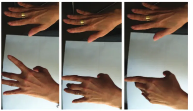
3A 57-year-old woman presented with a 4-years history of
progressive weakness, distal muscular atrophy and myotonia
in left hand (Figure 1). Her electromyography had a
myo-tonic pattern. Patient was first diagnosed as paraneoplasic
limbic encephalitis based on her brain MRI (Figure 2).
Diagnosis was genetically confirmed for myotonic dystrophy
type 1 (DM1).
DM1 or Steinert
’
s disease is an autosomal-dominant
dis-order characterized by muscle weakness and unusual features,
compared with other dystrophies, including myotonia,
anti-cipation, and multiple organ involvement
1,2. Anterior temporal
lobe subcortical white matter lesions are described in DM1,
but not in DM2 patients
3. Limbic encephalitis and CADASIL
are the most important imaging differential diagnosis.
References
1. Meola G, Sansone V. Cerebral involvement in myotonic dystrophies. Muscle Nerve. 2007;36(3):294-306. http://dx.doi.org/10.1002/mus.20800
2. Machuca-Tzili L, Brook D, Hilton-Jones D. Clinical and molecular aspects of the myotonic dystrophies: a review. Muscle Nerve 2005;32(1):1-18. http://dx.doi.org/10.1002/mus.20301
3. Kornblum C, Reul J, Kress W, Grothe C, Amanatidis N, Klockgether T et al. Cranial magnetic resonance imaging in genetically proven myotonic dystrophy type 1 and 2. J Neurol. 2004;251(6):710-4. http:// 10.1007/s00415-004-0408-1
1Departamento de Neurologia, Faculdade de Medicina, Universidade Federal de Goiás, Goiânia GO, Brazil; 2Departamento de Neurologia, Universidade Federal de Goiás, Goiânia GO, Brazil;
3Universidade Federal de Goiás, Goiânia GO, Brazil.
Correspondence: Leonardo Ferreira Caixeta; Avenida Cristo Rei 626, Setor Jaó; 74674-290 Goiânia GO, Brasil; E-mail: [email protected] Conflict of interest:There is no conflict of interest to declare.
Received 02 April 2014; Received in final form 22 July 2014; Accepted 01 August 2014.
Figure 1.
Dystrophy and sequence of myotonia in left hand.
Figure 2.
MRI (T2 weight coronal and axial slices) showing
bilateral anterior temporal hypersignal in our case. Notice that
mesial temporo-limbic structures are not affected.
DOI:10.1590/0004-282X20140139