Case 1 — Hypertensive 58-Year-Old Woman with Hepatic Nodules
Presented Dyspnea and Pleural Effusion One Week after an
Episode of Pulmonary Embolism
Tânia Marie Ogawa, Ricardo Mauro Ghetti Cabral, Vera Demarchi Aiello
Instituto do Coração (InCor) HC-FMUSP, São Paulo, BrazilThree years ago, woman aged 58 (March 2002) presented retrosternal pain unrelated to stress. At the time, she sought medical treatment, underwent clinical and laboratory evaluation and was diagnosed with arterial hypertension and hypercholesterolemia. She received dietary counseling and 20 mg of simvastatin daily was prescribed.
In June 2005, she presented vomiting and abdominal pain. She sought treatment in another Medical Service. At this Service, she took laboratory tests (June 222005), which revealed cholesterol 132 mg/dL (HDL-cholesterol 54 mg/dL, LDL-cholesterol 70 mg/dl), triglycerides 41 mg/dL; glucose 116 mg/dL. Magnetic resonance imaging (June 222005) revealed nodules in both hepatic lobes, one of which with centripetal enhancement and late homogenization compatible with hemangioma. The gall bladder was normal, there was no abnormality in the pancreas, spleen, kidneys, blood vessels or lymph. In the same hospitalization, colonoscopy (July 19 2005) revealed hemorrhoids and no other abnormalities in the colon and rectum. The patient was diagnosed with hepatic hemangioma and was discharged.
About two months later, there was swelling of the left lower limb and the patient sought emergency medical care at this hospital (August 312005).
Physical examination (August 312005) revealed blood pressure of 160/100 mmHg, heart rate 100 bpm. Examination of the lungs, heart and abdomen showed no changes. There was left lower limb edema.
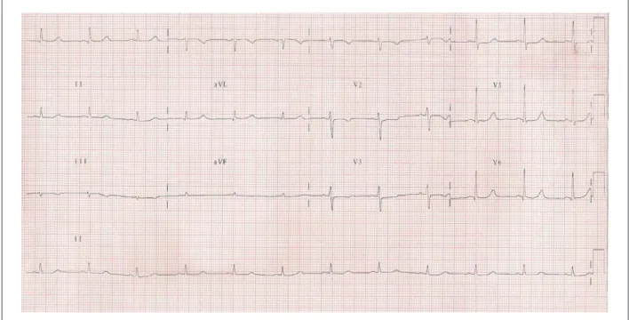
Electrocardiogram (August 312005) revealed sinus tachycardia, heart rate 116 bpm and mild changes in ventricular repolarization (fig. 1).
Laboratory tests (September 2 2005) revealed hemoglobin 12.4 g/dL, hematocrit 38%, leukocytes 20700/mm³ (2% metamyelocytes, 12% bats, 67% segmented, 1% basophils,
5% lymphocytes and 13% monocytes), platelets 244000/ mm³, creatinine 0.4 mg/dL, urea 17 mg/dL, potassium 4 mEq/L, sodium 133 mEq/L, INR 1.3 and activated partial thromboplastin time 1.39.
Chest radiography revealed elevation of the right hemidiaphragm.
Ultrasound examination (September 012006) was suggestive of recent thrombosis of common and superficial femoral veins and the left popliteal vein.
Deep venous thrombosis was diagnosed and enoxaparin was administered subcutaneously.
Echocardiogram (September 6 2005) revealed normal left ventricular dimensions (43 mm in diastole and 25 mm in systole), ejection fraction 73%; aorta, left and right atria and right ventricle were normal, no signs of pulmonary hypertension.
High resolution computed tomography (September 8 2005) revealed decreased vascularization in the topography of the right and left descending interlobar artery and right pleural effusion with irregular opacity of the adjacent parenchyma.
Pulmonary thromboembolism was diagnosed.
Oral anticoagulant treatment with warfarin was associated with antihypertensive treatment. The patient was discharged after 15 days, with prescribed elastic stockings, enalapril 10 mg, atenolol 50 mg and warfarin 2.5 mg daily.
Ultrasound of the abdomen (September 20 2006) revealed enlarged liver and blunt edges, heterogeneous texture. Multiple bilobar nodules were diagnosed. The largest one was about 1.4 cm. There was an extensive hypoechoic area in the right lobe, which could be secondary to perfusion disturbance.
Nine days later, the patient returned to the emergency room with signs of severe dyspnea, even at rest and abdominal pain.
On physical examination (September 23 2006) the patient was tachypneic, blood pressure 110/70 mmHg, heart rate 72 bpm. Examination of the lungs revealed pleural effusion semiology, affecting two thirds of the lower right hemithorax. Heart examination was normal. The examination also revealed distended and painful abdomen on palpation of right upper quadrant and left lower limb edema.
Electrocardiogram (September 26 2005) revealed sinus rhythm, 69 bpm, ventricular repolarization (fig. 2).
Chest radiography revealed large right pleural effusion. Abdominal ultrasound (September 26 2006) revealed hepatomegaly without other abnormalities.
Mailing Address: Vera D. Aiello •
InCor – Av. Dr. Enéas de Carvalho Aguiar, 44 – 05403-000 – São Paulo, SP E-mail: [email protected]
keywords
Venous thrombosis; pleural effusion; shock; heart arrest; pneumothorax
Section Editor: Alfredo José Mansur ([email protected])
Thorax tomography (26 September 2005) revealed massive right pleural effusion with subtotal atelectasis of the lung. The liver revealed increased dimensions and heterogeneous parenchymal density.
The right hemithorax was punctured (September 27 2005) with output of 1700 mL of bright yellow fluid. Biochemical tests in the pleural fluid revealed: glucose 51 mg/dL, lactate dehydrogenase 508 IU/L, albumin 1.3 g/dL.
Figure 1 – ECG - Sinus tachycardia and mild changes in ventricular repolarization.
There was jaundice, and two days later, the patient woke prostrate, with inaudible blood pressure and cyanosis of the extremities. Central venous catheter was inserted into the right subclavian vein.
A few hours later, the patient presented hypoxia followed by cardiac arrest in pulseless electrical activity during tracheal intubation. The patient had right pneumothorax, which had already been diagnosed on radiographs to control the installation of central venous catheter. When the pneumothorax was relieved by puncturing the second intercostal space, the arrest was reversed. Subsequently, the pneumothorax was treated with drainage in the fourth right intercostal space.
The laboratory evolution is shown in Table 1.
The patient developed shock and broad-spectrum antibiotic therapy was started, including anaerobic drugs (ampicillin, metronidazole and ceftriaxone). There was a need for increasing levels of norepinephrine to maintain blood pressure. The patient developed anuria and severe metabolic acidosis (lactate 100 mg/dL, pH 7.08,
PaCO2 19 mmHg, O2 saturation 95.5%, paO2 135 mm Hg, bicarbonate 5.3 mEq/L and base excess -23 mEq/L). Hemodialysis was started slowly, but there was worsening of hypotension and hemodialysis was discontinued.
Because of anemia (Table 1), the patient received several units of packed red blood cells. The next morning (October 1 2005), presented irreversible cardiac arrest in asystole.
Clinical aspects
Hypertensive woman aged 58 with dyslipidemia and findings of hepatic hemangioma in image analysis sought emergency service due to abdominal pain and vomiting.
Hepatic hemangioma is the most common benign liver tumor, with incidence ranging from 0.4% to 20% in autopsy series1. It is usually identified as an incidental finding of imaging or laparoscopy in women (60% to 80% of cases) between the third and fifth decades of life — an age slightly younger than the patient in question. Most hemangiomas are small, solitary and do not cause symptoms. Larger lesions of 4 cm and 10 cm are symptomatic, respectively 40% and 90% of cases. The pain in the upper abdomen is the most common symptom, resulting from infarction and necrosis, pressure on adjacent structures, distention of the hepatic capsule, or high blood flow. Compressive symptoms can occur in giant hemangiomas1.
Magnetic resonance imaging (MRI) in this patient revealed nodules in both hepatic lobes, one showing late centripetal enhancement and late homogenization, which is characteristic of hepatic hemangioma on imaging studies (magnetic resonance imaging, abdominal computed tomography and abdominal ultrasound with color Doppler), showing early, peripheral and progressive centripetal capture and venous phase delayed on contrast examination. In most patients with hemangioma, gastrointestinal symptoms are due to other causes such as gastroenteritis and dyspeptic syndromes.
Hemangiomas are usually solitary, but multiple tumors can occur in up to 10% of patients1. They are usually located in the right hepatic lobe and larger tumors may be pedunculated. In this patient, there are nodules in both hepatic lobes, but only one of them showed a classic pattern of hemangioma on imaging. However, as already explained, one cannot exclude the possibility that the other nodes correspond to multiples hemangiomas of different sizes.
Differential diagnosis is based on other benign liver tumors, especially: 1) Hepatocellular adenoma: unlikely for this patient, as it is clearly associated with the use of contraceptives and women of childbearing age, and MRI shows blood pickup pattern, unlike hemangioma; 2) Focal nodular hyperplasia: these are also single lesions, but in 7% to 20% of cases, they are multiple2. In 5% to 10% of cases, it is associated with hepatic hemangioma, usually occurring in women of the same age, and its relation to oral contraceptive use is not yet well established. The imaging shows arterial signals within the tumor, unlike peripheral
Table 1 - Laboratory tests
September 23 September 27 October 1
Hemoglobin (g/dL) 11.5 11.5 4.1 Hematocrit (%) 36 37 15 Leukocytes (/mm³) 19,700 18,500 15,900
Bats (%) 8 37
Segmented (%) 70 31
Neutrophils (%) 74 78 69 Eosinophils (%) 1 0 0 Lymphocytes (%) 11 12 20 Monocytes (%) 14 9 11 Platelets (/ mm³) 296,000 264,000 64,000 Creatinine (mg/dL) 0.5 0.6 1.4
Urea (mg/dL) 21 20
PT (INR) 3.7 1.5 1.93
APTT (rel) 0.99
Sodium (mEq/L) 131 130 Potassium (mEq/L) 4.1 4.5 Glucose (mg/dL) 65
Billirrubins (mg/dL) 8.34 7.33 Direct (mg/dL) 5.66 5.81 Indirect (mg/dL) 2.68 1.52
AST (IU/L) 89 394
ALT (UI/L) 38 81
Alkaline phosphatase (UI/L) 1112 875 Amilase (UI/L) 51
and centripetal uptake occurring in the hemangioma, which was the pattern shown by the NMR of the patient.
Other differential diagnoses are the primary malignant liver tumors (unlikely, at least at this stage of evolution, because the patient has no consumptive symptoms and more than 80% of cases are associated with liver cirrhosis) and liver metastasis of other malignancies3-7. Thinking about the possibility that the nodules in the liver could correspond to liver metastases associated with gastrointestinal symptoms of the patient, the patient underwent colonoscopy for colon cancer, which revealed only hemorrhoids.
In evolution, the patient was discharged with follow-up of the hemangioma. This approach is broadly supported by the literature, since most patients do not become symptomatic or present tumor growth or complications in the long-term follow-up. Surgical therapy is indicated only in highly symptomatic patients or in the presence of complications (2% of cases)1.
About two months later, the patient sought emergency care again due to swelling of the left lower limb. She had high blood pressure (160 x 100 mmHg) and tachycardia (HR 116 bpm), which showed sinus on electrocardiogram (ECG). He had no other significant changes on physical examination.
In this case, the diagnosis of deep venous thrombosis of the left lower limb is required, which was confirmed by venous Doppler ultrasound, and anticoagulant therapy with heparin was promptly started. The ECG showed sinus tachycardia (HR 116 bpm) with a slight alteration of ventricular repolarization on the inferior wall.
Complementary tests showed leukocytosis with left shift, metamyelocytes on blood count and chest radiography with elevation of right hemidiaphragm. By summing up previous data, investigation was carried out to detect Pulmonary Thromboembolism (PTE) with computed angiography, which revealed bilateral PTE. The echocardiogram showed no signs of overload of the right heart chambers or pulmonary hypertension8,9.
In cases of PTE, the ECG does not usually confirm the diagnosis. The most common finding was sinus tachycardia (a little specific finding). Even in cases of massive PTE, the ECG can be completely normal. Leukocytosis is a nonspecific finding that may occur in the PTE. There are no data in the literature on the level of acceptable increase of leukocytes in the PTE. It is known that in cases of stress or acute inflammatory reaction, blood counts can be as high as 25,000 leuc/mm3. Leukocytosis secondary to stress response occurs by mobilization of leucocytes from the periphery and increased release of leukocytes in the bone marrow due to catecholamines or glucocorticoids. The most important data here is the evolution with rapid decrease in white blood cell count - but that is not available in the case. However, an increase of 20,700 leukocytes/mm3 with the appearance of young forms in the periphery should always raise the suspicion of acute infectious process. The absence of other data to
support the hypothesis of infection such as fever and poor general condition, apart from good progress until hospital discharge virtually rule out the diagnosis of infection10.
Chest radiography is a widely used initial examination to rule out differential diagnoses of PTE. In this case, the elevation of the right hemidiaphragm, correlated with previous patient data, could be due to pulmonary infarction secondary to PTE (Hampton signal), or raise the suspicion of an expansive or inflammatory process in the right hypochondrium. However, without other abnormalities on physical examination or clinical findings we cannot definitively establish this diagnosis. The small right pleural effusion seen on tomography is perfectly compatible with an established diagnosis of PTE, but this is an unspecific finding10.
The PTE treatment was carried out with anticoagulant therapy, initially with low molecular weight heparin and later with warfarin. In the case of this patient, there was no indication of fibrinolytics, because she was hemodynamically stable, oligosymptomatic, and echocardiogram showed no dysfunction of the right heart chambers. Importantly, the presence of hepatic hemangioma is not a contraindication for the administration of thrombolytic drugs if needed; only cerebral vascular malformations are absolute contraindications9-15.
It is also interesting to note that this patient has not had, so far, classical risk factors for DVT and PTE, except for age older than 40 years. There is no description of use of contraceptives or use of hormone replacement therapy, common at this age. There is no relationship described in the literature between hepatic hemangiomas and thrombosis; therefore, these do not constitute a risk factor. At this point, one should think of two hypotheses: 1) The patient’s liver nodules correspond to malignant neoplasm; or 2) This patient has thrombophilia. As the clinical history and examination showed hepatic tumors, one should start searching for malignant neoplasm in the liver.
The patient underwent an abdominal ultrasound examination at the outpatient care, which revealed an enlarged liver with a heterogeneous aspect and multiple nodules. Moreover, there was an extensive hypoechoic area in the right hepatic lobe which may correspond to perfusion disturbance.
after diagnosis. The patient has no conditions classically associated with the development of hepatocellular carcinoma (virus infection of hepatitis B and hepatitis C, alcoholism, hemochromatosis, exposure to exogenous toxins or use of contraceptives)5.In non-endemic areas, the most common age of onset is 70 to 80 years of age, and male/female ratio is 2:1. Therefore, although it may present multiple liver nodules and vascular enhancement, epidemiology does not strengthen this hypothesis. In this situation, the collection of alpha-fetoprotein could help in the investigation — values greater than 500 are highly suggestive of hepatocellular carcinoma; 5) Fibrolamellar carcinoma: is a variant of hepatocellular carcinoma which can be found in younger people with a mean age of 29 years; it is not related to the use of oral contraceptives or virus infections B or C and almost always results in a non-cirrhotic liver. It usually presents lobulated and calcified solitary mass. Ultrasound shows hyperechoic lesions and MRI shows homogeneous vascular uptake as demonstrated in this case. In 55% of the cases, it presents a central scar. These data are not compatible with the examination of this patient; 6) Metastatic lesions are the leading cause of malignant hepatic tumor. The finding on ultrasound is variable and the lesions are usually multiple (98% of the cases), small and affect the two lobes in 77% of the cases. The main cancers that lead to liver metastases, in decreasing order of prevalence are: colon, stomach, pancreas, breast and lung. Some types of tumors may lead to liver metastases early, without symptoms related to the primary tumor.
Nine days after the ultrasound examination, the patient returned to the emergency room with severe dyspnea and abdominal pain. She had major pleural effusion and leukocytosis. Given this situation, pleural effusion was punctured in order to investigate the characteristics of the pleural fluid and provide respiratory relief to the patient.
Pleural effusions may occur secondarily to diseases of the pleura itself, adjacent lung diseases or systemic diseases. The transudates commonly arise secondarily to systemic diseases such as heart failure, nephrotic syndrome, diseases with hypoalbuminemia, among others. The exudates may be associated with neoplasms, infections, autoimmune diseases, diseases of the lymphatic system, movement of fluid from the abdomen to the pleural space, among others. Some parameters are used to differentiate between transudate and exudate, as the relationship between fluid proteins and serum proteins (if greater than 0.5, it suggests exudate), relationship between lactate dehydrogenase of the fluid and serum lactate dehydrogenase (if greater than 0.6, it suggests exudate) and isolated LDH value (if greater than 2/3 of the upper limit of normality for serum LDH, it suggests exudate). The analysis of glucose in the fluid is also important in the diagnosis: if the liquid glucose <60 or the ratio of liquid glucose and glucose <0.5, the diagnostic hypotheses are reduced to rheumatic pleurisy, lupus pleuritis, malignant effusion, empyema or complicated parapneumonic effusion; pleural tuberculosis and esophageal rupture16-20.
The patient’s pleural effusion is characterized as an
exudate with low glucose. She had no fever and had no prior history of rheumatic diseases, making the chances of lupus or lupus or rheumatoid pleuritis and parapneumonic effusion less likely. In this case, the pH, amylase and ADA (Adenosine Deaminase) in the liquid and cell analysis would help to further strengthen the differential diagnoses. We then stick to a probable diagnosis of neoplasic pleural effusion20.
Malignant pleural effusion is mostly caused by metastases. Associated primary tumors are, in decreasing order of prevalence, lung, breast, lymphoma, ovarian cancer and malignant mesothelioma.
The fact that the patient had abdominal distension and pain on palpation of right hypochondrium leads us to think of an acute process of abdominal origin, with an enlarged liver and distension of the capsule of Glisson. As the patient was taking oral anticoagulants and had extended prothrombin time (INR 3.7), we think of bleeding of hepatic origin; but computed tomography scans of the same day revealed no blood in the liver topography. Like other hypotheses, we have: 1) Liver congestion, but the patient had no other signs of cirrhosis or portal hypertension; 2) Hepatic infarction leading to increased inflammation and edema of the parenchyma, compatible with the tomography image, but we do not have any available data on entry transaminases, which are usually quite high. In evolution, however, the patient has low levels of AST and ALT, which makes the hypothesis unlikely; 3) Increase in tumor mass with vascular and biliary tract compression.
The patient evolved with shock, had a cardiac arrest secondary to pneumothorax, with reversion after resuscitation. She had refractory shock to vasoactive agents and fall of serum hemoglobin without manifestation of bleeding. She had leukocytosis with left shift, and thrombocytopenia. There was elevation of liver and canalicular enzymes, and increased billirubins.
T h e p a t i e n t p r e s e n t e d s h o c k t w o d a y s a f t e r pleurocentesis with removal of 1,700 mL of pleural fluid. In this situation, one of the reasons that should be suggested is the possibility of complications in of thoracic puncture. Central venous catheter was inserted, followed by Cardiopulmonary Resuscitation (CPR) and again the possibility of iatrogenic pneumothorax should be considered. After pleural puncture, patients return to spontaneous circulation. She evolved with shock which, at that time, could be caused by systemic inflammatory response post-CPR, in addition to other known causes of shock (septic, cardiogenic, etc.). The presence of anemia and thrombocytopenia associated with increased prothrombin time raises the possibility of Disseminated Intravascular Coagulation (DIC), liver bleeding or failure.
As a cause of DIC, three major hypotheses arise: 1) Kasabach-Merritt syndrome, which is an intratumoral consumption coagulopathy, presented in some cases of giant hepatic hemangiomas, which is not the case with this patient; 2) DIC secondary to infection/sepsis. Cholangitis should be considered for this patient because of increased billirubin and canalicular enzymes associated with abdominal pain, toxemia and leukocytosis, even without fever; 3) Systemic inflammatory response syndrome post-CPR, which leads to circulatory shock of distributive hemodynamic pattern and can justify all the changes presented by the patient, including hepatic failure21.
(Dr. Tânia Marie Ogawa; Dr. Ricardo Mauro Ghetti Cabral)
Diagnostic hypothesis
The most likely diagnosis is of liver metastases secondary to primary neoplasm of undefined focus. In addition to the fact that the patient presented pleural effusion, it is suggested that the primary site is lung or breast.
(Dr. Tânia Marie Ogawa; Dr. Ricardo Mauro Ghetti Cabral)
Necropsy
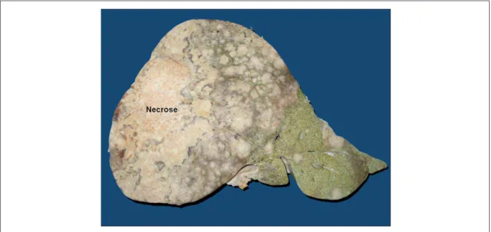
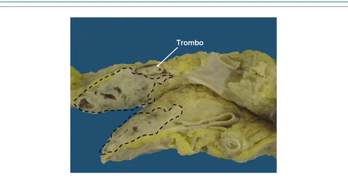
The main finding of the necropsy was a tumor that affected the liver, pancreas and lungs. Most neoplastic mass was located in the liver, which showed large necrotic nodule in the right lobe and multiple peripheral nodules measuring up to 2 cm in diameter (fig. 3). Such masses resulted in a marked increase in liver volume (weight = 4028 g) and firm adhesions with the diaphragm, and adrenal atrophy by compression of the right suprarenal artery. On the tail of the pancreas, there was irregular area of the parenchyma with a homogeneous and whitened
aspect (fig. 4) and multiple small subpleural nodules of 0.5 cm on average diameter were found in the lungs (fig. 5).
The histological pattern was adenocarcinoma, with moderately differentiated tubular aspect in the liver (fig. 6) and lungs (fig. 7) and signet ring cell pattern in the pancreas (fig. 8). There was metastatic adenocarcinoma to lymph nodes and tumor implantation in the gastric muscular wall in the region corresponding to the pylorus.
In particular, the gastric mucosa, the gallbladder, the papilla of Vater and extrahepatic bile ducts showed no neoplastic involvement.
We also found thrombosis of the splenic artery (fig. 4) and vein thrombosis in the lower limbs, in accordance with clinical data.
No recent or incipient pulmonary infarction were found and no signs of previous pulmonary thromboembolism.
Segments of small intestine (fig. 9) showed signs of recent ischemia and the kidneys showed bilateral cortical necrosis.
The heart weighed 320 g and showed no significant macro or microscopic abnormalities.
(Dr. Vera Demarchi Aiello)
Pathological diagnoses
Adenocarcinoma of undefined primary site (probable cholangiocarcinoma — see comments below). Probable metastatic involvement of the pancreas. Subpleural neoplastic metastases to mesenteric lymph nodes and gastric muscle wall. Deep venous thrombosis of lower limbs. Bilateral renal cortical necrosis and ischemic necrosis of segments of the small intestine, without identification of thrombosis and/or embolism to the respective arteries.
(Dr. Vera Demarchi Aiello)
Figure 4 –Macroscopic section of the pancreas tail with an area of the parenchyma replaced by tumor. Splenic artery thrombosis is also observed.
Figure 5 – Pleural surface with multiple nodular metastases (arrows).
Comments
The primary site of malignancy could not be clarified with certainty, but judging by the large liver mass it is more likely to be a cholangiocarcinoma. One cannot rule out completely the possibility of primary pancreatic carcinoma and multiple liver metastases. The immunohistochemical markers currently
available cannot distinguish between these two types of epithelial neoplasia.
Figure 6 – Photomicrograph of the liver showing left epithelial neoplasia (adenocarcinoma). Hematoxylin-eosin staining, objective lens magniication = 20X.
Figure 7 – Photomicrograph of the lung with subpleural metastatic adenocarcinoma. Hematoxylin-eosin staining, objective lens magniication = 5X.
diagnosed pulmonary embolism from the clinicopathologic point of view, it is also part of this syndrome.
In a study of over 500,000 cases of cancer, researchers from the California Cancer Registry identified and quantified venous thromboembolism events before and after diagnosis of malignancy22.They found that 0.11% of cancers were associated with this type of event within one year of diagnosis of malignancy. Among cases with
metastatic cancer, the standardized incidence ratio was 2.3, while for the other stages of cancer this ratio was 1.07. They also found that there were seven types of malignancy associated with a significantly elevated standardized incidence ratio of venous thromboembolism, namely: acute myeloid leukemia, non-Hodgkin lymphoma, renal, ovarian, pancreatic, gastric and lung carcinomas.
Figure 8 – Photomicrograph of the pancreas showing signet ring cell tumor (arrows in panel “a”). In panel b), there is malignant perineural invasion. Hematoxylin-eosin staining, objective lens magniication = 40X and 20X, respectively.
Figure 9 – Photomicrograph of small intestine with recent congestion and necrosis of the Wall. Hematoxylin-eosin staining, objective lens magniication = 10X.
References
1. Tait N, Richardson AJ, Muguti G, Little JM. Hepatic cavernous haemangioma: a 10 year review. Aust N Z J Surg. 1992;62(7):521-4.
2. Terminology of nodular hepatocellular lesions. International Working Party. Hepatology. 1995;22(3):983-93.
3. Befeler AS, Di Bisceglie AM. Hepatocellular carcinoma: diagnosis and treatment. Gastroenterology. 2002;122(6):1609-19.
4. Bruix J, Sherman M, Llovet JM, Beaugrand M, Lencioni R, Burroughs AK, et al; EASL Panel of Experts on HCC. Clinical management of hepatocellular carcinoma: conclusions of the Barcelona-2000 EASL
conference. European Association for the Study of the Liver. J Hepatol. 2001;35(3):421-30.
5. Chang MH. Decreasing incidence of hepatocellular carcinoma among children following universal hepatitis B immunization. Liver Int. 2003;23(5):309-14.
6. Hassoun Z, Gores GJ. Treatment of hepatocellular carcinoma. Clin Gastroenterol Hepatol. 2003;1(1):10-8.
8. Goldhaber SZ, Elliot CG. Acute pulmonary embolism: part I: epidemiology, pathophysiology and diagnosis. Circulation. 2003;108(22):2776-9.
9. Konstandinidis S, Geibel A, Heusel G, Heinrich F, Kasper W; Management Strategies ans Prognosis of Pulmonary Embolism-3 Trial Investigators. Heparin plus alteplase compared with heparin alone in patients with submassive pulmonary embolism. N Engl J Med. 2002;347(15):1143-50.
10. Torbicki A, Perrier A, Konstandinidis S, Agnelli G, Galie N, Pruszczyk P, et al; ESC Committee for Practice Guidelines (CPG). Guidelines on the diagnosis and management of acute pulmonary embolism: the Task Force for the Diagnosis and Management of Acute Pulmonary Embolism of the European Society of Cardiology (ESC). Eur Heart J. 2008;29(18):2276-315.
11. Konstandinidis S, Geibel A, Olschewski M, Heinrish F, Grosser K, Rauber K, et al. Association between thrombolytic treatment and the prognosis of hemodynamically stable patients with major pulmonary embolism: results of a Multicenter Registry. Circulation. 1997;96(3):882-92.
12. Goldhaber SZ, Visani L, De Rosa M. Acute pulmonary embolism: clinical outcomes in the International Cooperative Pulmonary Embolism Registry (ICOPER). Lancet. 1999;353(9162):1386-9.
13. Pineda LA, Hathwar VS, Grant BJ. Clinical suspicion of fatal pulmonary embolism. Chest. 2001;120(3):791-5.
14. Wells PS, Anderson DR, Rodger M, Ginsberg JS, Kearon C, Gent M, et al. Derivation of a simple clinical model to categorize patients’ probability of
pulmonary embolism: Increasing the models utility with the SimpliRED D-dimer. Thromb Haemost. 2000;83(3):416-20.
15. Kucher N, Goldhaber SZ. Cardiac biomarkers for risk stratification of patients with acute pulmonary embolism. Circulation. 2003;108(18):2191-4.
16. Light RW. Clinical practice: pleural effusion. N Engl J Med. 2002;346(25):1971-7.
17. Sahn SA. State of the art: the pleura. Am Rev Respir Dis. 1988;138(1):184-234.
18. Light RW, Macgregor MI, Luchsinger PC, Ball WC Jr. Pleural effusions: the diagnostic separation of transudates and exudates. Ann Intern Med. 1972;77(4):507-13.
19. Heffner JE, Brown LK, Barbieri CA. Diagnostic value of tests that discriminate between exudative and transudative pleural effusions. Primary Study Investigators. Chest. 1997;111(4):970-80.
20. Good JT Jr, Taryle DA, Sahn SA. The pathogenesis of low glucose, low pH malignant effusions. Am Rev Respir Dis. 1985;131(5):737-41.
21. Levi M, Toh CH, Thachil J, Watson HG. Guidelines for the diagnosis and management of disseminated intravascular coagulation. British Committee for Standards in Haematology. Br J Haematol. 2009;145(1):24-33.