333
Jornal Brasileiro de Patologia e Medicina Laboratorial
y
Lipoma-like hibernoma: an atypical lipoma/well-differentiated
liposarcoma mimicker
Hibernoma lipoma-símile: um mimetizador do lipoma atípico/lipossarcoma bem diferenciado
Jorge S. Reis-Filho1 João Cruz2 Carmen Ruiz de Valbueva3 Fernando C. Schmitt4
key words
unitermos
abstract
Hibernomas are benign lipomatous tumors which show differentiation toward brown fat. Recently, unusual variants have been described, including myxoid, spindle cell, and lipoma-like variants. Lipoma-lipoma-like hibernoma (LLH) is characterized by mature univacuolated adipocytic cells with rare admixed multivacuolated brown fat-like cells, which may resemble lipoblasts, leading to a misdiagnosis of atypical lipoma/well-differentiated liposarcoma (AL/ WDLS). We herein report a case of LLH arising on the anterior aspect of the left thigh of a 17-year-old female. A marginal excision was performed. The patient was discharged and remains well four months after surgery. Histological examination showed a lobulated neoplasm composed of univacuolated mature adipose cells admixed with small vessels and occasional mast cells. Scattered islands of brown fat-like cells accounting for less than 10% of the neoplasm were found. Sometimes these cells presented indented and scalloped nuclei, resembling lipoblasts. A final diagnosis of LLH was made based on the presence of focal areas with typical hibernoma morphology, and the lack of atypical hyperchromatic stromal cells. Pathologists must be aware of the typical histological findings of LLH, not to confuse it with AL/WDLS.
Lipoma Hibernoma Liposarcoma Adipocytic tumors
resumo
Hibernomas são tumores benignos que apresentam diferenciação similar à das células do tecido adiposo marrom. Recentemente, variantes incomuns desta neoplasia foram descritas, incluindo hibernomas mixóides, de células fusiformes e lipoma-símile. O hibernoma lipoma-símile (HLS) é caracterizado por adipócitos maduros entremeados por uma pequena quantidade de células com morfologia similar às do tecido marrom. Por vezes, tais células se assemelham a lipoblastos, criando dificuldades no diagnóstico diferencial com lipomas atípicos/lipossarcomas bem diferenciados lipoma-símile. Os autores relatam um caso de HLS acometendo a face anterior da coxa esquerda de uma adolescente de 17 anos. Uma exérese com margens livres de comprometi-mento neoplásico foi realizada. A paciente não apresenta evidências de recidiva quatro meses após o tratamento cirúrgico. Ao exame histológico, observou-se neoplasia predominantemente constituída por adipócitos maduros entremeados por agregados de células com morfologia similar às do tecido adiposo marrom, perfazendo menos de 10% da neoplasia. Tais células ocasional-mente apresentavam núcleos indentados, assemelhando-se a lipoblastos. O diagnóstico final de HLS foi realizado baseado na presença de áreas focais mas com morfologia típica de hibernoma, bem como na ausência de células estromais hipercromáticas bizarras. Patologistas devem estar familiarizados com a morfologia dos HLS para não confundi-los com tumores do tecido adiposo com potencial de malignidade.
Lipoma Hibernoma Lipossarcoma Tumores do tecido adiposo
1. Fellow of the Institute of Molecular Pathology and Immunology, Universidade do Porto (Ipatimup), Portugal. 2. Resident in Pathology, Hospital São João, Universidade do Porto, Portugal.
3. Surgical pathologist, Hospital São João, Universidade do Porto, Portugal.
4. Director of the Molecular Pathology Unit – Institute of Molecular Pathology and Immunology, Universidade do Porto (Ipatimup), Portugal
RELATO DE CASO
CASE REPORT
Recebido em 08/10/01 Aceito para publicação em 18/02/02
Rio de Janeiro, v
334
Rio de Janeiro, v
. 38, n. 4, 2002
Jornal Brasileiro de Patologia e Medicina Laboratorial
Introduction
Hibernoma is an unusual subcutaneous or intra-muscular lipomatous tumor, whose cells show differen-tiation toward brown fat (2, 3, 6). This entity was first recognized in 1906 (3). However, Gery was the first to coin the term hibernoma, in 1914 (6). It usually affects adults, with a slight male predominance and a peak of incidence between the 3rd and 5th decades (mean of 38 years) (2, 3, 6).
Recently, Furlong et al. described three unusual variants of this neoplasm (3): spindle cell, myxoid, and lipoma-like variants. According to their data, the most troublesome of them is the lipoma-like variant. This variant is characterized by abundant univacuolated cells and only scattered granu-lar or pale multivacuolated hibernoma cells. These cells may be mistaken as lipoblasts, and led to a diagnosis of atypical lipoma/well-differentiated liposarcoma in approximately one fourth of the Armed Forces Institute of Pathology consultation cases (3). To the best of our knowledge, only brief mentions addressing the existence of this rare variant of hibernoma in major surgical and soft tissue pathology textbooks were published so far (2, 4, 6), however, there were no illustrations of its misleading histological features. We describe herein a case of lipoma-like hibernoma (LLH), emphasizing the differential diagnosis with atypical lipoma/ well-differentiated liposarcoma (AL/WDLS).
Case report
A 17-year-old female presented at our hospital with a slow growing painless subcutaneous mass with reddish to brown surface in the anterior aspects of the left thigh, measuring 9.5 cm of diameter. The patient underwent surgery with clinical diagnostic hypotheses of lipoma or hemangioma. A marginal excision was carried out. No further adjuvant treatment was performed. The patient was discharged and remains well four months after surgery.
On gross examination, the specimen was composed of multiple fragments of a tan-to-brown, lobulated mass, measuring 10 cm of maximum diameter. The subcutaneous tumor was fixed in 10% neutral buffered formalin and paraffin embedded. Sixteen 4µm histological sections were
cut and routinely stained with hematoxylin-eosin for morphologic evaluation. The microscopic analysis showed a markedly lobulated tumor infiltrating the subcutaneous tissue and striated muscle with pushing margins. It was predominantly composed of a population of mature
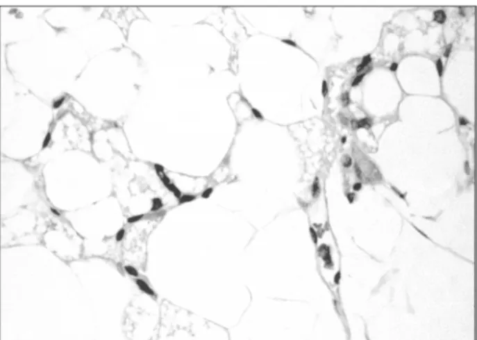
univacuolated adipocytes with discrete variation in cytoplasmic and nuclear dimensions, admixed with small vessels as well as occasional mast cells. There were scattered islands of moderate to large cells, with eosinophilic multiva-cuolated cytoplasm accounting for less than 10% of the neoplastic cell population. These cells showed two or more sharply outlined eosinophilic-to-clear fatty vacuoles that sometimes indented or scalloped the bland, round-to-oval, eccentrically located nuclei, which exhibited distinct nucleoli, sometimes resembling lipoblasts (Figure 1). In some of those islands, large cells with finely vacuolated eosinophilic granular cytoplasm, and small, central nuclei, very similar to typical brown fat cells, were depicted ( Figu-re 2 and inset). In several foci of the neoplasm, discrete branching small capillary vessels were observed, some of them with fibrin thrombi. Neither atypical spindle-shaped hyperchromatic stromal cells nor atypical mono or multivacuolated lipoblasts were observed. A final diagnosis of lipoma-like variant of hibernoma was made.
Figure 1 – High power magnification highlighting a lipoblast-like pale staining hibernoma cell (H&E 400x)
Figure 2 – Photomicrography showing a discrete island of typical eosinophilic hibernoma cells (H&E 100x). Inset. Typical hibernoma cell with finely vacuolated cytoplasm and slightly eccentric bland nucleus with distinct nucleolus (H&E 400x)
335
Rio de Janeiro, v
. 38, n. 4, 2002
Jornal Brasileiro de Patologia e Medicina Laboratorial
Discussion
Hibernomas are unusual benign subcutaneous or intramuscular adipose tissue tumors, which present an invaria-ble benign clinical course. They usually affect adults, with very rare bona fide examples described in children (6). Despite the initial belief that these neoplasms would be restricted to the areas of residual brown fat (6), such as neck, axilla, back, and mediastinum, the thigh was the most frequently affected body site in the largest series published so far (3).
Recently, Furlong et al.(3) reported the prototypic histological features of hibernomas, defining the classic pattern, as well as three histological variants: myxoid, lipoma-like and spindle cell variants (3). Only 12 cases of the lipoma-like hibernoma (LLH) were included in this series, accounting for 7% of the cases (3). According to Furlong et al., LLH is defined as a hibernoma composed predominantly of univacuolated fat cells with only scattered granular or pale hibernoma cells (3), without atypia in either the lipomatous or stromal components.
According to Furlong et al. (3), the lipoma-like variant is the most troublesome for surgical pathologists due to the presence of occasional multivacuolated adipocytic cells mimicking lipoblasts (3). In their series, the referral pathologist entertained a diagnosis of atypical lipoma or well-differentiated liposarcoma in 23% of the cases (3). Similar doubts were raised during the routine histological examination of the present case. However, the lack of atypia and the finding of rare foci of bona fide granular or pale brown fat cells supported the diagnosis of lipoma-like hibernoma. Some subtle histological findings might help in the differentiation of prototypic lipoblasts and brown fat cells: while lipoblasts of AL/WDLS are usually characterized by large cytoplasmic vacuoles that indent or scallop the atypical and hyperchromatic nucleus (2,
4, 6), the vacuolated cells of LLH may show a finely vacuolated clear or pink cytoplasm, and a central or slightly eccentric bland indented nucleus, often harboring a prominent nucleolus (3). In addition, LLH usually lacks the scattered bizarre, mono or multinucleated stromal cells and the fibrous septa composed of hyperchromatic spindle cells and bizarre multinucleated cells that are peculiar to AL/WDLS (2). Moreover, in difficult cases, cytogenetic analysis might play a role in the differentiation of LLH and well-differentiated liposarcoma/ atypical lipoma (1, 2, 5, 6), since the former usually shows rearrangements of 11q13 (MEN1 gene locus) or 10q22, while the latter presents ring or long marker chromosomes derived from the 12q13-15 (1, 2, 5).
For a long time, lipoblasts have been considered as a diagnostic hallmark of liposarcoma and looking for lipoblasts used to be a very common practice in surgical pathology. However, adopting the concepts proposed by Fletcher (2) and Dei Tos (1), lipoblasts are neither required nor sufficient to diagnose a AL/WDLS, since lipoblasts may not be found in several atypical lipomatous tumors harboring the AL/WDLS´ characteristic cytogenetical features (ring or long marker chromosomes – 12q13-15) (1, 2, 5). Conversely, lipoblasts or lipoblast-like cells may be observed in several benign tumors, including lipoblastoma/lipoblastomatosis, pleomorphic lipoma, chondroid lipoma, LLH, and even in reactive processes, such as silicon granuloma (1, 2).
In conclusion, we would like to stress that a correct diagnosis of LLH must be achieved in order to avoid an overtreatment, because while atypical lipomatous tumors frequently recur and might dedifferentiate to a dedifferentiated liposarcoma that harbors a metastatic potential in up to 5% of the recurrences (2, 6), hibernomas are benign tumors that do not recur with complete local excision (2, 3, 6).
References
1. Dei Tos, A.P. Lipomatous tumors. Curr. Diagn. Pathol., 7(1): 8-16, 2001.
2. Fletcher, C.D.M. Soft tissue tumors. In: Fletcher, C.D.M. Diagnostic histopathology of tumors, 2ndedition, Hong Kong: Churchill
Livingstone, 2000, v. 2, p. 1473-540.
3. Furlong, M.A.; Fanburg-Smith, J.C . & Miettinen, M. The morphologic spectrum of hibernoma: a clinicopathologic study of 170 cases. Am. J. Surg. Pathol., 25(6): 809-14, 2001. 4. Rosai, J. Soft tissues. In: Rosai, J. Ackermans surgical pathology, 8th
edition, Saint Louis: Mosby, 1996, v. 2, p. 2021-134. 5. Rubin, B.P. & Fletcher, C.D.M. The cytogenetics of lipomatous
tumours. Histopathology, 30(6): 507-11, 1997.
6. Weiss, S.W. & Goldblum, J.R. Benign lipomatous tumors. In: Weiss, S.W., Goldblum, J.R. Soft tissue tumors. 4th ed. St. Louis,
MI: Mosby, Inc., 2001, p. 571-640.
Correspondence to
Jorge Sergio Reis Filho Ipatimup R. Roberto Frias s/n 4200 – Porto – Portugal Fax: +351225570799 Phone: +351225570700 e-mail: [email protected]