273
Jornal Brasileiro de Patologia e Medicina Laboratorial
y
Laboratory assessment of iron status and reticulocyte parameters
in differential diagnosis of iron deficiency anemia and
heterozygous
β
-thalassemia
Avaliação laboratorial do estado do ferro e parâmetros reticulocitários no diagnóstico diferencial da anemia
ferropriva e
β
-talassemia heterozigótica
Gisélia A.F.M. de Lima1
Helena Z.W. Grotto2
key words
unitermos
abstract
Introduction:The soluble form of transferrin receptor (sTfR) has been pointed as a useful
parameter to assess the iron status and erythropoiesis activity. Immature reticulocytes present high concentration of membrane transferrin receptor. We tested the correlation between sTfR
and reticulocyte parameters in iron deficiency anemia (IDA) and heterozygous β-thalassemia
(hetero β-thal) patients. Laboratory parameters related to iron status and reticulocytes were
studied in order to establish their clinical value to distinguish both anemias. Material and Methods: Reticulocyte measurements were obtained using a semi-automated analyzer and serum concentration of sTfR was determined by an immunoenzymatic technique. Forty-nine
IDA and 43 hetero β-thal patients were studied. Results: Reticulocyte count and sTfR values
were significantly higher in IDA than in hetero β-thal group, but the best parameter to
distinguish both anemias was sTfR index, obtained by the ratio sTfR/ferritin level. Transport compartment was better evaluated by transferrin dosage than by transferrin iron binding capacity (TIBC) determination. The association of serum iron with transferrin measurements (transferrin index) improved the accuracy of the transferrin test. Discussion: The correlation between highly immature reticulocytes and sTfR level was observed only in IDA group, suggesting that cellular iron deprivation is the main responsible factor for up regulation of the
sTfR synthesis in immature red blood cells. High sTfR values in hetero β-thal patients reflect a
degree of ineffective erythropoiesis in this hemoglobinopathy. Conclusion: We concluded that sTfR, ferritin and transferrin measurements are useful and precise parameters to discriminate
IDA from hetero β-thal patients.
Soluble transferrin receptor
Iron metabolism
Microcytosis
Reticulocyte fractions
resumo
Introdução: A forma solúvel do receptor da transferrina (sTfR) tem sido indicada como um parâmetro útil na avaliação do estado do ferro e da atividade eritropoiética. Reticulócitos imaturos apresentam alta concentração dos receptores de transferrina na sua membrana. Estudamos a correlação entre sTfR e parâmetros reticulocitários em pacientes com anemia ferropriva (AF) e com
β-talassemia heterozigótica (β-tal hetero). Os parâmetros laboratoriais relacionados ao estado do ferro e reticulócitos foram estudados a fim de se estabelecer a utilidade clínica dos mesmos na distinção entre os dois tipos de anemia. Material e métodos: As medidas reticulocitárias foram obtidas usando-se um analisador hematológico semi-automático, e as concentrações de sTfR foram determinadas por técnica imunoenzimática. Foram estudados 49 pacientes com AF e 43 com β-tal hetero. Resultados: As contagens de reticulócitos e os valores de sTfR foram significativamente mais elevados na AF do que na β-tal hetero, mas o melhor parâmetro para diferenciar as duas anemias foi o índice de sTfR, obtido pela razão sTfR/ferritina. O compartimento de transporte foi mais bem avaliado pela dosagem de transferrina do que pela capacidade de ligação do ferro à transferrina (TIBC). A associação do ferro sérico à medida de transferrina (índice de transferrina) melhorou a acurácia do teste de transferrina. Discussão: A correlação entre reticulócitos imaturos e nível de sTfR foi observada apenas no grupo com AF, sugerindo que a falta de ferro intracelular seja o principal fator responsável pelo estímulo à síntese de sTfR nas células sangüíneas imaturas. Os valores elevados de sTfR nos pacientes com β-tal hetero refletem um certo grau de eritropoiese ineficaz nessa hemoglobinopatia. Conclusão: Concluímos que as medidas de sTfR, ferritina e transferrina são parâmetros úteis e precisos para diferenciar AF de β-tal hetero.
1. Post-graduate student. 2. Ph.D. Department of Clinical Pathology, School of Medical Sciences, State University of Campinas (Unicamp), Campinas, São Paulo, Brazil.
This work was supported by “Fundo de Apoio ao Ensino e à Pesquisa”, from Unicamp (Faep 1123/97).
Receptor solúvel da
transferrina
Metabolismo do ferro
Microcitose
Imaturidade de reticulócitos
Recebido em 14/11/01 Aceito para publicação em 05/03/02
Rio de Janeiro, v
274
Rio de Janeiro, v
. 38, n. 4, 2002
Jornal Brasileiro de Patologia e Medicina Laboratorial
Introduction
New and reliable laboratory measurements of iron status have been used for detecting and assessing different stages of iron deficiency. Furthermore, there is a consensus that iron status should be interpreted using a group of parameters and not only one single test. It is also pertinent to consider costs and time spent to achieve a correct diagnosis.
Some authors have suggested the use of reticulocyte parameters to distinguish iron deficiency anemia (IDA) from heterozygous β-thalassemia (hetero β-thal) (26), both presenting hypochromia and microcytosis of erythrocytes and both with a high incidence in Brazil (21). The clinical value of the reticulocyte quantitation was renewed by the automation of the reticulocyte count together with the possibility of determining the immaturity of reticulocyte population as erythropoiesis activity analysis (8). Yoldi et al. (26) evaluated samples from hetero β-thal and IDA using flux cytometry, and established that a value < 2% for highly fluorescent reticulocytes (HFR) would be used to discri-minate between these pathologies. Membrane reticulocyte presents transferrin receptor and the receptor concen-tration is higher in more immature cells. The soluble form of transferrin receptor (sTfR) was first described by Kohgo et al. (15) and corresponds to a truncated form lacking the cytoplasmic and transmembrane domains of the intact receptor (6). The sTfR determination has been pointed as a useful parameter to assess the iron status and erythro-poiesis activity (1).
The objective of this study was to test laboratory parameters related to reticulocytes, iron metabolism, including sTfR determination, in order to determine their use to distinguish hetero β-thal from IDA.
Material and methods
Subjects
A group of adult patients (n = 92) with hypochromic and microcytic anemia was studied. IDA (n = 49) was defined as serum ferritin (SF) level below 30ng/ml for men and 12ng/ml for women (minimum normal ferritin levels in our laboratory). Patients with SF levels higher than the above mentioned and Hb A2 level higher than 3.4 % were considered as hetero β-thal (n = 43). Fifty-seven nonanemic subjects were used as controls.
Methods
Hematological measurements including reticulocyte parameters (absolute reticulocyte count and high fluorescent reticulocytes – HFR) were obtained using a Cell Dyn 3500 analyzer (Abbott – USA). Reticulocyte was identified by a non-fluorescent method using New methylene blue as dye. Fractions of reticulocyte maturity were calculated depending on the absorption intensity and they were classified as: mature, midmature and highly immature reticulocytes fractions.
Hemoglobin A2 was quantified spectrophotometrically after cellulose acetate electrophoresis (16).
Determination of iron status: determination of serum iron (SI) and the transferrin iron binding capacity (TIBC) were done with a Mira Plus Cobas analyzer (Roche – Switzerland) using Unimate 5 Iron and Unimate 7 UIBC kits (Roche Diagnostic Systems – Switzerland). SF was determined by a chemiluminescence system (kit Immulite – Diagnostic Products Co. – USA)
Transferrin concentration (TFR) was quantified by nephelometric method (Beckman – Ireland). The serum concentration of sTfR was measured by an immuno-enzymatic technique (Quantikine – R&D Systems – USA).
Statistical analysis: In order to compare the variables between groups, the Kruskal-Wallis test was employed. The Spearman correlation coefficient test was used for assessing the association between variables, with level of significance set at < 0.05. The capacity of the tests to discriminate between the groups was studied by means of ROC curves that defined the optimal decision limit of each parameter and the accuracy of each method. The protocol of this study was approved by the Committee of Ethics of the School of Medical Sciences, University of Campinas, São Paulo, Brazil.
Results
275
Rio de Janeiro, v
. 38, n. 4, 2002
Jornal Brasileiro de Patologia e Medicina Laboratorial
Groups
Parameters
Control (n = 57) Hetero β-thal (n = 43) IDA (n = 49)Rtc* (x 106/l) 67.9 ± 17.6 (28.8-134.0) 160.19 ± 137.8 (64-808) 93.7 ± 52.43 (31.0-305.0)
HFR (%) 6.2 ± 2.9 (2.9-18.9) 7.33 ± 3.74 (6.6-23.6) 6.96 ± 3.55 (1.9-21.2) SI (µg/dl) 96.7 ± 29.3 (42.0-149.0) 86.8 ± 29.7 (24.0-163.0) 26.6 ± 17.45 (2.0-76.0) TIBC (µg/dl) 352.4 ± 58.5 (245.0-467.0) 402.9 ± 99.9 (264.0-689.0) 409.1 ± 105.8 (210.0-630.0) TS (%) 28.0 ± 11.0 (12.0-60.0) 24.0 ± 12.0 (10.0-60.0) 8.0 ± 9.0 (1.0-6.0) SF (ng/ml) 103.8 ± 72.4 (17.7-334.0) 142.24 ± 156.2 (24.0-682.0) 7.4 ± 4.75 (1.5-23.5) TRF* (mg/dl) 249.8 ± 28.7 (202.0-317.0) 231.1 ± 55.7 (125.0-366.0) 337.5 ± 61.3 (111.0-427.0) TI* 0.41 ± 0.11 (0.20-0.65) 0.39 ± 0.15 (0.18-0.76) 0.08 ± 0.05 (0.01-0.26) sTfR* (nmol/l) 16.88 ± 3.1 (12.5-27) 27.1 ± 15.82 (15.0-91.0) 50.26 ± 21.4 (23.5-108.5) sTfRI* 27.7 ± 23.60 (5.2-99.4) 46.51 ± 48.92 (3.0-279) 1287.5 ± 1329.2 (119.0-5812.0)
Rtc: reticulocyte count; HFR: high immature reticulocyte fraction; SI: serum iron; TIBC: transferrin iron binding capacity; TS: transferrin saturation; SF: serum ferritin; TRF: serum transferrin; TI: transferrin index = SI/TRF; sTfR: soluble transferrin receptor; sTfRI: sTfR index = sTfR/SF. *p < 0.05.
Hematological and iron parameters in different groups of patients and control.
Values are means
±
SD and range
Table 1
Variables
Control Hetero β-thal IDAHFR x sTfR r = 0.049 r = 0.007 r = 0.349
p = 0.795 p = 0.963 p = 0.025
sTfRI x SI r = - 0.081 r = 0.087 r = - 0.140
p = 0.670 p = 0.611 p = 0.381
TS x TI r = 0.904 r = 0.628 r = 0.675
p = 0.0001 p = 0.0001 p = 0.0001
TIBC x TRF r = 0.688 r = 0.169 r = 0.517
p = 0.0001 p = 0.322 p = 0.0005
sTfR x SF r = 0.178 r = 0.0117 r = - 0.384
p = 0.345 p = 0.945 p = 0.013
sTfR x Rtc r = 0.262 r = 0.2883 r = 0.170
p = 0.1615 p = 0.0881 p = 0.286
sTfR x Hb r = 0.121 r = - 0.4249 r = - 0.1117
p = 0.523 p = 0.0098 p = 0.4867
Hb: hemoglobin (g/dl).
Correlation between variables in patients with hetero
β
-thal, IDA and controls
Table 2
Groups
and hetero β-thal and also between IDA and control groups(p = 0.0001).
The correlation between HFR and sTfR was modest (r = 0.349), but significant (p = 0.025) only in IDA group. Transferrin saturation (TS) and TI indices measure iron transport compartment and the correlation coefficients were positive in all groups (Table 2). However, TI was more accurate than TS to distinguish IDA from hetero β-thal, with 100% of
276
Rio de Janeiro, v
. 38, n. 4, 2002
Jornal Brasileiro de Patologia e Medicina Laboratorial
Three of them could be identified as IDA by TI value. The other one presented TRF concentration below normal limit and sTfRI compatible with IDA. We did not investigate if the patient had some associated clinical condition that could explain a decreasing synthesis or loss of TRF. Ten of the 43 hetero β-thal patients presented TIBC values higher than the superior normal limit. One of them showed results compatible with iron deficiency, although ferritin level was normal (SF = 27 mg/l). Transferrin saturation, sTfR and sTfRI values (10%, 72nmol/l and 270, respectively) indicated an iron depletion, but TRF concentration, contrary to expected,
showed normal values (212mg/dl). The second patient was firstly classified as hetero β-thal, according to hematological values, HbA2 and ferritin determinations. However, the film examination revealed nucleated red blood cells, punctuate basophilic, poikilocytes and cell fragments. In fact, clinical features suggest that this individual is β-thalassemic intermedia patient. Medical records reported a splenectomy when the patient was 7 years old and repetitive transfusion proceedings from 7 to 22 years old, due to anemia. Nowadays the patient presents an associated chronic liver disease. The high TIBC value (476mg/dl), low transferrin
Parameter
Accuracy
Sensitivity
Specificity
Decision
(%)
(%)
(%)
limit
Rtc 76.2 68.0 74.4 ≤ 93 x 106/l
HFR 54.1 82.0 30.2 ≤ 8.3%
TIBC 53.6 60.0 62.8 ≥ 401µg/dl
TS 94.2 84.0 95.3 ≤ 10%
TI 99.2 95.1 100 ≤ 0.17
TRF 90.1 87.8 88.9 ≥ 278mg/dl
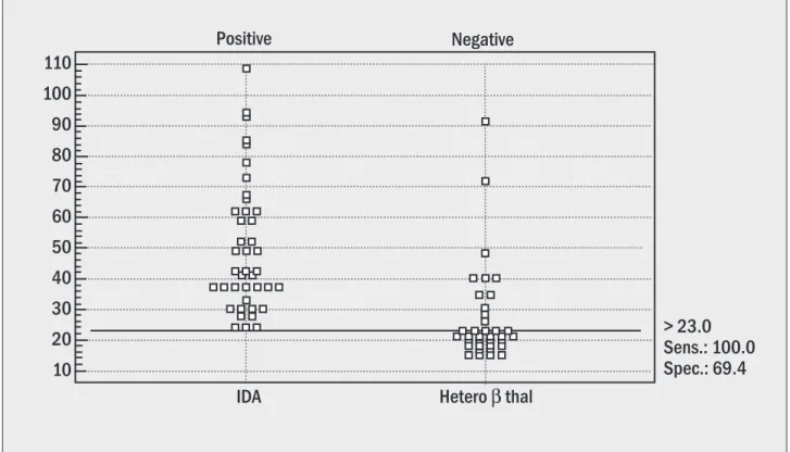
sTfR 87.6 100 69.4 ≥ 23nmol/l
sTfRI 99.7 97.6 97.2 ≥ 140
Accuracy, sensitivity, specificity and cutoff values to distinguish IDA from hetero
β
-thal
(ROC curve results)
Table 3
Figure 1 – Transferrin index (SI/TRF) in IDA and hetero β-thal. The discrimination limit is set at 0.17
Positive
Negative
0.8
0.7
0.6
0.5
0.4
0.3
0.2
0.1
0.0
< = 0.17
Sens.: 95.1
Spec.: 100.0
277
Rio de Janeiro, v
. 38, n. 4, 2002
Jornal Brasileiro de Patologia e Medicina Laboratorial
Figure 2 – sTfR in IDA and hetero β-thal. The discrimination limit is set at 23.0nmol/l
Positive
Negative
110
100
90
80
70
60
50
40
30
20
10
IDA
> 23.0
Sens.: 100.0
Spec.: 69.4
Hetero
β
thal
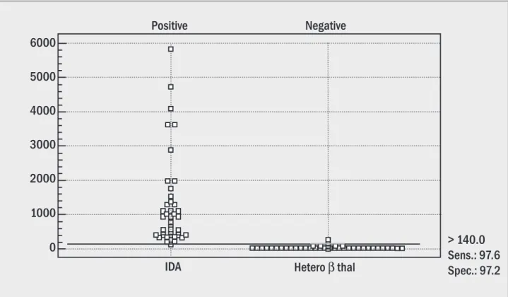
saturation and serum iron determinations suggest an associated iron deficiency anemia, a diagnosis which has been reinforced by an elevated sTfR value (41nmol/l). The other eight patients did not present additional laboratorial results that could explain high TIBC values. sTfR measurement showed an accuracy of 0.876 (Figure 2) and this result was improved by the ratio sTfRI (accuracy = 0.997) (Figure 3). Only one IDA and one hetero β-thal showed values out of limits, providing 97.6% sensitivity and 97.2% specificity for sTfRI parameter.
Discussion
Transferrin receptors are membrane glycoproteins responsible for binding transferrin during the endocytosis of iron. The density of surface transferrin receptor is proportional to the iron requirement of the cell. Then, the transferrin is abundant in cells of organs with the highest iron requirements, such as the erythron marrow and the placenta. After internalization by endocytosis, the endosome containing the transferrin-receptor complex becomes acidified and iron loses its affinity for transferrin. Receptor and transferrin return to the cell surface, where the transferrin is released while the iron is transported to the cytosol (7). The initial study about circulating transferrin
receptor was reported by Kohgo et al. (15). They demons-trated that the concentration of this protein was significantly elevated in enhanced erythropoiesis and iron deficiency. The clinical use of the sTfR has been reported in several situations, especially to distinguishing iron deficiency from the hypoproliferative anemia that is associated with chronic diseases (10). The serum receptor increases with the severity of the iron deficiency anemia, but not in chronic diseases. In addition, sTfR provides an assessing of erythropoiesis status, because an increase in the sTfR concentration is proportional to the expansion of the erythroid marrow (13). In hemolytic anemias with efficient erythropoiesis there is a close parallel rise in sTfR and absolute reticulocyte count (7). On the other hand, in disorders in which there is an ineffective erythropoiesis, such as myelodysplasia, an increase in sTfR is observed, whereas discret reticulocyte count occurs (3). Patients with hetero β-thal have increased erythroid marrow activity, although they present various degrees of an ineffective erythropoiesis (4).
278
Rio de Janeiro, v
. 38, n. 4, 2002
Jornal Brasileiro de Patologia e Medicina Laboratorial
Figure 3 – sTFRI index in IDA and hetero β-thal. The discrimination limit is set at 140.0
Positive
Negative
6000
5000
4000
3000
2000
1000
0
> 140.0
Sens.: 97.6
Spec.: 97.2
IDA
Hetero
β
thal
hetero β-thal associated to IDA and observed that sTfR was significantly higher in hetero β-thal than in normal controls, but significantly lower than in IDA. sTfR was not useful in diagnosing associated IDA in hetero β-thal patients.
In our study we tested parameters related to sTfR, transferrin and reticulocyte counts in patients with IDA and hetero β-thal. Reticulocytes are good indicators of erythropoiesis activity. Automated methods using flow cytometry and RNA dyes resurrected the confidence in reticulocyte counts and introduced new approaches concerning bone marrow response by the quantification of reticulocyte maturity (19). Thus, immature reticulocyte fraction has been reported as an early predictor of the regenerative activity of bone marrow after bone marrow transplantation (8, 12).
Yoldi et al. (26) described high fluorescent reticulocytes as a good discriminant between hetero β-thal and IDA, since the value in IDA is higher than in hetero β-thal. Such difference has not been found in our study. An explanation for that fact could be that the used methodology has not included fluorescence to identify reticulocyte maturity. However, we carried out a similar study using flow cytometry and thiazole orange as dye and the results did not change (HFR mean = 5.11 ± 4.47 to hetero β-thal and
279
Rio de Janeiro, v
. 38, n. 4, 2002
Jornal Brasileiro de Patologia e Medicina Laboratorial
References
1. Beguin, Y. The soluble transferrin receptor: biological aspects and clinical usefulness as quantitative measure of erythropoiesis. Haematologica, 77: 1-10, 1992.
2. Beilby, J. et al. Transferrin index: an alternative method for calculating the saturation of transferrin. Clin. Chem., 38: 2078-81, 1992
3. Bowen, D.T. et al. Estimation of effective and total erythropoiesis in myelodysplasia using serum transferrin receptor and erythropoietic concentrations, with automated reticulocyte parameters. Leukemia, 8: 151-5, 1994.
4. Centis, F. et al. Soluble transferrin receptor following bone marrow transplantation from donors heterozygous for beta thalassemia. Haematologica, 79: 448-51, 1994.
5. Cermak, J. & Brabec, V. Transferrin receptor-ferritin index: a useful parameter in differential diagnosis of iron deficiency and hyperplastic erythropoiesis. Eur. J. Haematol., 61: 210-12, 1998.
6. Cook, J.D.; Skikne, B.S. & Baynes, R.D. Serum transferrin recep-tor. Ann. Rev. Med., 44: 63-74, 1993.
7. Cook, J.D. The measurement of serum transferrin receptor. Am. J. Med. Sci.,318(4): 269-76, 1999.
8. Corberand, J.X. Reticulocyte analysis using flow citometr y.
Hematol. Cell. Ther., 38: 487-94, 1996.
9. Fairbanks, V.F. Laboratory testing for iron status. Hosp. Pract. off Ed., 26(3): 17-24, 1991.
10. Ferguson, B.J. et al. Serum transferrin receptor distinguishes the anemia of chronic disease from iron deficiency ane-mia. J. Lab. Clin. Med., 119: 385-90, 1992.
11. Gimferrer, E.; Ubeda, J. & Remacha, A.F. Serum transferrin re-ceptor levels are “physiologically” high in heterozygous
β-thalassemia. Haematologica, 82: 728-34, 1997.
12. Grotto, H.Z.W. et al. Immature reticulocyte fraction as a criterion for marrow engraftment. Evaluation of a semi-automated reticulocyte counting method. Clin. Lab. Haem., 21: 285-87, 1999.
13. Huebers, H.A. et al. Intact transferrin receptors in human plas-ma and their relation to erythropoiesis. Blood, 75: 102-7, 1990.
14. Intragumtornchai, T. et al. In vivo transferrin-iron receptor relationships in erythron of rats. Am. J. Physiol.,255: R236-31, 1988.
15. Kohgo, Y. et al. Circulating transferrin receptor in human serum.
Brit. J. Haematol.,64: 277-281, 1986.
16. Marengo-Rowe, A.J. Rapid electrophoresis and quantitation of haemoglobin on cellulose acetate. J. Clin. Pathol.,18: 790-92, 1965.
17. Mullner, E.W. & Kuhn, L.C. A stem-loop in 3’ untranslated region mediates iron-dependent regulation of transferrin receptor mRNA stability in cytoplasma. Cell, 53: 815-25, 1988.
evolution of iron deficiency in a phlebotomy program. They demonstrated that there was a progressive elevation in sTfR when serum ferritin fell below the normal range. Before this stage there was no significant change in sTfR levels. All of our patients had serum ferritin below the normal range and due to this fact we could observe an inverse correlation between sTfR and ferritin. In the phlebotomy study the mean sTfR/ferritin ratio was 100 at baseline stage and a ratio of 500 identified the point at which iron stores were fully exhausted. Our minimum sTfR/ ferritin ratio value was 110.0. Up to 140.0 hetero β-thal patients could be identified.
Plasma iron transport is made by transferrin, a monomeric glycoprotein that binds two atoms of Fe3+ with high affinity (23). The TIBC is a measurement of transferrin concentration and was described by Ramsay forty years ago. Despite TIBC being indispensable for determining iron status, technical problems found by Bill Ramsay have not been solved until now (9, 24). An alternative approach would be measuring transferrin directly by immunological assay. In our study, the nephelometry was used to measure the concentration of transferrin, which showed to be a
more accurate method than TIBC. It also has the advantages of requiring a small sample and being nonsusceptible to iron contamination (22). We could notice the apparent limitation of TIBC estimation when some of hetero β-thal patients showed high TIBC values, although transferrin determination was normal. Probably a spuriously high value was due to a non-transferrin iron measurement (25).
We conclude that the interpretation of iron status is based on a group of laboratory findings and, according to our results, the association of sTfR and ferritin measu-rements is more precise to distinguish hetero β-thal from IDA. However, in a screening survey where it is pertinent to consider costs, transferrin evaluation may be adopted, since it showed a good performance to discriminate both microcytic hypochromic anemias.
Acknowledgments
280
Rio de Janeiro, v
. 38, n. 4, 2002
Jornal Brasileiro de Patologia e Medicina Laboratorial
Correspondence to
Helena Z. W. Grotto
Departamento de Patologia Clínica Faculdade de Ciências Médicas – Unicamp CEP 13083-970 – Campinas-SP–Brasil Caixa Postal 6111
Fax: (55) (19) 3289-3273 Phone: (55) (19) 788-7064 e-mail: [email protected]
18. Rouault, T. et al. Hemin, chelatable iron and the regulation of transferrin receptor biosynthesis. J. Biol. Chem.,260: 14862-66, 1985.
19. Rowan, R.M.; Cavill, I. & Corberand, J.X. The reticulocyte count: progress toward the resurrection of a useful clinical test.
Clin. Lab. Haem., 18: 3-8, 1996.
20. Skikne, B.S.; Flowers, C.H. & Cook, J.D. Serum transferrin re-ceptor : a quantitative measure of tissue iron deficiency.
Blood,75: 1870-76, 1990.
21. Sonati, M.F. et al. Hereditar y hemoglobinopaties in a population from southest Brazil. Hemoglobin,20(2): 175-79, 1996.
22. Woo, J. & Henry, J.B. Metabolic intermediates and inorganic ions. In: Henr y, J.B. (ed.). Clinical diagnosis methods. Philadelphia: WB. Saunders Company, 1996. p. 190-91. 23. Workwood, M. Influence of disease on iron status. Proc. Nutr.
Soc., 56: 409-19, 1997.
24. Workwood, M. The laboratory assessment of iron status: an update. Clin. Chim. Acta, 259: 3-23, 1997.
25. Workwood, M. Iron deficiency anaemia. In: Dacie, J.V. & Lewis, S.M. (ed.). Practical Haematology. London: Churchill Livingstone, 1995: 437-44.
26. Yoldi, F. et al. Automatización del recuento de reticulócitos.