THERAPEUTIC CHALLENGE
Endovascular treatment of endotension with dacron stent
graft reinforcement and femorofemoral crossover bypass
-
therapeutic challenge
Tratamento endovascular da endotensão com endoprótese de dácron e derivação
femoro-femoral cruzada - desafio terapêutico
Edwaldo Edner Joviliano1, Marcelo Bellini Dalio2, José Geraldo Ciscato Junior2, Nei Rodrigues Alves Dezotti2,
Takachi Moriya1, Carlos Eli Piccinato3
Part I - Clinical case
A 54 year-old man with an asymptomatic 5.9 cm AAA was treated with an Excluder® 26 mm/14.5 mm/16 cm endoprosthesis (Gore, USA). Initial aortic morphology showed a proximal neck of 2.0 cm of extension by 23 mm of diameter. he common iliac arteries measured 13 mm in right diameter and 12 mm in let diameter. No endoleak was detected immediately ater the procedure.
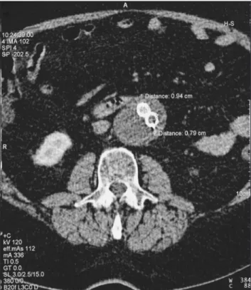
One month after the procedure, computed tomog-raphy (CT) scan showed maintenance of aneurysmal di-ameters without evidence of endoleak (Figure 1). After one year, follow-up duplex ultrasound and CT scan showed no endoleak, but a small growth of the aneurys-mal sac to 6.2 cm was observed (Figure 2). Aortography with selective visceral and hypogastric arteries showed no endoleak (Figure 3). We decided to observe the out-come of the condition with a second CT scan within six months. The patient only returned after one year, when CT scan found the aneurysmal diameter had increased to 7.5 cm, but without evidence of endoleak. The case was interpreted as endotension, and intervention was considered. Since the patient presented class II heart fail-ure, poorly controlled cardiac arrhythmia and a previous
left pneumonectomy for a neoplasm, an endovascular approach was chosen.
Part II - What was done?
Endovascular intervention was performed using a right aorto-mono-iliac Zenith® endoprosthesis (Cook, USA) 28 mm/125 mm with and extension 12 mm/88 mm deployed inside the previous one and an occluder 14 mm/20 mm for the let iliacartery. Pre-procedure aortography showed no evidence of endoleak. Ater endograt deployment, a fem-oro-femoral crossover let-right bypass grat with 8 mm PTFE was carried out. No complication was noted and the patient was discharged ater two days.
Ater six months, a new CT scan showed an aneurys-mal diameter of 6.5 cm, i.e., a reduction of 1 cm compared with the pre-intervention exam (Figure 4).
CT scan studies were all performed using a 16-slice he-lical scanner. All series, including noncontrast images, were obtained at 5.0 mm thickness. Arterial phase images were obtained at 3.0 mm thickness and later reconstructed at 1.5 mm thickness.
he mechanism of persistent sac pressurization without endoleak has yet to be explained. It seems to be the result of Keywords: Aortic aneurysm, blood vessel prosthesis, vascular surgical procedures endotension.
Palavras-chave: Aneurisma aórtico, prótese vascular, procedimentos cirúrgicos vasculares, endotensão.
1 Professor Associado, Divisão de Cirurgia Vascular e Endovascular, Departamento de Cirurgia e Anatomia, Faculdade de Medicina de Ribeirão Preto (FMRP), Universidade de São Paulo (USP),
Ribeirão Preto, SP.
2 Médico Assistente, Divisão de Cirurgia Vascular e Endovascular, Departamento de Cirurgia e Anatomia, FMRP, USP, Ribeirão Preto, SP. 3 Professor Titular, Divisão de Cirurgia Vascular e Endovascular, Departamento de Cirurgia e Anatomia, FMRP, USP, Ribeirão Preto, SP.
Endovascular treatment of endotension - Joviliano EE et al. J Vasc Bras 2010, Vol. 9, Nº 2
82
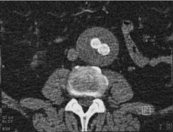
Figure 1 - Computed tomography angiography at one month without
evidence of endoleak Figure 2 aneurysm sac diameter- Computed tomography angiography showing increase in
Figure 3 - A) Aortography and B) selective angiography showing no endoleak
altered the design of the Excluder® with the aim of decreas-ing porosity; the efect of this change on reducdecreas-ing the in-cidence of endotension is still under investigation. Recent work has showed that sac behavior ater EVAR with this newer low-permeability Excluder® endoprosthesis is signif-icantly improved compared with the previous one. his new design may be a solution for the endotension problem.11
Treatment for patients with endotension remains con-troversial. Mennander et al.12 proposed a nonoperative ap-proach for endotension. In their study, three patients pre-sented ruptured aneurysm during the follow-up, without associated hemorrhage, conirming a continuous increase in sac size and aneurysm wall tension in the presence of an efective endograt seal. Evidently, sac enlargement is not always associated with blood low inside the sac, and continuous iltration of transudative material through the pressure transmission and luid accumulation facilitated by
Endovascular treatment of endotension - Joviliano EE et al. J Vasc Bras 2010, Vol. 9, Nº 2 83
grat may result in sac rupture. he fact that the aneurys-mal sac can burst because of endotension and the poorly understood consequences of this rupture make conserva-tive treatment fail as a better choice for such patients. A recent consensus recommended that aneurysmal sac en-largement associated with endotension needs to be treat-ed.13 Open surgery can be a deinitive solution, but it can be diicult to recommend it because of the surgical risk of most of these patients.
Diferent authors have described techniques with-out aortic cross clamping and other less invasive options. Risberg et al.5 reported that puncture, fenestration or resec-tion of the sac did not seem to be adequate. Mory et al.14 suggested an open surgical approach based on decompres-sion and fenestration of the aneurysm sac, proximal aortic neck banding, and transmural endograt ixation with good results in three cases. However, this technique did not ob-viate major laparotomy and did not exclude the endograt that retained its porosity.
Various authors have proposed treatment with en-dograt relining with satisfactory results. Goodney & Fillinger15 reported nine cases using another Excluder® device with a mean follow-up of 16 months, in which mean diameter decreased 2.0 mm/year and mean sac vol-ume decreased 2.6% per year. hey argued for a selective strategy in three ways: reline limbs only; reline limbs only and advance the bifurcation; and reline with aortic cuf and limbs. It depends on the parts in contact with the sac. Kougias et al.6 also reported cases of endograt relin-ing with another Excluder® component. he use of aortic cufs and limbs clearly does not totally eliminate contact between the relatively porous PTFE grat and lowing blood. he authors believe that the dynamic process of transudation and resorption would be tipped in favor of resorption. Sac expansion appeared to be arrested by re-lining the high-permeability device with low-permeability Excluder® endoprosthesis components. Conversely, the nature of the original expansion pattern makes it diicult to declare that the relining procedure with another PTFE is a deinitive solution for endotension.
In our report we did not detect any previous endole-ak and the patient was not submitted to any intervention other than aortouniiliac endograft with femorofemoral crossover bypass. We describe a technique with total ex-clusion of the previous components using a graft with known low permeability, such as dacron from Zenith® graft. Smith et al. showed a rapid sac diameter decrease of 41 mm in six months.7 In our case, the sac decreased by 10 mm in six months. This case reports a valuable minimally invasive option for patients with endotension
and an alternative to open surgical conversion or relin-ing with another PTFE graft.
In conclusion, an increase in aneurysmal sac diam-eter by endotension ater EVAR is an indication for inter-vention. For patients with advanced age or poor clinical conditions, a minimally invasive technique may be more suitable than open surgical conversion. Endovascular treatment of endotension with dacron stent grat seems to be a safe and eicient alternative to relining with another PTFE stent grat.
References
1. EVAR trial participants. Endovascular aneurysm repair versus open repair in patients with abdominal aortic aneurysm (EVAR trial 1): randomized controlled trial. Lancet. 2005;365:2179-86.
2. EVAR trial participants. Endovascular aneurysm repair and out-come in patients unit for open repair of abdominal aortic aneurysm (EVAR trial 2): randomized controlled trial. Lancet. 2005;365:2187-92.
3. Gilling-Smith G, Brennan J, Harris P, Bakran A, Gould D, McWilliams R. Endotension after endovascular aneurysm repair: deinition, classiication, and strategies for surveillance and intervention. J Endovascular Surg. 1999;6:305-7.
4. Fillinger M. hree-dimensional analysis of enlarging aneurysms af-ter endovascular abdominal aortic aneurysm repair in the Gore Excluder Pivotal clinical trial. J Vasc Surg. 2006;43:888-95.
Figure 4 - Computed tomography angiography at six months after new
Endovascular treatment of endotension - Joviliano EE et al. J Vasc Bras 2010, Vol. 9, Nº 2
84
5. Risberg B, Delle M, Lönn L, Syk I. Management of aneurysm sac hygroma. J Endovasc her. 2004;11:191-5.
6. Kougias P, Lin PH, Dardik A, Lee WA, El Sayed HF, Zhou W. Successful treatment of endotension and aneurysm sac enlarge-ment with endovascular stent graft reinforceenlarge-ment. J Vasc Surg. 2007;46:124-7.
7. Smith ST, Clagett GP, Arko FR. Endovascular conversion with femo-rofemoral bypass as a treatment of endotension and aneurysm sac enlargement. J Vasc Surg. 2007;45:395-8.
8. Melissano G, Bertoglio L, Esposito G, Civilini E, Setacci f, Chiesa R. Midterm clinical success and behavior of the aneurysm sac af-ter endovascular AAA repair with the Excluder graft. J Vasc Surg. 2005;42:1052-7.
9. Kottke-Marchant K, Anderson JM, Miller KM, Marchant RE, Lazarus H. Vascular graft associated complement activation and leukocyte adhesion in an artiicial circulation. J Biomed Mater Res. 1987;21:379-97.
10. Trocciola SM, Dayal R, Chaer RA, et al. he development of en-dotension is associated with increased transmission of pressure serous components in porous expanded polytetraluoroethylene stent-grafts: characterization using a canine model. J Vasc Surg. 2006;43:109-16.
11. Tanski W 3rd, Fillinger M. Outcomes of original and low-permea-bility Gore Excluder endoprosthesis for endovascular abdominal aortic aneurysm repair. J Vasc Surg. 2007;45:243-9.
12. Mennander A, Pimenof G, Heikkinen M, Partio T, zeitlin R, Salenius JP. Nonoperative approach to endotension. J Vasc Surg. 2005;42:194-9.
13. Veith FJ, Baum RA, Ohki T, et al. Nature and signiicance of en-doleaks and endotension: summary of opinions expressed at an international conference. J Vasc Surg. 2002;35:1029-35.
14. Mory M, Allenberg JR, Schumacher H, von Tengg-Kobligk H, Böckler D. Open decompression, proximal banding, and aneurysm sac fenestration as an alternative to conversion in the manage-ment of endotension after EVAR. J Endovasc her. 2008;15:449-52.
15. Goodney PP, Fillinger MF. he efect of endograft relining on sac expansion after endovascular aneurysm repair with the original-permeability Gore Excluder abdominal aortic aneurysm endo-prosthesis. J Vasc Surg. 2007;45:686-93.
Correspondence:
Marcelo Bellini Dalio Departamento de Cirurgia e Anatomia, FMRP, USP Av. Bandeirantes, 3900, 90 andar, CEP 14048-900 – Ribeirão Preto, SP, Brazil Tel.: +55 16 3602.2406 Fax: +55 16 3633.0836 E-mail: [email protected]
Author contributions: