Endovascular repair of abdominal aortic aneurysm with lumbar
vertebral erosion in Behçet’s disease: case report
Tratamento endovascular de aneurisma de aorta abdominal com erosão de vértebra
lombar associada à doença de Behçet: relato de caso
Nathalia Leslie Albanez Rodrigues de Souza1
*
, Daniel Emílio Dalledone Siqueira1, Alex Aparecido Cantador1,
Leandro Pablos Rossetti1, Giovani José Dal Poggetto Molinari1, Ana Terezinha Guillaumon1
Abstract
Behçet’s disease is an autoimmune, multifactorial, systemic condition with several clinical manifestations, including vascular disorders. An aortic aneurysm with vertebral erosion is rare in association with this pathology and there are only four case reports listed on the PubMed database. his article reports the case of a female patient with a long-standing diagnosis of Behçet’s Disease who developed a saccular infrarenal abdominal aortic aneurysm with lumbar vertebral erosion. Her surgical treatment consisted of endovascular repair with a monoiliac endoprosthesis and a femorofemoral crossover bypass, because of limitations imposed by the anatomy of the aortic bifurcation. his paper discusses the rarity of this presentation of the disease and treatment outcomes and ofers a brief review of the relevant literature.
Keywords: Behçet’s Disease; aneurysm; spinal column; vertebral lesions.
Resumo
A doença de Behçet é uma doença sistêmica, multifatorial e autoimune com diversas manifestações clínicas, entre elas o acometimento vascular. Aneurisma de aorta associado a erosão de vértebra lombar é condição rara na literatura, existindo apenas quatro relatos de caso nas bases de dados da PubMed. O presente artigo relata o caso de paciente do sexo feminino com diagnóstico de Doença de Behçet de longa data e aneurisma sacular de aorta abdominal infrarrenal com erosão de vértebra lombar. O caso foi tratado por meio de técnica endovascular com colocação de endoprótese monoilíaca e enxerto fêmoro-femoral cruzado, devido a limitações anatômicas da bifurcação aórtica. O artigo aborda a raridade desse tipo de apresentação da doença e o desfecho do tratamento e apresenta revisão da literatura sobre esse tema.
Palavras-chave: Doença de Behçet; aneurisma; coluna vertebral; lesões de coluna vertebral.
1Universidade Estadual de Campinas – UNICAMP, Departamento de Cirurgia, Campinas, SP, Brazil.
Financial support: None.
Conlicts of interest: No conlicts of interest declared concerning the publication of this article. Submitted: January 08, 2017. Accepted: April 11, 2017.
INTRODUCTION
Behçet’s disease (BD) is a systemic, multifactorial
disease of unknown etiology that was irst described by the Turkish dermatologist Hulusi Behçet in
1937.1 The syndrome was initially characterized
by the triad of oral ulcers, genital ulcers, and uveitis. It was subsequently recognized that clinical
manifestations also include synovitis, cutaneous vasculitis, involvement of the gastrointestinal and urogenital systems, meningoencephalitis, and cardiovascular involvement.2,3 Vascular manifestations include
stenoses and occlusions, thrombosis, or formation
of aneurysms and pseudoaneurysms, with incidence rates ranging from 25 to 30% of patients, the most
common of which is deep venous thrombosis of the lower limbs.3,4 In isolation, arterial involvement is rare,
but is associated with potentially fatal complications,
primarily due to development of aneurysms.3-5
The vessel most often involved is the abdominal aorta, followed by the femoral artery and the pulmonary
arteries, so the risk of surgical complications is high
and morbidity and mortality rates are elevated.2,3
Aneurysms secondary to BD do not respond well
to drug-based treatment and surgery is mandatory.5
Conventional open surgery is the modality that has
traditionally been used to treat arterial damage in these patients. This can be challenging because of technical dificulties and postoperative morbidity. Furthermore, outcomes are negatively impacted by the presence of
disease activity and are prone to complications such as occlusion of grafts and formation of anastomotic
pseudoaneurysms, which are the most common and
the most devastating of these possibilities.
Formation of a saccular abdominal aortic aneurysm with erosion of lumbar vertebra is a rare condition in
BD and, up to 2017, there are only four case reports
indexed on PubMed.2,6-8
The patient described in this case report signed
a free and informed consent form and the need for Institutional Research Ethics Committee approval was waived.
CASE DESCRIPTION
The patient was a 35-year-old female who had been diagnosed with BD 20 years previously on the basis of oral and genital ulcers and recurrent
episodes of posterior uveitis and was attending regular consultations with a rheumatology team. She also had a history of smoking, systemic arterial
hypertension, dyslipidemia, and chronic obstructive pulmonary disease. One year previously she had begun to suffer from lumbar pain that did not improve with
standard analgesics or opioid derivatives. A simple
abdominal X-ray revealed calciication in the area of the abdominal aorta and bone erosion in the region of the third lumbar vertebra. She was referred to
a Lymphedema and Angiodysplasia Clinic and supplementary investigation work up was initiated with
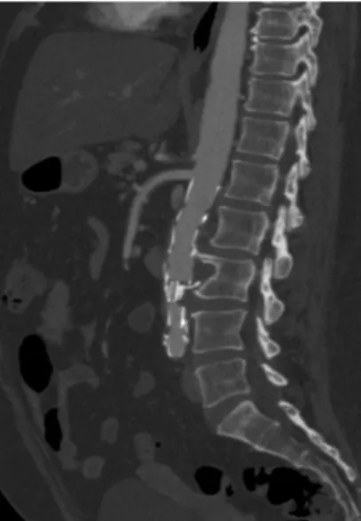
angiotomography of the aorta for diagnostic conirmation and treatment planning. The angiotomography showed an infrarenal, saccular abdominal aortic aneurysm
with a maximum diameter of 3.6 cm and erosion of
the third lumbar vertebra (Figures 1 and 2) and an
aortic bifurcation with 9 mm diameter. both clinical and laboratory parameters showed that the patient was in remission from BD. The treatment chosen was endovascular aneurysm repair using a customized
Braile Biomédica monoiliac endoprosthesis with dimensions of 20 mm x 14 mm x 150 mm (proximal diameter, distal diameter, and length, respectively)
and a femorofemoral crossover bypass with a number 6 dacron prosthesis for revascularization of the left lower limb. This strategy was adopted because of a discrepancy between the proximal and distal diameters
of the aorta and the small diameter of the distal aorta (9 mm), which was too small to safely implant a
bifurcated endoprosthesis. No occlusion device was
used on the left external iliac artery. During construction of the crossover graft, the appearance of the femoral
arteries was normal and no parietal abnormalities that
Figure 2. Preoperative sagittal angiotomography showing aortic aneurysm with erosion of lumbar vertebra.
Figure 3. Postoperative axial angiotomography showing endoprosthesis within the aneurysm sac, excluding it, with no evidence
of endoleaks.
would compromise the quality of the anastomoses were observed. No leakage into the aneurysm sac was observed on the intraoperative control angiography.
After the procedure, the patient exhibited good
clinical recovery and was discharged on the third day after the operation. She is attending outpatients follow-up with satisfactory progress and full resolution
of the lumbar pain. She is also being seen by the
DISCUSSION
Behçet’s disease is a systemic inflammatory
disease, autoimmune in nature, that is characterized by vasculitic lesions, in the majority of cases manifest as oral and genital ulcers and uveitis. The disease also involves the skin, joints, vessels of all calibers,
genitourinary and gastrointestinal tracts, and the central
nervous system. Its pathophysiology is unknown, but it is currently believed that there is an interaction between genetic factors – such as presence of HLA
B51 – and environmental factors – such as infection by bacteria of the Streptococcus genus. Behçet’s
disease manifests in young adults, with incidence at
20 to 30 years of age and with no difference between
the sexes. Although vascular involvement is not one of the diagnostic criteria for BD, it is present
in 25 to 30% of cases. The disease causes vasculitis of the vasa vasorum which, in conjunction with inlammatory activity and a hypercoagulable state, leads to thrombosis and formation of aneurysms. Early diagnosis of BD is essential because of its potentially fatal complications. The possibility of arterial aneurysms should be investigated during the acute phase of the disease, but they can also form during the chronic phase of the disease. The treating team should therefore be alert for signs and symptoms that
suggest this complication. Involvement of the aorta in BD is different from atherosclerotic aneurysmal
disease, because in the latter there is accentuated
destruction and weakening of the artery wall.3,5
Options for surgical treatment include open surgery
and endovascular techniques. The classical method
for surgical treatment of aneurysms associated with
BD is open surgery. However, this is subject to challenges related to technical dificulties and the high
risk of formation of anastomotic pseudoaneurysms.
Kalko et al. analyzed 16 BD patients with 18 arterial aneurysms, six aortic, ive of which had ruptured.5
All of the surgeries were conducted using expanded
polytetraluoroethylene prosthetic grafts. The mean
follow-up period was 17 months, and during this
period two patients were reoperated because of
development of anastomotic pseudoaneurysms and
one patient developed an arterial aneurysm. Twelve
of the 16 patients were in remission from the disease.5
Erentuğ et al.1 reported two cases of ruptured aortic
aneurysms associated with BD that were repaired
with aortobifemoral and aortoaortic bypasses and
followed-up for 30 months without postoperative
complications. Hosaka et al.9 reported a cases series
of 10 patients with BD who were treated for arterial
involvement with open surgery, observing ive graft occlusions and ive pseudoaneurysms during the follow-up period. Balcioglu et al. analyzed nine patients with
BD and aortic aneurysms (six infrarenal and three suprarenal), who were treated with endovascular surgery after immunosuppression with methylprednisolone and cyclophosphamide to achieve remission of BD
inlammatory activity.3 Three patients required a
hybrid procedure with visceral debranching and endovascular repair on the same day. The follow-up
period was 40 months, with 100% survival over 1 year
and 88% over 2 years. There were no occlusions of
Figure 4. Postoperative sagittal angiotomography showing
endoprosthesis within the aneurysm sac, excluding it, with no evidence of endoleaks.
Figure 5. 3-D reconstruction produced with Osirix software,
endoprostheses or pseudoaneurysms, although one
patient developed a istula between the duodenum and the endoprosthesis, which was corrected by
resection of the duodenum and an omentum patch to the endoprosthesis.3 Park et al. treated seven
patients with aortic aneurysms using endovascular
surgery and observed one case of degeneration of
the distal endoprosthesis anchor site.10 Nitecki et al.11
operated on 55 patients, in two groups: either open
or endovascular surgery. Their results showed that length of hospital stay was shorter and morbidity and
mortality rates were lower in the group treated with the endovascular method compared with the open surgery group.
Cases of aortic aneurysms with vertebral body
erosion are rare in the literature and even rarer when
vertebral erosion is secondary to a BD-associated abdominal aortic aneurysm (AAA).2,6-8,11-15 Vertebral
lytic lesions are generally associated with fractures,
osteoporosis, neoplasms, infections, or inlammatory states. Possible factors associated with lumbar pain in patients with AAA are size of aneurysm, incorrect control of arterial blood pressure, aortic dissection, and erosion of a vertebral body.2,16,17
The diagnostic criteria for BD are presence of
oral ulcers plus two of the following: genital ulcers, typical ocular lesions, typical skin lesions, or positive pathergy test.18 It is current opinion that due to the
high rate of aneurysmal disease recurrence in BD patients, open or endovascular surgical treatment is
insuficient without additional immunosuppressant
therapy.3 Postoperative follow-up of aneurysms in
BD should be regular and should involve assessment
of all arteries.5,19
In conclusion, in Behçet’s disease, vascular
involvement increases morbidity and mortality and should always be considered and investigated in this
patient population. Aortic aneurysms with erosion of
lumbar vertebrae are rare, but should be considered in patients who have been diagnosed with BD and present with dificult-to-treat lumbar pain. Endovascular treatment is proving to be a promising alternative to open surgery for treating these patients, but additional studies with longer follow-up are needed to enable adequate evaluation of the results.
REFERENCES
1. Erentuğ V, Bozbuğa N, Ömeroğlu SN, et al. Rupture of Abdominal Aortic Aneurysms in Behçet’s Disease. Ann Vasc Surg. 2003;17(6):682-5. PMid:14738093. http://dx.doi.org/10.1007/s10016-003-0076-0. 2. Örücü M, Keleş D, Peker E, et al. Abdominal aortic aneurysm causing lumbar vertebral erosion in Behçet’s disease presenting by
low back pain. Rheumatol Int. 2015;35(2):367-70. PMid:24957970. http://dx.doi.org/10.1007/s00296-014-3077-0.
3. Balcioglu O, Ertugay S, Bozkaya H, Parildar M, Posacioglu H. Endovascular Repair and adjunctive immunosuppressive therapy of aortic involvement in Behçet’s Disease. Eur J Vasc Endovasc Surg. 2015;50(5):593-8. PMid:26321000. http://dx.doi.org/10.1016/j. ejvs.2015.07.011.
4. Ulusan Z, Karadag A, Tasar M, Kalender M, Darcin O. Behcet’s disease and cardiovascular involvement: our experience of asymptomatic Behcet’s patients. Cardiovasc J Afr. 2014;25(2):63-6. PMid:24844550. http://dx.doi.org/10.5830/CVJA-2014-003. 5. Kalko Y, Basaran M, Aydin U, Kafa U, Basaranoglu G, Yasar T. The
surgical treatment of arterial aneurysms in Behçet disease: a report of 16 patients. J Vasc Surg. 2005;42(4):673-7. PMid:16242553. http://dx.doi.org/10.1016/j.jvs.2005.05.057.
6. El Maghraoui A, Tabache F, Bezza A, et al. Abdominal aortic aneurysm with lumbar vertebral erosion in Behçet’s disease revealed by low back pain: a case report and review of the literature. Rheumatology. 2001;40(4):472-3. PMid:11312389. http://dx.doi. org/10.1093/rheumatology/40.4.472.
7. Roeyen G, Van Schil P, Vanmaele R, et al. Abdominal aortic aneurysm with lumbar vertebral erosion in Behçet’s dis- ease. A case report and review of the literature. Eur J Endovasc Surg. 1997;13(2):242-6. PMid:9091166. http://dx.doi.org/10.1016/S1078-5884(97)80030-5. 8. Ahn H, Kwon S, Park H. Abdominal aortic aneurysm rupture with vertebral erosion presenting with severe refractory back pain in Behçet’s disease. Ann Vasc Surg. 2010;24(2):254. PMid:19900780. http://dx.doi.org/10.1016/j.avsg.2009.05.011.
9. Hosaka A, Miyata T, Shigematsu H, et al. Long-term outcome after surgical treatment of arterial lesions in Behçet disease. J Vasc Surg. 2005;42(1):116-21. PMid:16012460. http://dx.doi.org/10.1016/j. jvs.2005.03.019.
10. Park JH, Chung JW, Joh JH, et al. Aortic and arterial aneurysms in behçet disease: management with stent-grafts: initial experience. Radiology. 2001;220(3):745-50. PMid:11526277. http://dx.doi. org/10.1148/radiol.2203001418.
11. Nitecki S, Ofer A, Karram T, Schwartz H, Engel A, Hoffman A. Abdominal aortic aneurysm in Behçet’s Disease: new treatment options for an old and challenging problem. Isr Med Assoc J. 2004;6(3):152-5. PMid:15055270.
12. Güler K, Kırali K, Erentug V, et al. An Abdominal aneurysm causing vertebral destruction in a patient with BD. Turk J Vasc Surg. 1998;7:155-7.
13. Geng L, Conway D, Barnhart S, Nowatzky J. Behçet’s disease with major vascular involvement. BMJ Case Rep. 2013;2013:bcr2013200893. PMid:24214153. http://dx.doi.org/10.1136/bcr-2013-200893. 14. Belczak SQ, Aun R, Valentim L, Sincos IR, Nascimento LD, Puech-Leão
P. Tratamento endovascular de aneurismas da aorta em pacientes com doença de Behçet: relato de dois casos. J Vasc Bras. 2010;9(2):89-94. http://dx.doi.org/10.1590/S1677-54492010000200014. 15. Camargo PAB, Bertanha M, Moura R, et al. Endovascular repair
of a thoracoabdominal pseudoaneurysm in a patient with Behçet’s disease. J Vasc Bras. 2015;14(4):351-5. http://dx.doi. org/10.1590/1677-5449.01115.
16. Tsuchie H, Miyakoshi N, Kasukawa Y, et al. High prevalence of abdominal aortic aneurysm in patients with chronic low back pain. Tohoku J Exp Med. 2013;230(2):83-6. PMid:23759898. http:// dx.doi.org/10.1620/tjem.230.83.
18. Kwon TW, Park SJ, Kim HK, Yoon HK, Kim GE, Yu B. Surgical treatment result of abdominal aortic aneurysm in Behçet’s disease. Eur J Vasc Endovasc Surg. 2007;35(2):173-80. PMid:17964825. http://dx.doi.org/10.1016/j.ejvs.2007.08.013.
19. Cho SB, Kim T, Cho S, Shim WH, Yang MS, Bang D. Major arterial aneurysms and pseudoaneurysms in Behçet’s disease: results from a single centre. Scand J Rheumatol. 2011;40(1):64-7. PMid:20840016. http://dx.doi.org/10.3109/03009742.2010.497161.
*
Correspondence
Nathalia Leslie Albanez Rodrigues de Souza Universidade Estadual de Campinas – UNICAMP, Faculdade de Ciências Médicas, Departamento de Cirurgia Rua Tessália Vieira de Camargo, 126 - Cidade Universitária Zeferino Vaz CEP 13083-887 - Campinas (SP), Brazil E-mail: [email protected]
Author information
NLARS, AAC - Vascular surgeons, Members of Sociedade Brasileira de Angiologia e de Cirurgia Vascular (SBACV); Former residents in Vascular Surgery at Faculdade de Ciências Médicas, Universidade Estadual de Campinas (UNICAMP). DEDS - Vascular surgeon, Member of Sociedade Brasileira de Angiologia e de Cirurgia Vascular (SBACV); Graduate student of Vascular Surgery at Faculdade de Ciências Médicas, Universidade Estadual de Campinas (UNICAMP). LPR - Resident in Vascular Surgery at Universidade Estadual de Campinas (UNICAMP). GJDPM - PhD in Surgery from Universidade Estadual de Campinas (UNICAMP), Vascular surgeon; Full member of Sociedade Brasileira de Angiologia e de Cirurgia Vascular (SBACV); Hired physician at Hospital das Clínicas (HC), Universidade Estadual de Campinas (UNICAMP). ATG - Full professor, chief, Disciplina de Moléstias Vasculares, Hospital das Clínicas (HC), Universidade Estadual de Campinas (UNICAMP); Full member of Sociedade Brasileira de Angiologia e de Cirurgia Vascular (SBACV) and member of SVS.
Author contributions
Conception and design: NLARS, DEDS, AAC, GJDPM, ATG Analysis and interpretation: NLARS, DEDS, AAC, LPR, GJDPM, ATG Data collection: NLARS, LPR Writing the article: NLARS Critical revision of the article: NLARS, DEDS, AAC, GJDPM, ATG Final approval of the article*: NLARS, DEDS, AAC, LPR, GJDPM, ATG Statistical analysis: N/A. Overall responsibility: NLARS