Management and conduct of vascular diseases
of the portal system
Manejo e conduta das doenças vasculares do sistema porta
Lenon Cardoso1, hiago Cerizza Pinheiro1, Maissa Marçola Scandiuzzi2, Fernanda Soares Simoneti1, Daniel Ilias1, Marlon Moda1, Ronaldo Antônio Borghesi1
Abstract
Aneurysms and thromboses of the portal vein are rare pathologies of the portal system that commonly follow an asymptomatic course. he vast majority of cases are diagnosed as incidental indings during imaging studies. Symptoms of aneurysms are the result of mass efects, while thrombosis symptoms are a function of the liver’s ability to form a collateral circulation network in the thrombosis. he scant experience with such cases poses a dilemma for patient management and so the vast majority of authors choose an expectant approach with rigorous patient surveillance and only intervene in symptomatic patients. We report one case of an aneurysm of the portal vein and one case of portal vein thrombosis and discuss management and observation of these patients.
Keywords: thrombosis; aneurysm; portal system.
Resumo
O aneurisma e a trombose de veia porta são doenças raras do sistema porta, que comumente cursam sem sintomas. A grande maioria dos pacientes é diagnosticada com achados em exames de imagem. Os sintomas são atribuídos ao efeito de massa, no caso do aneurisma, e relativos à capacidade hepática de formar uma rede de circulação colateral, no caso da trombose. A escassa experiência nesses casos representa um dilema na abordagem desses pacientes e, portanto, a grande maioria dos autores opta por seguimento rigoroso e a intervenção é indicada apenas para os pacientes sintomáticos. Neste trabalho, relatamos um caso de aneurisma de veia porta e outro de trombose da veia porta, propondo o manejo e o acompanhamento desses pacientes.
Palavras-chave: trombose; aneurisma; sistema porta.
1Pontifícia Universidade Católica de São Paulo – PUC-SP, School of Medical and Health Sciences, São Paulo, SP, Brazil. 2Universidade Federal do Triângulo Mineiro – UFTM, Uberaba, MG, Brazil.
Financial support: None.
Conlicts of interest: No conlicts of interest declared concerning the publication of this article. Submitted: July 28, 2014. Accepted: December 02, 2014.
INTRODUCTION
Vascular diseases of the portal system include aneurysms and thromboses. Unlike arterial aneurysms, venous aneurysms are rare. They are generally found in the popliteal, jugular and saphenous veins and rarely occur at other sites.1 Aneurysms of the portal
vein (APV) account for 3% of venous aneurysms.2
They are most commonly seen in the extrahepatic
region, at the conluence between the splenic and superior mesenteric veins, followed by the portal vein
and its branches.3 Barzilai and Kleckner4 published
the irst report of a case of APV in 1956, describing and aneurysm of the portal vein with thrombosis in
its interior that had ruptured into the biliary system, found during an autopsy on a cirrhotic patient.4 Since
then, around 200 cases of the condition have been described in the literature.5
The physiopathogenesis of aneurysm formation has
not yet been established, but two theories exist: the irst
is a congenital condition, resulting from a failure of the distal vitelline vein to completely regress or from
weakness of the internal vein wall, while the second
is an acquired condition, secondary to chronic liver disease, portal hypertension, trauma, pancreatitis or surgery.3 It is believed that the increased intraluminal
pressure observed in portal hypertension may cause
dilation of the portal vein’s relatively thin walls. However, some authors believe that there must be some other explanation in view of the low incidence of portal vein aneurysms among patients with portal
hypertension.6
Thrombosis of the portal vein (TPV) describes
complete or partial obstruction of blood low through the portal vein caused by a thrombus within the vessel
lumen.7 The irst description of TPV was published
in 1869 by Balfour and Stewart,8 and in the Western
world the condition is considered the primary cause
of extrahepatic portal hypertension among patients
with normal livers.9 Although it is a rare event in the general population, its prevalence among cirrhotic
patients ranges from 4.4 to 15%, and it is responsible for 5 to 10% of cases of portal hypertension.10
If the portal vein becomes obstructed, the liver
loses two thirds of its blood supply. Notwithstanding, the condition is well-tolerated and patients are generally asymptomatic, in contrast with acute arterial obstruction, which always causes severe acute liver
dysfunction and can be fatal.11,12 Obstruction of the
portal vein triggers two compensatory mechanisms:
vasodilation of the hepatic artery and development
of collateral circulation to divert blood low away
from the obstruction.11 This process leads to loss of
hepatic tissues and, over the long term, a consequent loss of liver function.13
The etiology of TPV is generally multifactorial,
and risk factors can be classiied as localized or systemic:11,14
- Localized risk factors: cancer; focal abdominal inlammatory lesions; lesions of the portal venous
system (surgery, trauma and iatrogeny), and
cirrhosis;
- Systemic: congenital thrombophilias (factor V Leiden, prothrombin gene mutations,
hyperhomocysteinemia, C and S protein
deiciencies, and antithrombin deiciency);
acquired thrombophilias (antiphospholipid
antibody syndrome and hyperhomocysteinemia); myeloproliferative diseases; paroxysmal nocturnal hemoglobinuria; oral contraceptive use, and
puerperium.
Children can also develop TPV after an episode of omphalitis or after umbilical catheterization.14
Objective
To describe one case of portal vein aneurysm and one case of thrombosis of the portal vein and their clinical and surgical management.
CASE REPORTS
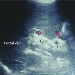
Case 1. A 27-year-old male patient with chronic pain in the right hypochondrium was admitted to the emergency room, where abdominal ultrasonography was
conducted (Figure 1), showing chronic cholelithiasis
Figure 1. Ultrasonography showing the portal vein with a
and TPV. An abdominal computed tomography scan
with intravenous contrast conirmed TPV (Figure 2).
Laboratory tests did not reveal indings that could explain etiology and so the thrombosis was considered idiopathic. The decision was taken to employ clinical management with 100 mg of acetylsalicylic acid per day, which resulted in clinical improvement, and outpatients follow-up.
Case 2. A 60-year-old asymptomatic male patient was referred to the tertiary healthcare service
because of a family history of chronic liver disease.
Investigation with abdominal ultrasonography found
hepatic steatosis and an APV (Figure 3). Computed
tomography showed an aneurysmal dilatation of the proximal portal vein with a maximum diameter of 5.5 cm and a 1.1 cm neck, with no signs of thrombosis
(Figure 4). Expectant management was also chosen
for this patient, with outpatients monitoring.
DISCUSSION
The clinical manifestations of vascular diseases of the portal system vary as a function of the degree
to which the hepatic parenchyma is damaged and
of the development of compensatory mechanisms.
The great majority of patients with APV are
asymptomatic and diagnosis is generally the result of
an incidental inding on an imaging exam requested
for other reasons. When present, the symptoms are
caused by mass effects: abdominal pains, due to
compression of neighboring structures, and jaundice, caused by compression of bile ducts. Rupture of the biliary system can also occur, causing gastrointestinal bleeding.3,6
In view of this, the great majority of physicians opt
for conservative management, monitoring the patient
with sequential imaging exams. In 2009, a systematic review was conducted of 93 reports published in the literature, describing a total of 176 patients with 198
visceral venous aneurysms. The authors concluded that careful observation is the most appropriate conduct,
except when complications occur.15 Recently, a case
was described in which a male patient diagnosed with asymptomatic APV was followed for 8 years
Figure 2. Portal vein without low and with foci of calciication
(chronic thrombosis). Exuberant collateral circulation in the hepatic hilum (cavernomatous transformation). Splenomegaly.
and enjoyed full regression of the aneurysm, with
no explanation.16
In 2012, Ma et al.5 proposed a interesting algorithm
for management of patients with APV (Figure 5).5 With regard to TPV, the appearance and extent of the thrombus are determinant factors of the
resulting clinical manifestations, which in turn can
be divided into acute and chronic, and differentiated
by emergence of cavernoma (a network of collateral
vessels connecting proximal and distal potions of
the thrombus, which deines the chronicity of the
process).10,17 Acute manifestations include intestinal
congestion and ischemia, which can cause pain and/
or abdominal distension; diarrhea; rectal bleeding; nausea; vomiting; anorexia; fever; lactic acidosis;
splenomegaly, and sepsis. If the obstruction is not resolved rapidly, intestinal perforation, peritonitis,
shock and death may follow. On the other hand, chronic
TPV can be asymptomatic, except for hypersplenism and consequent pancytopenia, esophageal varices, abdominal collateral circulation and ascites. This fact means that it is necessary to conduct an upper digestive
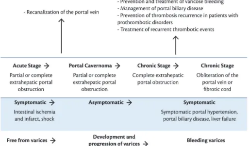
endoscopy on all patients with TPV.10Figure 6 illustrates
an adaptation of a diagram originally proposed by Chinese researchers that relates the course of TPV to progression of symptoms.17
Since patients with TPV in the chronic phase are
very often asymptomatic, diagnosis is commonly an
incidental imaging exam inding. Once clinical suspicion has been aroused, the irst examination requested is usually abdominal ultrasonography, which may show solid hyperechoic material in the portal vein; distension of the portal vein and/or tributaries, and a network of collateral vessels or cavernoma. Next, abdominal ultrasonography with Doppler, computed
tomography or magnetic resonance imaging may be
used to conirm diagnosis.10
Although resolution of TPV in the acute phase has
been described, speciic therapeutic management is
mandatory to resolve portal obstruction and avoid serious complications, primarily to impede progression to the chronic phase of the process.18 The objective
Figure 4. View of aneurysm on abdominal computed tomography
with contrast.
of treatment is correction of risk factors, prevention
of thrombosis growth and achieving patency of the
portal vein.10 The great majority of authors recommend
anticoagulant treatment in the acute phase of the
disease; but this becomes controversial with respect
top the chronic phase, because it could encourage unfavorable progression to esophageal varices.11,19 Other authors recommend that anticoagulant treatment
should only be prescribed for patients with conirmed
thrombotic disorders or a family history of venous thrombosis.20 Other treatments that do not enjoy
consensus among authors include: thrombolysis,
transjugular intrahepatic portosystemic shunts (TIPS) and surgical shunts (distal splenorenal).10,11,19
Prognosis is more related to the presence of comorbidities, rather than to presence of bleeding esophageal varices.21 A study conducted in the city
of Bologna conducted a prospective investigation
of patients with hepatocellular carcinoma who were
candidates for liver transplantation and concluded that TPV that are not caused by tumoral invasion should not be considered as an absolute contraindication to
transplantation in patients with hepatocellular carcinoma
and cirrhosis of the liver.22 Another study, conducted at
the University of Michigan, concluded that presence
of TPV may be associated with reduced short-term survival after transplantation, which should be taken into account when taking decisions on indications
for transplantation and on the best time to attempt transplantation in these patients.23
We conclude that vascular diseases of the portal
system are rare entities, the great majority of which
Figure 6. Progression of thrombosis of the portal vein and treatment strategy for patients free from cancer and cirrhosis. Adapted
from Qi et al.17
can be managed by watchful waiting. Rigorous
surveillance is necessary to enable assessment of any
emerging need for intervention in cases in which the
disease progresses.
REFERENCES
1. Gillespie DL, Villavicencio JL, Gallagher C, Chang A, Hamelink JK, Fiala LA, et al. Presentation and management of venous aneurysms. J Vasc Surg. 1997;26(5):845-52. http://dx.doi.org/10.1016/S0741-5214(97)70099-5. PMid:9372824
2. Mucenic M, Rocha MS, Laudanna AA, Cançado ELR. Treatment by splenectomy of a portal vein aneurysm in hepatosplenic schistosomiasis. Rev Inst Med Trop Sao Paulo. 2002;44(5):261-4. http://dx.doi.org/10.1590/S0036-46652002000500005. PMid:12436166
3. Vyas S, Mahajan D, Sandhu MS, Duseja A, Khandelwal N. Portal vein aneurysm: is it an incidental finding only? Ann Hepatol. 2012;11(2):263-4. PMid:22345345.
4. Barzilai R, Kleckner MS Jr. Hemocholecyst following ruptured aneurysm of portal vein: report of a case. AMA Arch Surg. 1956;72(4):725-7. http://dx.doi.org/10.1001/archsurg.1956.01270220173023. PMid:13301133
5. Ma R, Balakrishnan A, See TC, Liau SS, Praseedom R, Jah A. Extra-hepatic portal vein aneurysm: A case report, overview of the literature and suggested management algorithm. Int J Surg Case Rep. 2012;3(11):555-8. http://dx.doi.org/10.1016/j.ijscr.2012.07.009. PMid:22922358
6. Andraus W, Amico EC, Machado MA, Bacchella T, Machado MCC. Portal vein aneurysm. Clinics (Sao Paulo). 2007;62(2):203-5. http:// dx.doi.org/10.1590/S1807-59322007000200018. PMid:17505708
7. Bayraktar Y, Harmanci O. Etiology and consequences of thrombosis in abdominal vessels. World J Gastroenterol. 2006;12(8):1165-74. PMid:16534866.
9. Belli L, Romani F, Riolo F, Rondinara G, Aseni P, Di Stefano M, et al. Thrombosis of portal vein in absence of hepatic disease. Surg Gynecol Obstet. 1989;169(1):46-9. PMid:2787060.
10. Ponziani FR, Zocco MA, Campanale C, Rinninella E, Tortora A, Di Maurizio L, et al. Portal vein thrombosis: insight into physiopathology, diagnosis, and treatment. World J Gastroenterol. 2010;16(2):143-55. http://dx.doi.org/10.3748/wjg.v16.i2.143. PMid:20066733 11. Valla DC, Condat B. Portal vein thrombosis in adults: pathophysiology,
pathogenesis and management. J Hepatol. 2000;32(5):865-71. http://dx.doi.org/10.1016/S0168-8278(00)80259-7. PMid:10845677
12. Harmanci O, Bayraktar Y. Portal hypertension due to portal venous thrombosis: etiology, clinical outcomes. World J Gastroenterol. 2007;13(18):2535-40. http://dx.doi.org/10.3748/wjg.v13.i18.2535. PMid:17552000
13. Alves RLJ, Macedo FA, Latorre MV, de Paula BHR, Barradas F, Tavares M. Trombose de veia porta: revisão de literatura e relato de caso. Cadernos UniFOA. 2012;7(18):101-8.
14. Schettino GCM, Fagundes EDT, Roquete MLV, Ferreira AR, Penna FJ. Trombose de veia porta em crianças e adolescentes. J Pediatr. 2006;82(3):171-8.
15. Sfyroeras GS, Antoniou GA, Drakou AA, Karathanos C, Giannoukas AD. Visceral venous aneurysms: clinical presentation, natural history and their management: a systematic review. Eur J Vasc Endovasc Surg. 2009;38(4):498-505. http://dx.doi.org/10.1016/j. ejvs.2009.05.016. PMid:19560947
16. Lall P, Potineni L, Dosluoglu HH. Complete spontaneous regression of an extrahepatic portal vein aneurysm. J Vasc Surg. 2011;53(1):206-8. http://dx.doi.org/10.1016/j.jvs.2010.07.063. PMid:20869190
17. Qi X, Han G, Bai M, Fan D. Stage of portal vein thrombosis. J Hepatol. 2011;54(5):1080-2, author reply 1082-3. http://dx.doi. org/10.1016/j.jhep.2010.10.034. PMid:21145872
18. Simão A, Correia L, Carvalho A, Costa N. Trombose da veia porta: importância do diagnóstico e tratamento precoces. Liver Today: Boletim Informativo APEF. 2009;7:4-5.
19. Boyer TD. Management of portal vein thrombosis. Gastroenterol Hepatol. 2008;4(10):699-700. PMid:21960890.
20. Condat B, Pessione F, Helene Denninger M, Hillaire S, Valla D. Recent portal or mesenteric venous thrombosis: increased recognition and frequent recanalization on anticoagulant therapy. Hepatology. 2000;32(3):466-70. http://dx.doi.org/10.1053/jhep.2000.16597. PMid:10960436
21. Janssen HL, Wijnhoud A, Haagsma EB, van Uum SH, van Nieuwkerk CM, Adang RP, et al. Extrahepatic portal vein thrombosis: aetiology and determinants of survival. Gut. 2001;49(5):720-4. http://dx.doi. org/10.1136/gut.49.5.720. PMid:11600478
22. Piscaglia F, Gianstefani A, Ravaioli M, Golfieri R, Cappelli A, Giampalma E, et al. Criteria for diagnosing benign portal vein thrombosis in the assessment of patients with cirrhosis and hepatocellular carcinoma for liver transplantation. Liver Transpl. 2010;16(5):658-67. PMid:20440775.
23. Englesbe MJ, Kubus J, Muhammad W, Sonnenday CJ, Welling T, Punch JD, et al. Portal vein thrombosis and survival in patients with cirrhosis. Liver Transpl. 2010;16(1):83-90. http://dx.doi. org/10.1002/lt.21941. PMid:20035521
Correspondence
Lenon Cardoso Av. Barão de Tatuí, 859/24 CEP 18030-000 - Sorocaba (SP), Brazil E-mail: [email protected]
Author information
LC is a medical student at the School of Medical and Health Sciences, Pontifícia Universidade Católica de São Paulo (PUC-SP). TCP is a resident physician of General Surgery, School of Medical and Health Sciences, Pontifícia Universidade Católica de São Paulo (PUC-SP). MMS is a physician from Universidade Federal do Triângulo Mineiro (UFTM). FSS is a medical student at the School of Medical and Health Sciences, Pontifícia Universidade Católica de São Paulo (PUC-SP). DI is a resident physician of General Surgery, School of Medical and Health Sciences, Pontifícia Universidade Católica de São Paulo (PUC-SP). MM is a resident physician of General Surgery, School of Medical and Health Sciences, Pontifícia Universidade Católica de São Paulo (PUC-SP). RAB is a full professor at the Department of Surgery, School of Medical and Health Sciences, Pontifícia Universidade Católica de São Paulo (PUC-SP).
Author contributions