J Bras Pneumol. 2007;33(3):351-354
351
Case Report
Trauma-related thoracoplasty: case report*
Gabriela Addor1, Andreia Salarini Monteiro2, David Henrique Nigri1, Luiz Felippe Judice3, Rui Haddad4, Carlos Alberto de Barros Franco5
Abstract
Trauma primarily affects young people and is the leading cause of death in the first three decades of life. Flail chest is observed in approximately 10% of all patients with severe chest trauma, and the mortality rate among such patients is 10-15%. We report herein the case of a car accident victim with chest trauma causing hemopneumothorax and multiple rib fractures, intense pain and deformity of the chest wall. Surgical stabilization was performed, with good results. Therapeutic options are also discussed.
Keywords: Thoracic injuries; Flail chest; Thoracoplasty.
* Study carried out at the Graduate School of Medicine of the Pontifícia Universidade Católica do Rio de Janeiro – PUCRJ, Pontifical Catholic University of Rio de Janeiro – Rio de Janeiro (RJ) Brazil.
1. Assistant Professor in the Pulmonology Department of the Graduate School of Medicine of the Pontifícia Universidade Católica do Rio de Janeiro – PUCRJ, Pontifical Catholic University of Rio de Janeiro – Rio de Janeiro (RJ) Brazil.
2. Pulmonologist in the Intensive Care Unit of the Miguel Couto Municipal Hospital, Rio de Janeiro (RJ) Brazil.
3. Full Professor in the Surgery Department of the Universidade Federal Fluminense – UFF, Fluminense Federal University – School of Medicine, Niterói (RJ) Brazil.
4. Full Professor in the Thoracic Surgery Department of the Graduate School of Medicine of the Pontifícia Universidade Católica do Rio de Janeiro – PUCRJ, Pontifical Catholic University of Rio de Janeiro – Rio de Janeiro (RJ) Brazil.
5. Full Professor in the Pulmonology Department of the Graduate School of Medicine of the Pontifícia Universidade Católica do Rio de Janeiro – PUCRJ, Pontifical Catholic University of Rio de Janeiro – Rio de Janeiro (RJ) Brazil.
Correspondence to: Gabriela Addor. Rua Sorocaba, 464/402, Botafogo, CEP 22271-110, Rio de Janeiro, RJ, Brasil. Phone 55 21 2266-7392. E-mail: [email protected]/[email protected]
Submitted: 26 April 2005. Accepted, after review, 23 June 2006.
Introduction
Trauma primarily affects young people and is the leading cause of death in the first three decades of life. It is estimated that chest trauma is responsible for 25% of such deaths in the United States. An analysis of the various causes of chest injury has shown that traffic accidents and falls are the principal causes of the increased incidence of
chest injuries that result in rib cage instability.(1)
Flail chest is observed in approximately 10% of the cases of severe chest trauma, with 10-15% mortality. This high mortality rate is, in part, related to the accompanying severe extrathoracic injuries (skull-brain trauma, damage to the abdominal viscera, and vascular lesions). However, in patients who survive the initial trauma, the principal
causes of death are pneumonia and sepsis, together with the prolonged intubation and mechanical ventilation that are frequently required in such cases. Bilateral flail chest
and being over 50 years of age are aggravating factors.(2)
Despite the quantity of pertinent data in the literature, the treatment for severe flail chest with multiple fractures and respiratory failure remains controversial.
Case report
352 Addor G, Monteiro AS, Nigri DH, Judice LF, Haddad R, Franco CAB
J Bras Pneumol. 2007;33(3):351-354
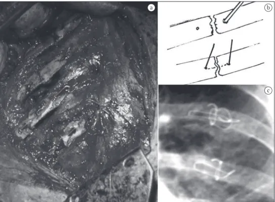
the third to the eighth left costal arches, as seen on chest X-rays and computed tomography scans of the chest (Figures 1 and 2). Tomography of the skull, cervical spine, abdomen, and pelvis, were normal, as were the electrocardiogram and echocardiogram, as well as the results of tests for muscle enzymes and markers of myocardial necrosis. Water-sealed thoracic drainage was performed, and the implan-tation of an epidural catheter was inserted in order to provide continuous analgesia using an infusion pump. The patient presented significant chest defor-mation and pain, despite high doses of painkillers. It was expected that this profile, unless surgically treated, would evolve to complex deformation of the chest wall, with possible respiratory impairment. The patient did not require mechanical ventilation. In view of this clinical profile, surgical treatment was indicated in order to stabilize the fractures. The procedure was performed using left posterior lateral thoracotomy. We found double fractures extending from the third to the eighth left costal arches, with complete rupture of various intercostal pedicles, herniation, and impaction of the fractured segment (six arches) within the pleural cavity, with signifi-cant compression of the pulmonary parenchyma (traumatic thoracoplasty). We performed reduc-tion of the fractures and fixareduc-tion of the ribs with number 5 steel wires, perforating the extremities of the ribs with a nº 2 drill, passing the steel wire from one rib segment to another, and tying it. A chest tube was inserted and left in place until the third day. The patient evolved to excellent pain control
and improved respiratory dynamics. Postoperative X-rays and tomography scans confirmed the favo-rable result of the surgical treatment (Figure 3).
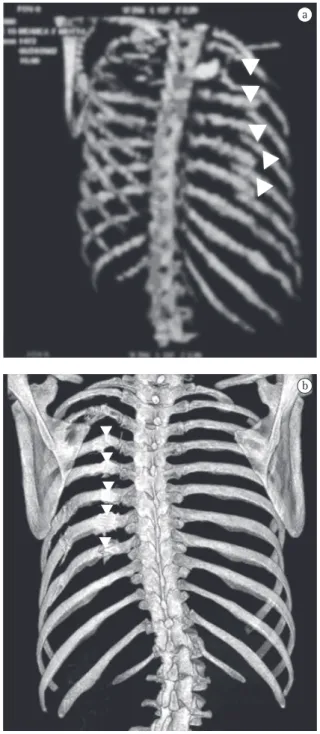
Figure 1 - Computed tomography (CT) of the chest showing ‘herniated’ rib segment within the cavity compressing the lung.
Figure 2 - a) Computed tomography (CT) with three-dimensional reconstruction showing double fractures (arrows); and b) CT with reconstruction of the costal arches 3 months after surgical procedure showing satisfactory alignment of the fractured arches and reduction of the deformation.
a
Trauma-related thoracoplasty: case report
J Bras Pneumol. 2007;33(3):351-354
353
Discussion
Closed chest trauma is an important cause of morbidity and mortality, principally when accompa-nied by flail chest. It occurs when there are multiple fractures of the chest cavity, more specifically when we observe fractures of three or more ribs, at two or more points, together with fluctuation,
paradox-ical breathing, or both.(3) Traumatic thoracoplasty
can be defined as a condition in which the frac-tured segment migrates into the chest and becomes lodged within the segments of the fixed costal arches, causing impaction of this segment within the pleural cavity. This causes intractable pain (perma-nent traction of intercostal nerves) and compresses the pulmonary parenchyma, considerably wors-ening the clinical profile of the patient. Severe flail chest is associated with respiratory failure, due to the retention of secretions, atelectasis, pneumonia,
and, later, restricted mobility of the chest cavity.(2)
Thoracic instability can be efficiently treated with analgesia, preferably epidural, and physical therapy, with proper secretion mobilization.
Prior to 1956, the treatment was carried out through external stabilization of the chest wall. From 1956 to 1975, early intubation and mechanical ventilation were used, resulting in good stabiliza-tion (‘internal pneumatic stabilizastabiliza-tion’), although there was no reduction in the mortality rate. Studies carried out since 1975, one in particular that was
conducted in 1983,(4) emphasized a conservative
strategy, with efficient analgesia and intensive physical therapy, without mechanical ventilation,
resulting in decreased mortality.The treatment for
thoracic instability that does not respond well to conservative treatment is an extremely controversial
issue.(2) Some authors opt for prolonged
intuba-tion and mechanical ventilaintuba-tion. Others, however, advocate surgical stabilization of the fractures with osteosynthesis and fixation of the unstable segments of the thorax, reducing the duration of the ventilatory support phase and that of the stay in the intensive care unit, as well as reducing morbidity, especially in
cases presenting no pulmonary contusion.(2) In one
a b
c
354 Addor G, Monteiro AS, Nigri DH, Judice LF, Haddad R, Franco CAB
J Bras Pneumol. 2007;33(3):351-354
study,(2) surgical stabilization was indicated for the
following patients: those who required thoracotomy to correct other injuries; those who presented exten-sive anterolateral injury and progresexten-sive dislocation of the fractured ribs (anterolateral dislocation is more prone to develop restriction and pseudo-arthrosis, with future professional limitation); nonintubated patients with persistent respiratory failure, despite continuous peridural analgesia and intense treat-ment for mobilization of secretions; and those who were intubated and ventilated exclusively due to flail chest, without concomitant conditions such as severe pulmonary contusion or brain injury, for which prolonged mechanical ventilation would be indicated. In 1997, one group of authors defended surgical fixation, used in conjunction with proper analgesia, for patients with refractory respiratory failure and for whom intubation was not indicated for any other reason and for young patients with severe impaction, such as the case presented here, in order to prevent future restriction. The use of surgical fixation has been shown to be efficient in select populations of patients, principally in those who have been submitted to surgery for another cause, as well as in the following: those who remain in pain, despite proper analgesia; those who present paradoxical chest movement, despite mechanical ventilation; those who present severe deformation of the chest wall; and those presenting flaccidity of the thorax, without significant thoracic contusion
and with difficulty in breathing.(1)
Another group of authors(5) obtained better
results in the operated group, in terms of length of hospitalization and clinical evolution, than in the clinically treated group (mean length of
hospi-talization, 9 vs. 21 days; mortality, 85 vs. 29%).
This strongly favors the operated group. However, controversy remains as to whether surgery is indi-cated in such cases. When indiindi-cated, the surgical treatment is typically aimed at avoiding accen-tuated deformations of the chest wall, thereby preventing difficult-to-control chronic pain and respiratory failure, both of which were observed in the case described herein and were considered as evidence that the surgical fixation of the fractures was indicated.
References
1. Zelenak J, Kutarna J, Hutan M, Kalig K. Stabilisation of thoracic wall in patients with chest injury. Bratisl Lek Listy. 2002;103(4-5):176-8.
2. Lardinois D, Krueger T, Dusmet M, Ghisletta N, Gugger M, Ris HB. Pulmonary function testing after operative stabilisation of the chest wall for flail chest. Eur J Cardiothorac Surg. 2001;20(3):496-501.
3. Bub RF, Campos M, Ghiotto JL. Traumatismo torácico. In: Knobel E, editor. Condutas no paciente grave. Rio de Janeiro: Atheneu; 1998. p. 891-907.
4. Sivaloganathan M, Stephens R, Grocott M. Management of flail chest. Hosp Med. 2000;61(11):811.