68
Forearm Compartment Syndrome from Mannitol Extravasation Case Report
International Braz J Urol Vol. 33 (1): 68-71, January - February, 2007
Mannitol Extravasation during Partial Nephrectomy Leading to
Forearm Compartment Syndrome
Bradley A. Erickson, Ronald L. Yap, Joseph F. Pazona, Brian J. Hartigan, Norm D. Smith
Departments of Urology and Orthopaedics, Feinberg School of Medicine, Northwestern University,
Chicago, Illinois, USA
ABSTRACT
We present the first known complication of forearm compartment syndrome after mannitol infusion during partial nephrectomy. We stress the importance of excellent intravenous catheter access and constant visual monitoring of the intravenous catheter site during and after mannitol infusion as ways to prevent this complication. Prompt recognition of compartment syndrome with appropriate intervention can prevent long-term sequelae.
Key words: carcinoma, renal cell; nephrectomy; mannitol; compartment syndrome
Int Braz J Urol. 2007; 33: 68-71
INTRODUCTION
Surgeons commonly use mannitol for partial nephrectomies that entail renal hilar clamping. We report a case of mannitol extravasation during a partial nephrectomy that led to forearm compartment syndrome requiring emergent fasciotomies.
CASE REPORT
A 36-year-old female underwent open partial nephrectomy for an incidentally found, enhancing 2.5 cm left lower pole mass (Figure-1). The patient had a medical history significant for IV drug abuse, hypertension and asthma. In the preoperative holding area, the anesthesia team noted that intravenous access was extremely difficult to obtain; this was thought to be secondary to the patient’s prior IV drug abuse. The team made multiple attempts at IV access before ultimately placing two large bore (16G) peripheral
IVs, both of which were used for infusion for the case duration.
The procedure proceeded without complications, with a cold ischemia time of approximately 30 minutes and a total OR time of 3.5 hours. Per usual for this type of case, we infused 12.5 grams of mannitol 5 minutes before hilar occlusion and 5 minutes after removing the clamps through the right peripheral forearm IV.
69
Forearm Compartment Syndrome from Mannitol Extravasation
hand surgery consult was obtained and forearm compartment pressures were found to be > 120 mmHg (normal < 30 mmHg). The patient was emergently brought to the operating room by the hand surgery team where fasciotomies were performed. The compartment syndrome was later felt to be secondary to mannitol extravasation from the right forearm peripheral IV.
Fasciotomies were closed 7 days later when the swelling had diminished sufficiently. At two-month follow-up, the patient displayed no residual forearm or hand weakness and fasciotomy incisions were well-healed. Final pathology of the partial nephrectomy specimen showed a 2.5 cm grade-I clear cell renal cell carcinoma with negative margins.
COMMENTS
Intravenous infusions of mannitol expand intravascular volume and decrease cellular edema by minimizing the large intracellular fluid shifts that normally occur during periods of organ ischemia. Researchers theorize that a reduction in cellular edema
more promptly restores blood flow to the ischemic organ after the insult is removed, as the vessels do not collapse from the surrounding engorged cells (1). These properties make it a useful renoprotective agent during partial nephrectomies.
Though mannitol itself is a relatively benign substance, its potent osmotic properties can be detrimental when it has extravasated into a closed space. For every 50 g of mannitol infused, a 1L intracellular to extracellular fluid shift is expected to occur (2). This fluid shift is easily accommodated intravascularly, but the relatively small forearm compartment is not as forgiving.
Compartment syndrome develops when interstitial pressures of a given muscular compartment
Figure 1 – CT scan showing enhancing 2.5 cm lower pole mass.
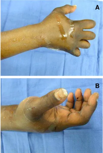
Figure 2A and B – Elevated forearm compartment pressures lead to this typical flexed appearance.
A
70
Forearm Compartment Syndrome from Mannitol Extravasation
surpass those of the capillary perfusion pressures. When this occurs, the capillaries collapse, resulting in local hypoxia and eventual necrosis of the intercompartmental musculature. Common causes include bone fractures, extensive soft tissue injuries, reperfusion of ischemic tissue, high pressure and hypertonic intravenous fluid administration and medication extravasation (3). In this particular case, mannitol extravasation likely led to a large volume shift from the vascular space to the interstitium, critically raising compartmental pressures.
Mannitol extravasation leading to forearm compartment syndrome has been reported before (3-5) but not during partial nephrectomy. Many things about this case make it unique. First, as previously noted, obtaining IV access was difficult, and though two large bore IVs were ultimately placed, the venous integrity in a patient with a long-standing IV drug abuse history and who experienced multiple failed peripheral IV sticks prior to surgery could have been questioned. Though intravenous infusion through the right forearm IV remained constant throughout the case, small injuries to the proximal vasculature probably allowed for seepage of the mannitol into the interstitium. The phenomenon resembles the high incidence of IV infiltration observed in patients with long hospital stays requiring multiple venous punctures. Second, the fact that anesthesiologists usually obtain intravenous access prior to the positioning of the patient for partial nephrectomy can make it quite difficult to monitor the site during the case. When patients are put in a modified flank position, as often required for partial nephrectomy, surgeons must secure the arm to the armboards in order to help stabilize the patient. Placing the IV site without regard to the eventual location of the arm can often render the IV site inaccessible. Though this particular IV site was visible, it was not immediately accessible to the anesthesia team. It remains unclear whether the inability to continually monitor the IV site contributed to the eventual complication; however, had we diagnosed problems with the IV sooner, we might have infused mannitol at an alternate location. Finally, unconscious patients run a particular risk for extravasation injury (3). The 4 Ps of compartment syndrome (pain, paresthesia, pallor,
pulselessness) are difficult to monitor without patient feedback and the diagnosis must be made by observation of the IV site alone. Constant monitoring of the IV site is, therefore, of utmost importance in unconscious patients. Early recognition by the intensive care nurse undoubtedly bears responsibility for preserving the underlying musculature in this patient. Though this unfortunate case did not prompt us to abandon our use of mannitol during partial nephrectomies, we did adopt some new practices with the goal of preventing a repeat of this complication. First, if venous integrity seems questionable, we ask that mannitol be infused through a central venous catheter. This requires communication with the anesthesia team early in the procedure and again before infusion of mannitol. Second, we ensure that the IV site intended for mannitol instillation remains visible to the anesthesia team throughout the case. Though this sometimes requires creative positioning and IV placement, none of these minor adjustments have compromised any parts of our subsequent cases. Finally, in unconscious patients, we stress monitoring of the peripheral IV site and surrounding soft tissue to all involved in postoperative care, as it is unclear over what time period compartment syndrome occurs (In this particular case, the nurse noticed symptoms approximately 3 hours after instillation).
CONFLICT OF INTEREST
None declared.
REFERENCES
1. Collins GM, Green RD, Boyer D, Halasz NA: Protection of kidneys from warm ischemic injury. Dosage and timing of mannitol administration. Transplantation. 1980; 29: 83-4.
2. Kumar MM, Sprung J: The use of hyaluronidase to treat mannitol extravasation. Anesth Analg. 2003; 97: 1199-200.
71
Forearm Compartment Syndrome from Mannitol Extravasation
4. Eroglu A, Uzunlar H: Forearm compartment syndrome after intravenous mannitol extravasation in a carbosulfan poisoning patient. J Toxicol Clin Toxicol. 2004; 42: 649-52.
5. Edwards JJ, Samuels D, Fu ES: Forearm compartment syndrome from intravenous mannitol extravasation during general anesthesia. Anesth Analg. 2003; 96: 245-6.
Accepted after revision: September 30, 2006
Correspondence address:
Dr. Bradley A Erickson
Dept Urology, Feinberg Sch Medicine
Northwestern University, Tarry Building #16-703 303 East Chicago Avenue
Chicago, IL, 60611, USA Fax: + 1 312 908-7275
E-mail: [email protected]
EDITORIAL COMMENT
Compartment syndrome is a rare but well known complication observed in trauma patients or may be due to incorrect positioning of patients during surgery. In urology, it may occur after time consuming procedures performed in lithotomy position (like radical prostatectomy or cystectomy) and is usually localized in the lower legs. Compartment syndrome of the forearm due to infusion of various medications is described as a rare phenomenon in the literature, as the authors pointed out.
Even if this particular complication is rather an anesthesiological pitfall than a urological complication this case is remarkable, because the use of mannitol is common in renal surgery, especially in partial nephrectomy or kidney transplantation. In the described case, the awareness of the personnel led to an early recognition of the complication and an immediate successful intervention.
Two facts are noteworthy in this case. There is no doubt about the indication for mannitol infusion with nephroprotective goals during partial
nephrectomy. However, considering the problems of the intravenous access, caused by the patient’s history of IV drug abuse, the attention should have been directed to the risk earlier and precautions should have been taken. I agree with the authors, that in this case a central venous access would have been suitable to avoid further complications. General anesthesia for partial nephrectomy, especially when there is a significant risk of major bleeding, as in the present case, should never been managed with a peripheral venous access alone. A central venous access should always be the first choice in terms of patient’s safety.