Energy Sources for Laparoscopic Partial Nephrectomy
-Critical Appraisal
Mauricio Rubinstein, Alireza Moinzadeh, Jose R. Colombo Jr, Luciano A. Favorito, Francisco
J. Sampaio, Inderbir S. Gill
Urogenital Research Unit, State University of Rio de Janeiro, Rio de Janeiro, Brazil, Section of
Laparoscopic and Robotic Surgery, State University of New York, Syracuse, New York, USA, and
Section of Laparoscopic and Robotic Surgery, Glickman Urological Institute, The Cleveland Clinic,
Cleveland, Ohio, USA
ABSTRACT
Laparoscopic partial nephrectomy (LPN) has emerged as a viable alternative for the conventional open nephron-sparing surgery (NSS). So far, an adequate renal parenchymal cutting and hemostasis, as well as caliceal repair remains technically challenging. Numerous investigators have developed techniques using different energy sources to simplify the technically demanding LPN. Herein we review these energy sources, discussing perceived advantages and disadvantages of each technique.
Key words: laparoscopy; surgical procedures, minimally invasive; nephrectomy; energy sources
Int Braz J Urol. 2007; 33: 3-10
INTRODUCTION
The majority of renal tumors are now inci-dentally diagnosed and smaller than 4 cm (1). The treatment of choice for most small renal masses is the NSS. Fergany et al. have demonstrated similar results comparing partial and radical nephrectomy for 10 year follow-up (2).
Although some different techniques of LPN have been described (3-7), in the senior author insti-tution (ISG), this technique include complete kidney exposure, hilar clamping, cold cut with laparoscopic scissors, precise collecting system closure, reconstruc-tion of partial nephrectomy bed over surgical bolsters, and the use of biological haemostatic agent (Floseal®).
Widespread application of LPN has been limited given the challenges associated with
intracorporeal suturing for hemostasis and collecting system closure. To simplify the procedure, several reports have been published using various energy modalities to replace the need for intracorporeal suturing (6-8).
In this review, we describe and evaluate several energy sources used to achieve cutting and hemostasis during LPN, as well as ablative tissue energies, outlining the advantages and disadvantages of each one.
LASERS: CUTTING AND HEMOSTATIC
ENERGY
vaporize tissue while leaving a coagulated field. Their efficacy to coagulate or excise tissue is regulated by specific wavelength, energy or power setting and mode of operation (continuous or pulsed) (8). Appli-cations in urology include lithotripsy, ablation of blad-der tumors, transurethral resection of prostate, and partial nephrectomy (9).
Several kinds of laser energy have been tested for parenchymal transection during LPN (8-11). The use of laser fibers with the specific application of tis-sue welding is based on delivering energy to the tar-get lesion, with heat absorption resulting in thermo-coagulation. This modality avoids needle trauma and suture reaction, may allow shorter operative time and less bleeding, although it presents thermal damage in tissue with indirect contact (12-15). The search for the ideal hemostatic method still continues since no single laser was proven to have ideal results.
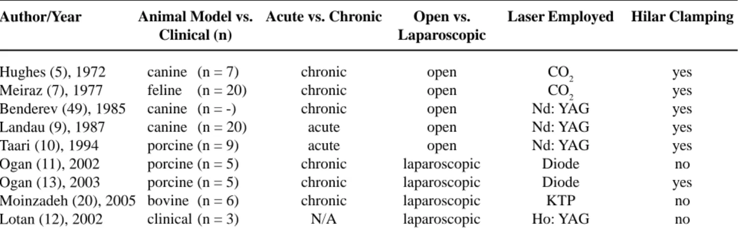
Descriptions of open laser partial nephrectomy using the CO2, Nd: YAG and holmium lasers have previously been published (10,16). Since this early experience, several authors have reported the use of lasers for LPN in animal model, as well as in the clinical field (Table-1). Lotan et al. (16) studied the use of holmium laser for partial nephrectomy in the porcine model. The authors performed transperitoneal lower pole laparoscopic partial nephrectomy in 5 pigs. Fibrin glue was applied to the nephrectomy bed to seal the collecting system. All
cases were performed with adequate hemostasis and without the need of further hemostatic devices.
Lotan et al. (17) described the first clinical report of laser during LPN, using the holmium: YAG laser in three patients. Indications included complex cyst, nonfunctioning lower pole, and renal mass. There was minimal blood loss and no need for hilar clamping. Although the laser alone was hemostatic, the authors used fibrin glue in two cases and oxidized cellulose in one case to reinforce the tissue against delayed bleeding. There were no perioperative com-plications and the average hospitalization was 3 days. The authors in this study concluded that with high power settings (0.2J/pulse at 60 pulses/sec and 0.8J/ pulse at 40 pulses/sec), the Ho: YAG laser can be used as an effective hemostatic tool in LPN.
The advantages of this laser are simplicity of use and relative low cost. The Ho: YAG laser is able to cut and coagulate tissues, with minimal damage to the adjacent renal parenchyma, preserving as much normal tissue as possible. The disadvantages include the smoke created and the splashing of blood on the camera, particularly when transecting larger vessels. The use of Diode laser in LPN was reported by Ogan et al. (18). They performed transperitoneal lower pole laparoscopic partial nephrectomy in 5 pigs without the need for hilar occlusion using a 980-nm diode laser. The laser hemostasis was insufficient in 3 cases, requiring adjunctive measures, as hemostatic
Open vs. Laparoscopic
open open open open open laparoscopic laparoscopic laparoscopic laparoscopic
Hilar Clamping
yes yes yes yes yes no yes
no no
*Adapted from Moinzadeh et al.: Potassium-titanyl-phosphate laser laparoscopic partial nephrectomy without hilar clamping in the survival calf model. J. Urol. 2005, 174: 1110-4.
Table 1 – Use of laser during laparoscopic partial nephrectomy.
Author/Year
Hughes (5), 1972 Meiraz (7), 1977 Benderev (49), 1985 Landau (9), 1987 Taari (10), 1994 Ogan (11), 2002 Ogan (13), 2003 Moinzadeh (20), 2005 Lotan (12), 2002
Animal Model vs. Clinical (n)
canine (n = 7) feline (n = 20) canine (n = -) canine (n = 20) porcine (n = 9) porcine (n = 5) porcine (n = 5) bovine (n = 6) clinical (n = 3)
Acute vs. Chronic
chronic chronic chronic acute acute chronic chronic chronic N/A
Laser Employed
clips to stop bleeding. The mean operative time was 126 minutes, the mean blood loss was 150 mL (50-300 mL), and no urinary extravasation was observed on retrograde pyelogram at 2 weeks. The authors con-cluded that the diode laser is feasible on the porcine model and limited its use in humans to small periferic tumors. The limitation of this laser was observed in controlling large vessels. Fibrin glue was applied to all partial nephrectomies, resulting in selling of the collecting system in all cases. It was unknown if the selling occurred as result of the glue or the laser. Fur-ther studies are necessary to achieve success with this kind of energy in LPN.
The same group has utilized an 810-nm pulsed diode laser (20W) plus a 50% liquid albumin-indocyanine green solder in 5 pigs demonstrating the tissue welding qualities of lasers (19). All surgeries were performed without complications with mean operative time of 82 minutes. Average blood loss was 43.5 mL and mean warm ischemia time was 11.7 min-utes. There was no evidence of urinoma formation or delayed hemorrhage in any of the animals. Histologic studies showed good preservation of renal paren-chyma beneath the solder.
The main advantage of this soldering tech-nique includes the ability to close the collecting sys-tem and control of bleeding during LPN, with short warm ischemia time (< 12 minutes). In this study, the laser was able to control large vessels, mimicking human LPN, and the violated collecting system was fixed with the solder without problems. Further stud-ies are required to confirm this fact.
The KTP laser has been recently tested for LPN in the calf model (20). Using 6 calves, the au-thors successfully completed the operation without hilar clamping in 11/12 procedures. One animal re-quired temporary occlusion of the hilum for hemor-rhage not controlled with the laser. The histological analysis revealed minimal effect on the adjacent area to the excision. The unique feature of the KTP laser includes the 532-nm wavelength, with specific up-take by hemoglobin. The authors believed that this aspect yielded excellent hemostasis in the robust calf model. In addition, minimal blood splatter was noted given decreased bleeding and thermomechanical ejec-tion when compared to the Ho:YAG laser.
HIDRO-JET DEVICE: MAINLY
CUTTING ENERGY
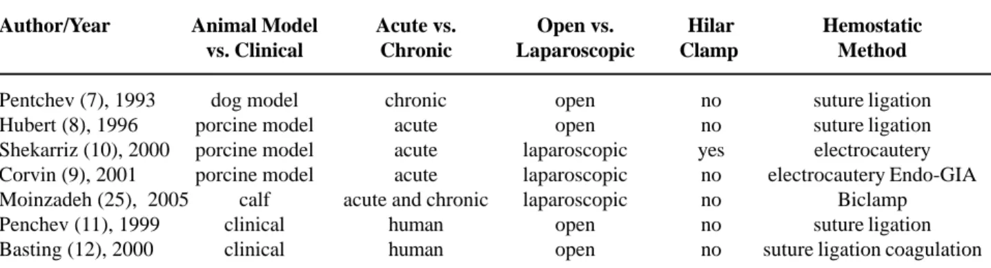
Hydro-jet technology has been established for surgery of the liver and other parenchymatous organs, using the principle of high-pressure water flow to cut tissues (21,22). The delivery probe allows dissection with both water high pressure and blunt dissection. Coagulation is applied usually with a bipolar thermo-applicator if needed. The first report in the urologic field was done by Pentchev et al. for open renal sur-gery in the canine model (23).
Shekarriz et al. published the first experimen-tal laparoscopic study in LPN. The authors performed the procedure with hilar clamping in the porcine model, using 5 animals (24). In this study, the mean warm ischemia time was 17 minutes. Moinzadeh et al. evaluated the feasibility of hydro-jet assisted LPN without renal hilar vascular control in the larger size and more robust calf model, to better reproduce the human kidney (25). The authors performed bilateral LPN using the Helix Hydro-Jet® (ERBE Tubingen,
Germany) without hilar control in 10 survival calves. All procedures were completed successfully without open conversion, and the hilar clamping was not needed in 18 (90%) cases. The mean operative time was 173 minutes (60-240), kidney section time was 63 minutes (13-150), and estimated blood loss was 174 cc (20-750). Histological studies showed a thin (1 mm) layer of adherent coagulum beneath the re-section area with minimal thermal artifact.
Clinically, Penchev et al. used the hydro-jet without hilar clamping in open partial nephrectomy for a low pole tumor (n = 1) and open anatrophic neph-rotomy of a staghorn calculi (n = 1) (26). The hydro-jet dissection time was 25 and 12 minutes, with blood loss of 150 and 100 mL, respectively. The procedures were done without vascular clamping or local hypo-thermia (27).
water jet device was useful for renal parenchymal transection.
To date, no clinical report of hydro-jet LPN has been published, but the suitability of the LPN tech-nique to improve hemostasis and dissection has been proven (Table-2). This use of kinetic energy has the advantage of dissect selective parenchyma while pre-serving vessels and the collecting system during the surgery. With this technology, the procedure may be easier, faster, avoiding the warm ischemia and the tech-nically challenging intracorporeal suturing during LPN. Since there is no cautery tissue damage, the Hydro-jet device preserves the renal parenchyma. Limitations include the theoretical spread of cancer cells with the use of the high pressure saline flow. In addition, cur-rent rigid instruments lacking flexibility make the laparoscopic angles of dissection challenging.
BIPOLAR ELECTRICAL CURRENT:
CUTTING AND HEMOSTATIC ENERGY
The bipolar needle electrode is composed of two 5 cm long needles, in parallel, that connects to a bipolar energy source. By electric current, it dissects and cauterizes the tissue that lies between the needles. This technique facilitates the procedure, creating a regional ischemia, without hilar occlu-sion. It is very efficient to coagulate deep parenchy-mal vessels before cutting out the renal tissue. Its linear shape can be a limitation to different tumors locations.
Barret et al. compared the efficacy and mor-bidity between three hemostatic techniques: high-fre-quency bipolar, high-frehigh-fre-quency unipolar, and ultra-sound during LPN in a porcine model without vascu-lar control. In this study, the authors evaluated perioperative complications, blood loss, renal func-tion, and histological findings in the parenchyma. There was a significantly decrease in blood loss when the ultrasound was employed (p = 0.0026). One pig developed hemorrhage in day 6. There was no differ-ence in histological results (28).
In another porcine study, Ong et al. demon-strated the use of the bipolar needle device in LPN with comparable results to those reported by Barret et al. (28). In this series, the blood loss was decreased (29). Janetschek et al. and Guillonneau et al. re-ported the use of bipolar hemostatic coagulation in LPN showing the clinical feasibility of this energy to achieve good hemostasis (30,31).
Some modifications in the future, including curved shape or articulating head may expand the use of this device for midpole and hilar renal masses. It seems that the damage to the remaining tissue is mini-mal. Nevertheless, more clinical studies are required to define the proper role in LPN.
FLOATING BALL: CUTTING AND
HEMOSTATIC ENERGY
The TissueLink Floating Ball (Tissuelink Medical, Inc., Dover, NH) comprises a monopolar
Table 2 – Use of water jet during laparoscopic partial nephrectomy.
Author/Year
Pentchev (7), 1993 Hubert (8), 1996 Shekarriz (10), 2000 Corvin (9), 2001 Moinzadeh (25), 2005 Penchev (11), 1999 Basting (12), 2000
Animal Model vs. Clinical
dog model porcine model porcine model porcine model
calf clinical clinical
Acute vs. Chronic
chronic acute acute acute acute and chronic
human human
Open vs. Laparoscopic
open open laparoscopic laparoscopic laparoscopic
open open
Hilar Clamp
no no yes
no no no no
Hemostatic Method
suture ligation suture ligation electrocautery electrocautery Endo-GIA
Biclamp suture ligation suture ligation coagulation
current that combines water-cooled with radio fre-quency for blunt dissection and coagulation purposes. The technology uses the radio frequency close to the instrument tip, sealing small blood vessels, achiev-ing good hemostasis prior to parenchymal resection. The electrical energy is transmitted by the saline irri-gation and converted into thermal energy on the tar-get tissue. Scar formation is prevented by the saline since the coagulated area remains cool, maintaining the temperature at or bellow 100o C (32).
Sundaram et al. first reported the feasibility of LPN with the Floating Ball without vascular control in 3 patients (33). The mean estimated blood loss was 275 mL and one patient had a urine leak that resolved spontaneously. Urena et al. retrospectively reviewed 10 LPN where this energy source was used to achieve hemostasis. Mean tumor size was 3.9 cm and mean blood was 352 mL. All margins were negative (32).
Stern et al. reported the largest series avail-able (34). The authors performed 14 LPN using the Floating Ball. The mean operative time in this series was 124 minutes and mean blood loss was 168 mL. The argon-beam coagulator, Fibrilartm (Ethicon,
Somerville, NJ) and fibrin glue were used for control minor bleeding.
The parenchymal resection is slower when performed without hilar control. To minimize the bleeding, the renal tissue can be coagulated prior to resection, and the scar produced does not affect the pathological analysis of tumor margin status. Vascu-lar structures up to 3 mm can be sealed by the use of the floating ball device. The depth of tissue penetra-tion is correlated to the type and durapenetra-tion of contact between the kidney surface and the device.
HARMONIC SCALPEL: CUTTING AND
HEMOSTATIC ENERGY
The harmonic scalpel (LaparoSonic Coagu-lating Shears; Ethicon Endo-Surgery, Cincinati, OH) has the potential to vibrate its jaws at a rate of 55,000 Hz, generating heat in the range of 50ºC TO 100ºC, coagulating and cutting the tissue simultaneously. This device forms a protein coagulum between the jaws of the shear resulting in minimal spread of energy laterally (2 mm).
Jackman et al. showed the ability of the har-monic scalpel to perform LPN in a porcine model without control of the hilar vessels (35). Additional hemostatic measures were necessary in 25% of the cases when a polar nephrectomy was performed. The authors concluded that the use of harmonic scalpel in hemiphrectomies is not recommended because the high risk of substantial hemorrhage.
Harmon et al. reported the use of harmonic scalpel in 15 patients undergoing LPN (36). All pro-cedures were completed without complications. The mean tumor size was 2.3 cm and mean blood loss was 368 mL. The renal bed hemostasis was accom-plished by using oxidized cellulose and argon beam coagulator. All resection margins were negative at the pathology results.
Although this device may aid in dissection of small superficial renal tumors, it is not sufficient to perform LPN particularly for larger and centrally located tumors. Overall, the use of this technology has not shown good results when used without others hemostatic device/agents.
ELECTRICAL SNARE: CUTTING AND
HEMOSTATIC ENERGY
This device was designed as a combination of an electrosurgical snare electrode (Cook Urologi-cal Inc., Spencer, IN) with an electrosurgiUrologi-cal genera-tor (ERBE USA, Inc., Marietta, GA), to produce re-nal transection and parenchymal hemostasis simulta-neously.
Elashry et al. compared the effectiveness of this snare during LPN, comparing it to ultrasonic dis-sectors in the porcine model. The electrical snare was faster and produced less intraoperative bleeding than the ultrasonic dissectors (37).
In the study from Washington University re-porting the use of the electrosurgical snare in LPN without occlusion of hilar vessels (38), the hemostasis was successfully achieved in all but one case, where it was necessary to use the argon-beam coagulator to stop the bleeding after parenchymal resection.
of renal pelvis injury. Clinical trials are still being awaited to confirm its applicability in LPN.
RADIO FREQUENCY ABLATION: TISSUE
HEMOSTATIC - ABLATIVE ENERGY
The Radio Frequency Ablation (RFA) creates a good parenchymal zone of coagulative necrosis usu-ally visible after 24 to 48 hours post procedure. This treated tissue is finally replaced by inflamation and fibrosis (39). In animal studies, Gill et al. demon-strated the renal parenchyma trombosis and coagula-tion noted after RF ablacoagula-tion (40,41).
The first clinical report on RFA assisted LPN was published by Gettman et al. (42). The RFA was used in 10 patients mainly to coagulate the tumor, facilitating the tumor excision with minimal bleed-ing. The Texas University group published the initial series of RFA assisted LPN (43) with 13 patients un-dergoing surgery. A total of 5 tumors were completely excised and 7 tumors were left in situ after treatment. There was one focal positive margin in a patient sub-mitted to RFA assisted LPN, but this patient remained disease-free after 1 year treatment.
In these studies the authors reserved the use of this RFA technique for polar, small, exophytic le-sions. There are some advantages related to complete tumor removal instead of only ablation, providing bet-ter oncologic approach for the patient. There is also minimal blood loss and good visualization during tu-mor excision. The limitations are concerned about the need for a learning curve with the RFA probes, the challenge to perform centrally located tumor excisions with high risk of collecting system injury. An addi-tional clinical experience with larger diameter tumors and long-term follow-up is necessary to confirm the real value of this technique.
ARGOM-BEAM COAGULATOR:
HEMOSTATIC ENERGY
The argon beam coagulator (ABC) provides hemo-stasis by delivering radiofrequency electrical energy to tissue across a jet of argon gas. The device uses a non-contact, monopolar, electrothermal type of he-mostasis (44).
The first report using the ABC was from Daniell et al. when they reported its use in cholecys-tectomies in animals and humans. They concluded that the ABC allowed a safely hemostasis and effec-tive controlled tissue electrocoagulation (45).
These techniques have been used associated to others kind of hemostatic agents, and with differ-ent approaches by another authors with relatively success (46-48).
The ABC allows a good visualization with-out smoke, safe hemostatic tissue electrocoagulation, with a rapid non-touching technique. The lack of smoke and the non-touching technique facilitate laparoscopic application. In the authors’ opinion, this kind of energy is well used to coagulate cortex ves-sels after closing the parenchymal defect but has lim-ited application for larger, infiltrating tumors. The use of the device must be done with caution because of the risk of gas embolism caused by intra-abdominal overpressurization during a laparoscopic procedure; to minimize the associated risks we must leave one instrument cannula open to drain the gas and have a good patient monitorization (e.g., end-tidal CO2, Dop-pler flow).
CONCLUSIONS
LPN has emerged and gained popularity in selected centers worldwide, and new energy sources have been employed to minimize the level of diffi-culty of the procedure. The key to achieve an ideal procedure remains in simplify the technique as re-gards closure of collecting system and minimal blood loss without the need for hilar occlusion. The im-proved energy sources may further decrease opera-tive time, warm ischemia time, and morbidity. The different devices presented are evolving but until to-day, no one has been totally superior and only the future will show us which of these instruments will stand the test of time.
CONFLICT OF INTEREST
REFERENCES
1. Jayson M, Sanders H: Increased incidence of serendipitously discovered renal cell carcinoma. Urology. 1998; 51: 203-5
2. Fergany AF, Hafez KS, Novick AC: Long-term results of nephron sparing surgery for localized renal cell carcinoma: 10-year followup. J Urol. 2000; 163: 442-5.
3. Gill IS, Desai MM, Kaouk JH, Meraney AM, Murphy DP, Sung GT, et al.: Laparoscopic partial nephrectomy for renal tumor: duplicating open surgical techniques. J Urol. 2002; 167: 469-7; discussion 475-6.
4. Janetschek G, Jeschke K, Peschel R, Strohmeyer D, Henning K, Bartsch G: Laparoscopic surgery for stage T1 renal cell carcinoma: radical nephrectomy and wedge resection. Eur Urol. 2000; 38: 131-8.
5. Gill IS, Matin SF, Desai MM, Kaouk JH, Steinberg A, Mascha E, et al.: Comparative analysis of laparoscopic versus open partial nephrectomy for renal tumors in 200 patients. J Urol. 2003; 170: 64-8.
6. McDougall EM, Elbahnasy AM, Clayman RV: Laparoscopic wedge resection and partial nephrectomy the Washington University experience and review of the literature. JSLS. 1998; 2: 15-23.
7. Rassweiler JJ, Abbou C, Janetschek G, Jeschke K: Laparoscopic partial nephrectomy. The European experience. Urol Clin North Am. 2000; 27: 721-36. 8. Landau ST, Wood TW, Smith JA Jr: Evaluation of
sapphire tip Nd:YAG laser fibers in partial nephrectomy. Lasers Surg Med. 1987; 7: 426-8. 9. Meiraz D, Peled I, Gassner S, Ben-Bassat M, Kaplan
I: The use of the CO2 laser for partial nephrectomy: an experimental study. Invest Urol. 1977; 15: 262-4. 10. Taari K, Salo JO, Rannikko S, Nordling S: Partial
nephrectomy with a combined CO2 and Nd:YAG laser: experimental study in pigs. Lasers Surg Med. 1994; 14: 23-6.
11. Dalsing MC, Packer CS, Kueppers P, Griffith SL, Davis TE: Laser and suture anastomosis: passive compliance and active force production. Lasers Surg Med. 1992; 12: 190-8.
12. Maragh H, Hawn RS, Gould JD, Terzis JK: Is laser nerve repair comparable to microsuture coaptation? J Reconstr Microsurg. 1988; 4: 189-95.
13. Chikamatsu E, Sakurai T, Nishikimi N, Yano T, Nimura Y: Comparison of laser vascular welding, interrupted sutures, and continuous sutures in growing vascular anastomoses. Lasers Surg Med. 1995; 16: 34-40. 14. Kopchok GE, White RA, White GH, Fujitani R, Vlasak
J, Dykhovsky L, et al.: CO2 and argon laser vascular
welding: acute histologic and thermodynamic comparison. Lasers Surg Med. 1988; 8: 584-8. 15. Johnson DE, Cromeens DM, Price RE: Use of the
holmium:YAG laser in urology. Lasers Surg Med. 1992; 12: 353-63.
16. Lotan Y, Gettman MT, Lindberg G, Napper CA, Hoopman J, Pearle MS, et al.: Laparoscopic partial nephrectomy using holmium laser in a porcine model. JSLS. 2004; 8: 51-5.
17. Lotan Y, Gettman MT, Ogan K, Baker LA, Cadeddu JA: Clinical use of the holmium: YAG laser in laparoscopic partial nephrectomy. J Endourol. 2002; 16: 289-92.
18. Ogan K, Wilhelm D, Lindberg G, Lotan Y, Napper C, Hoopman J, et al.: Laparoscopic partial nephrectomy with a diode laser: porcine results. J Endourol. 2002; 16: 749-53.
19. Ogan K, Jacomides L, Saboorian H, Koeneman K, Li Y, Napper C, et al.: Sutureless laparoscopic heminephrectomy using laser tissue soldering. J Endourol. 2003; 17: 295-300.
20. Moinzadeh A, Gill IS, Rubenstein M, Ukimura O, Aron M, Spaliviero M, et al.: Potassium-titanyl-phosphate laser laparoscopic partial nephrectomy without hilar clamping in the survival calf model. J Urol. 2005; 174: 1110-4.
21. Papachristou DN, Barters R: Resection of the liver with a water jet. Br J Surg. 1982; 69: 93-4.
22. Rau HG, Buttler E, Meyer G, Schardey HM, Schildberg FW: Laparoscopic liver resection compared with conventional partial hepatectomy—a prospective analysis. Hepatogastroenterology. 1998; 45: 2333-8. 23. Pentchev R, Damyanov C, Kavardjikova V:
Experimental application of the jet scalpel in renal surgery in the dog. Ann Urol (Paris). 1993; 27: 84-6. 24. Shekarriz H, Shekarriz B, Upadhyay J, Burk C, Wood
DP Jr, Bruch HP: Hydro-jet assisted laparoscopic partial nephrectomy: initial experience in a porcine model. J Urol. 2000; 163: 1005-8.
25. Moinzadeh A, Hasan W, Spaliviero M, Finelli A, Kilciler M, Magi-Galluzzi C, et al.: Water jet assisted laparoscopic partial nephrectomy without hilar clamping in the calf model. J Urol. 2005; 174: 317-21.
26. Basting RF, Djakovic N, Widmann P: Use of water jet resection in organ-sparing kidney surgery. J Endourol. 2000; 14: 501-5.
pig: comparison of three hemostasis techniques. J Endourol. 2001; 15: 307-12.
29. Ong AM, Bhayani SB, Hsu TH, Pinto PA, Rha KH, Thomas M, et al.: Bipolar needle electrocautery for laparoscopic partial nephrectomy without renal vascular occlusion in a porcine model. Urology. 2003; 62: 1144-8.
30. Janetschek G, Daffner P, Peschel R, Bartsch G: Laparoscopic nephron sparing surgery for small renal cell carcinoma. J Urol. 1998; 159: 1152-5.
31. Guillonneau B, Bermudez H, Gholami S, El Fettouh H, Gupta R, Adorno Rosa J, et al.: Laparoscopic partial nephrectomy for renal tumor: single center experience comparing clamping and no clamping techniques of the renal vasculature. J Urol. 2003; 169: 483-6.
32. Urena R, Mendez F, Woods M, Thomas R, Davis R: Laparoscopic partial nephrectomy of solid renal masses without hilar clamping using a monopolar radio frequency device. J Urol. 2004; 171: 1054-6. 33. Sundaram CP, Rehman J, Venkatesh R, Lee D, Rageb
MM, Kibel A, et al.: Hemostatic laparoscopic partial nephrectomy assisted by a water-cooled, high-density, monopolar device without renal vascular control. Urology. 2003; 61: 906-9.
34. Stern JA, Simon SD, Ferrigni RG, Andrews PE: TissueLink device for laparoscopic nephron-sparing surgery. J Endourol. 2004; 18: 455-6.
35. Jackman SV, Cadeddu JA, Chen RN, Micali S, Bishoff JT, Lee BR, et al.: Utility of the harmonic scalpel for laparoscopic partial nephrectomy. J Endourol. 1998; 12: 441-4.
36. Harmon WJ, Kavoussi LR, Bishoff JT: Laparoscopic nephron-sparing surgery for solid renal masses using the ultrasonic shears. Urology. 2000; 56: 754-9. 37. Elashry OM, Wolf JS Jr, Rayala HJ, McDougall EM,
Clayman RV: Recent advances in laparoscopic partial nephrectomy: comparative study of electrosurgical snare electrode and ultrasound dissection. J Endourol. 1997; 11: 15-22.
38. Collyer WC, Landman J, Olweny EO, Andreoni C, Kibel A, Andriole GL, et al.: Laparoscopic partial nephrectomy with a novel electrosurgical snare in a porcine model. J Endourol. 2002; 16: 673-9.
39. Schulman C, Zlotta A: Transurethral needle ablation of the prostate (TUNA): pathological, radiological and clinical study of a new office procedure for treatment of benign prostatic hyperplasia using low-level radiofrequency energy. Arch Esp Urol. 1994; 47: 895-901.
40. Hsu TH, Fidler ME, Gill IS: Radiofrequency ablation of the kidney: acute and chronic histology in porcine model. Urology. 2000; 56: 872-5.
41. Gill IS, Hsu TH, Fox RL, Matamoros A, Miller CD, Leveen RF, et al.: Laparoscopic and percutaneous radiofrequency ablation of the kidney: acute and chronic porcine study. Urology. 2000; 56: 197-200. 42. Gettman MT, Bishoff JT, Su LM, Chan D, Kavoussi
LR, Jarrett TW, et al.: Hemostatic laparoscopic partial nephrectomy: initial experience with the radiofrequency coagulation-assisted technique. Urology. 2001; 58: 8-11.
43. Jacomides L, Ogan K, Watumull L, Cadeddu JA: Laparoscopic application of radio frequency energy enables in situ renal tumor ablation and partial nephrectomy. J Urol. 2003; 169: 49-53; discussion 53. 44. Go PM, Goodman GR, Bruhn EW, Hunter JG: The argon beam coagulator provides rapid hemostasis of experimental hepatic and splenic hemorrhage in anticoagulated dogs. J Trauma. 1991; 31: 1294-300. 45. Daniell J, Fisher B, Alexander W: Laparoscopic
evaluation of the argon beam coagulator. Initial report. J Reprod Med. 1993; 38: 121-5.
46. Gill IS, Delworth MG, Munch LC: Laparoscopic retroperitoneal partial nephrectomy. J Urol. 1994; 152: 1539-42.
47. Winfield HN, Donovan JF, Godet AS, Clayman RV: Laparoscopic partial nephrectomy: initial case report for benign disease. J Endourol. 1993; 7: 521-6. 48. Yoshikawa Y, Ono Y, Hattori R, Gotoh M, Yoshino Y,
Katsuno S, et al.: Laparoscopic partial nephrectomy for renal tumor: Nagoya experience. Urology. 2004; 64: 259-63.
49. Benderev TV, Schaeffer AJ: Efficacy and safety of the Nd:YAG laser in canine partial nephrectomy. J Urol. 1985; 133: 1108-11.
Accepted after revision: October 29, 2006
Correspondence address: Dr. Inderbir S. Gill
Section of Laparoscopic and Robotic Surgery Glickman Urological Institute, A100
9500 Euclid Avenue