VII
Radiol Bras. 2008 Jul/Ago;41(4):VII–IX
Marcelo Souto Nacif1
, Amarino Carvalho de Oliveira Junior2
, Lucia Brandão de Oliveira3
, Wolney
de Andrade Martins4
, Denise Madeira Moreira5
, Carlos Eduardo Rochitte6
Study developed in the Unit of Radiology and Imaging Diagnosis at Hospital Pró-Cardíaco, Rio de Janeiro, RJ, and at Centro Universitário Serra dos Órgãos (Unifeso), Teresópolis, RJ, Brazil. 1. Fellow PhD degree in Radiology (Cardiac MRI), Universidade Federal do Rio de Janeiro (UFRJ), Rio de Janeiro, RJ, Professor at Centro Universitário Serra dos Órgãos (Unifeso), Teresópolis, RJ, Brazil. 2. Titular Member of Colégio Brasileiro de Radiologia e Diagnóstico por Imagem (CBR), Coordinator for Unit of Radiology and Imaging Diagnosis (SRDI) at Hospital Pró-Cardíaco, Rio de Janeiro, RJ, Brazil. 3. Master, Professor at Centro Universitário Serra dos Órgãos (Unifeso), Teresópolis, RJ, Brazil. 4. PhD, Professor at Centro Universitário Serra dos Órgãos (Unifeso), Teresópolis, RJ, Brazil. 5. PhD, Professor at Universidade Federal do Rio de Janeiro (UFRJ), Rio de Janeiro, RJ, Brazil. 6. Private Docent, Professor, Unit of Cardiovascular Magnetic Resonance Imaging and Computed Ttomography at Instituto do Coração do Hospital das Clínicas da Faculdade de Medicina da Universidade de São Paulo (InCor/HC-FMUSP), São Paulo, SP, Brazil. Mailing address: Dr. Marcelo Souto Nacif. Rua Tavares de Macedo, 136, ap. 1503, Bl. A, Icaraí. Niterói, RJ, Brazil, 24220-211. E-mail: [email protected]; www.msnacif.med.br
0100-3984 © Colégio Brasileiro de Radiologia e Diagnóstico por Imagem
Which is your diagnosis?
•
Qual o seu diagnóstico?
Female, 45-year-old patient weighting 68 kg, with 165 m in height, cardiac fre-quency of 80 bpm, blood pressure of 110
Nacif MS, Oliveira Jr AC, Oliveira LB, Martins WA, Moreira DM, Rochitte CE. Which is your diagnosis? Radiol Bras. 2008;41(4):VII–IX.
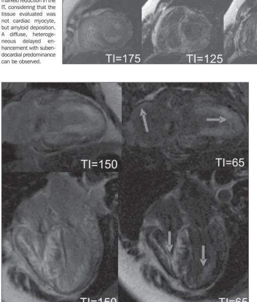
Figure 2. Images acquisition with ECG-gating, delayed enhancement, medial and basal short-axis views, with different inversion times.
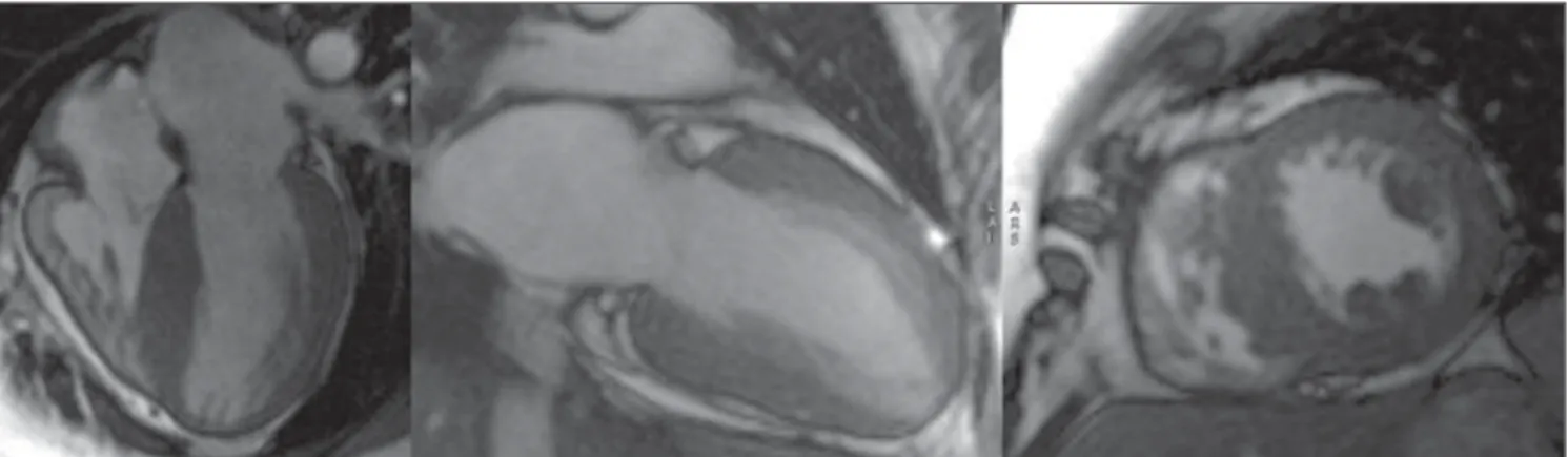
Figure 1. Images acquisition with ECG-gating, in cine-MRI at diastole, four-chamber and two-chamber planes and short-axis view.
× 70 mmHg, with a history of two sudden deaths in the family (a brother with 45 years, and her father with 54 years of age),
VIII Radiol Bras. 2008 Jul/Ago;41(4):VII–IX
Images description
Figure 1. Images acquisition with ECG-gating, in cine-MRI at diastole, four-cham-ber and two-chamfour-cham-ber planes and short-axis view. A significant diastolic dysfunction with a diffuse myocardial thickening is observed.
Figure 2. Images acquisition with ECG-gating, delayed enhancement, medial and basal short-axis views, with different inver-sion times. Note the difficulty in determin-ing an inversion time (IT) where, as usual, the cardiac muscle signal is suppressed (black). Diffuse delayed enhancement, with circumferential or widespread suben-docardial predominance, without respect-ing coronary territories.
Diagnosis: cardiac amyloidosis.
COMMENTS
Amyloidosis is a complex disease char-acterized by an abnormal deposition of amyloid protein in the heart tissue. Amy-loid substances can be typically found in al-most all organs, but the clinical evidences of the disease only can be detected upon an extensive deposition. Most frequently, the nervous system, heart and kidneys are in-volved(1–3).
Cardiac amyloidosis is not frequently diagnosed, and the actual incidence of this disease remains unknown.
It is estimated that in the majority of cases of primary cardiac amyloidosis, amy-loid proteins deposition is found in about 25% of patients with familial amyloidosis, generally late in the course of the disease. Different forms of presentation are de-scribed, and clinical manifestations can be observed in about one third of cases, so patients can be divided into four groups according to the main clinical manifesta-tion as follows: restrictive cardiomyopathy, systolic dysfunction (low cardiac output), orthostatic hypotension and conduction disorder (isolated atrial involvement)(2–6).
The origin of amyloid proteins repre-sents a true enigma for investigators. It is proposed that this deposition is a direct result from immunocompetent cells with some abnormality in their immune re-sponse. It seems to be clear that these pro-teins are locally produced by action of munological system cells, because of
im-munological intolerance or suppression, suggesting the participation of inactive lymphocytes in the final product(7,8).
Several types of amyloidosis may affect humans, but cardiac involvement is most frequent in the primary amyloidosis that involves deposition of insoluble mono-clonal immunoglobulin light chains pro-duced by plasma cells in the myocardium, most commonly as a result of multiple myeloma. This light chains deposition must demonstrate affinity for Congo red stain, or otherwise in case of a negative histopathol-ogy result, a diagnosis of light chain depo-sition disease (LCDD) should be consid-ered. This particular aspect is relevant, con-sidering that LCDD presents a better clini-cal progression and therapeutic response as compared with amyloidosis(2,7,8).
On the other hand, secondary amyloido-sis is caused by other proteins deposition and may be familial, senile or resulting from chronic inflammatory processes, with typically small and perivascular deposits. Extensive cardiac involvement may rarely occur(2,7–9).
In practice, it is impossible to define a pattern of clinical presentation for amyloi-dosis. The clinician should suspect of car-diac amyloidosis when untreatable chronic cardiac failure develops in patients with 50 or more years of age, or in the presence of cardiac failure with an initially restrictive nature, progressing with progressively untreatable low cardiac output and hy-potension(8,9).
The heart may be the end-organ, or be-ing involved along the progression of pri-mary or secondary systemic amyloidosis. In this context, cardiac amyloid infiltration may occur in four circumstances: 1) as a part of primary systemic amyloidosis or as-sociated with multiple myeloma, caused by AL amyloid protein (immunoglobulin) deposition; 2) as a part of secondary sys-temic amyloidosis, associated with chronic inflammatory diseases (Crohn´s disease, rheumatoid arthritis), caused by AA amy-loid protein deposition, with minor in-volvement of the heart; 3) as a manifesta-tion of an autosomal dominant inherited disease caused by AF amyloid protein (a prealbumin variant or transthyretin) depo-sition; 4) as a localized phenomenon in the elder patient, by SSA amyloid protein (also
an abnormal prealbumin or transthyretin) deposition(10,11).
Finally, cardiac transplantation can be mentioned as a treatment for cardiac amy-loidosis, but the poor global experience demonstrates recidivation of the disease in the transplanted heart. Additionally, cardiac transplantation does not contemplate the other organs affected by amyloidosis(2).
Cardiac magnetic resonance imaging (CMR)
CMR has become a relevant ancillary method in the diagnosis of amyloidosis. Rarely this method presents a false-nega-tive result in cases of significant clinical suspicion and, because of its high specific-ity, the diagnosis exclusion can be charac-terized in case of a negative result(2).
MRI can identify myocardial, interatrial septum thickening, signs of diastolic dys-function and the typical pattern of delayed subendocardial enhancement in the left ventricle, although all the cardiac chamber may be involved(2,12,13).
Amyloid tissues present short T1 and T2 relaxation times, besides alteration in the pattern of delayed myocardial enhancement after gadolinium injection. The myocardial signal intensity changes with focal or dif-fuse infiltration/replacement by amyloid tissue. Sometimes, the interatrial septum will be thickened (Figures 3 and 4)(2,12,13).
Numberless suggestive findings are de-scribed, and the positive result must be evi-denced by in vivo endomyocardial biopsy utilizing Congo red staining(2). However,
according to some authors, such as Abdel-Aty & Friedrich(13), MRI findings are so
accurate that biopsy may not be required. According Vogelsberg et al.(2), as far as
the above mentioned criteria are taken into consideration, the non-invasive diagnosis of cardiac amyloidosis by magnetic reso-nance imaging presents 80% sensitivity and 94% specificity, with a 92% positive predictive value and 85% negative predic-tive value.
Final considerations
IX
Radiol Bras. 2008 Jul/Ago;41(4):VII–IX
2. Vogelsberg H, Mahrholdt H, Deluigi CC, et al. Cardiovascular magnetic resonance in clinically suspected cardiac amyloidosis: noninvasive im-aging compared to endomyocardial biopsy. J Am Coll Cardiol. 2008;51:1022–30.
3. Nacif MS, Oliveira Jr AC, Moreira DM, et al. Qual o seu diagnóstico? Endomiocardiofibrose. Radiol Bras. 2006;39(6):ix–xii.
4. Nacif MS, Oliveira Jr AC, Moreira DM, et al. Qual o seu diagnóstico? Cardiomiopatia hipertrófica apical (CMHA). Radiol Bras. 2006;39(5):v–vii. 5. Barretto ACP, Precoma D, Serro-Azul JB, et al. Amiloidose cardíaca. Uma doença de muitas fa-ces e diferentes prognósticos. Arq Bras Cardiol. 1997;69:89–93.
6. Arima T, Ando Y, Okamura R, et al. Secondary amyloidosis with severe autonomic dysfunctions. J Auton Nerv Syst. 1995;52:77–81.
7. Hesse A, Altland K, Linke RP, et al. Cardiac amyloidosis: a review and report of a new transthyretin (prealbumin) variant. Br Heart J. 1993;70:111–5.
8. Talwar KK, Kumar V, Agarwal R, et al. Cardiac amyloidosis: hemodynamic, echocardiographic and endomyocardial biopsy studies. Indian Heart J. 1992;44:387–90.
9. Petersen EC, Engel JA, Radio SJ, et al. The clini-cal problem of occult cardiac amyloidosis. Foren-sic implications. Am J ForenForen-sic Med Pathol. 1992;13:225–9.
10. Mendes RGG, Evora PRB, Mendes JAM, et al. Comprometimento cardíaco na amiloidose sistê-mica. Diagnóstico in vivo. Arq Bras Cardiol. 1998;70:119–23.
11. Dubrey S, Mendes L, Skinner M, et al. Resolu-tion of heart failure in patients with AL amyloi-dosis. Ann Intern Med. 1996;125:481–4. 12. Klein AL, Hatle LK, Burstow DJ, et al. Doppler
characterization of left ventricular diastolic func-tions in cardiac amyloidosis. J Am Coll Cardiol. 1989;13:1017–26.
13. Abdel-Aty H, Friedrich MG. Magnetic resonance of cardiomyopathies and myocarditis. In: Kwong RY, editor. Cardiovascular magnetic resonance imaging. New Jersey: Humana Press; 2008. p. 399–414.
Figure 4. Images acquisition with ECG-gating, delayed enhancement, long-axis view and four-chamber plane demonstrating biatrial and biventricular (diffuse) subendocardial amyloid deposition. The interatrial septum thickening can also be identified at the four-chamber plane.
Usually, in cases of amyloid restrictive cardiopathy, the typical presentation will be related to the cardiac muscle involvement, leading to a decrease in walls compliance and restricted ventricular filling, in the absence of cavities dilatation or significant systolic dysfunction.
The most significant CMR weakness is the difficulty in defining an appropriate in-version time for acquisition of sequences with delayed myocardial enhancement.
REFERENCES
1. Kushwaha SS, Fallon JT, Fuster V. Restrictive car-diomyopathy. N Engl J Med. 1997;336:267–76.