ABSTRACT
http://dx.doi.org/10.1590/1678-775720140049
Analysis of the reaction of subcutaneous tissues
in rats and the antimicrobial activity of calcium
hydroxide paste used in association with different
substances
Raquel Zanin MIDENA1, Roberto Brandão GARCIA1, Bruno Cavalini CAVENAGO1, Marina Angélica MARCIANO1, Paloma Gagliardi MINOTTI1, Ronald ORDINOLA-ZAPATA1, Paulo Henrique WECKWERTH2, Flaviana Bombarda de ANDRADE1, Marco Antonio Hungaro DUARTE1
1- Universidade de São Paulo, Faculdade de Odontologia de Bauru, Departamento de Dentística, Endodontia e Materiais Dentários, Bauru, SP, Brazil. 2- Universidade do Sagrado Coração, Departamento de Ciências da Saúde, Bauru, SP, Brazil.
Corresponding address: Raquel Zanin Midena - Alameda Octávio Pinheiro Brisolla, 9-75 - 17012-901 - Bauru - SP - Brazil - Phone/Fax number: 55
14-32358344 - e-mail: [email protected]
6XEPLWWHG)HEUXDU\0RGL¿FDWLRQ2FWREHU$FFHSWHG2FWREHU
T
he aim of this study was to evaluate the subcutaneous tissue response in rats and theantimicrobial activity of intracanal calcium hydroxide dressings mixed with different
substances against E. faecalis. Fifty four rats were divided into three experimental groups
according to the vehicle in the calcium hydroxide treatment: 0.4% chlorohexidine in
propylene glycol (PG), Casear ia sylvest r is Sw in PG and calcium hydroxide+PG (control
group). The pastes were placed into polyethylene tubes and implanted into the subcutaneous tissue. After 7, 14 and 30 days, the samples were processed and histologically evaluated (hematoxylin and eosin). The tissue surface in contact with the material was analyzed, and the uantitative analysis determined the volume density occupied by the in ammatory in ltrate (giant cells, polymorphonuclear cells and mononuclear cells), broblasts, collagen
bers and blood vessels. For the antimicrobial analysis, 0 dentin bloc s infected with E.
faecalis were treated with calcium hydroxide pastes in different vehicles; 0.4% chlorhexidine
in PG, PG, extract from Casear ia sylvest r is Sw in PG and a positive control (infection and
without medication) for 7 days. The ef ciency of the pastes was evaluated by the live dead technique and confocal microscopy. The results showed that 0.4% chlorhexidine induced
a higher in ammatory response than the other groups. The Casear ia sylvest r is Sw extract
showed satisfactory results in relation to the intensity of the in ammatory response. n the microbiological test, there were no statistical differences between the evaluated intracanal dressings and the percentage of bacterial viability was between 33 and 42%. The control
group showed an 86% viability. Antimicrobial components such as chlorhexidine or Casearia
sylvest r is Sw did not improve the antimicrobial activity against E. faecalis in comparison
to the calcium hydroxide+PG treatment. In addition, the incorporation of chlorhexidine in the calcium hydroxide paste promoted the highest in ammatory response.
Key words: Calcium hydroxide. Chlorhexidine. Endodontics. Materials testing.
INTRODUCTION
In the presence of pulp necrosis, the whole root canal system presents contamination by microorganisms and their products16. The removal
of these irritants is usually performed with a biomechanical preparation and antimicrobial
solutions4,28. Due to the limitations of this technique
in consistently eliminating the root canal infection, the use of an intracanal medication is necessary to continue the antisepsis process4. The use of
of CH occurred due to its high pH level, which provides microbial inhibition8. Another important
factor is related to lipopolysaccharide inhibition24.
In addition, the calcium release plays an important role in the formation of mineralized tissue20.
Several substances have been mixed with calcium hydroxide to improve some of its properties such as viscosity, radiopacity, antimicrobial spectrum action and ionic dissociation rate3,9,15. The
alkaline pH level is approximately 12.6, however, some types of bacteria such as En t er ococcu s faecalis is considered resistant to the antimicrobial action of calcium hydroxide inside the root canals18.
Chlorhexidine digluconate is a bisguanide which is known for its antimicrobial action against bacteria26. The association of chlorhexidine to
calcium hydroxide seems to increase the ef ciency of calcium hydroxide paste against Ent er ococcus faecalis11. However, chlorhexidine used alone has caused damage to the DNA in oral mucosa cells and leukocytes14, and the association of calcium
hydroxide with chlorhexidine increases the production of reactive oxygen2, which can result in
increased tissue aggression28.
The search for herbal medicines for use in medicine and dentistry is intense, including its uses as an intracanal dressing13. One of the herbs
currently being studied is Casear i a sy l v est r i s Swart (Sw). It is a plant that originates from Latin America and can be found from Mexico to Argentina. In Brazil, it is found in abundance, it is a species very common in the State of São Paulo25.
This plant extract has shown anti in ammatory7
and antimicrobial actions22. In relation to its
chemical composition, essential oils (terpenes and triterpenes), saponins, fatty acids, tannins, resins, anthocyanosides and flavonoids were found25.
Some chemical components in particular can take direct action against E. faecalis, such as tannins,
avonoids and essential oils.
The addition of Casear ia sy lv est r is extract and chlorhexidine to calcium hydroxide does not interfere with its physico-chemical properties6. The
aim of this study was to analyze the biocompatibility and the antimicrobial action of Casear ia sylvest r is Sw (Guaçatonga) and chlorhexidine mixed with calcium hydroxide paste.
MATERIAL AND METHODS
This study evaluated three calcium hydroxide pastes: Group 1 – calcium hydroxide (Merck KGaA, Darmstadt, Germany) mixed with 0.4% chlorhexidine in propylene glycol (Specific Pharmacy, Bauru, SP, Brazil); Group 2 – calcium hydroxide (Merck KGaA, Darmstadt, Germany) mixed with the extract from Casear ia sylvest r is Sw in propylene glycol (Max Pharma Trade and Import
of Pharmaceutical Ingredients/Inputs, São Paulo, SP, Brazil); Group 3 (control) – calcium hydroxide (Merck KGaA, Darmstadt, Germany) mixed with propylene glycol (Speci c Pharmacy, Bauru, SP, Brazil).
The Casear ia sy lv est r is Sw extract utilized in this study was obtained from the Max Pharma pharmaceutical laboratory (Max Pharma Trade and Import of Pharmaceutical Ingredients/Inputs, São Paulo, SP, Brazil) and was produced in accordance with the Brazilian pharmacopeia. The Casear ia sy lv est r is Sw leaves were subjected to a drying process in a circulating air, anatomical oven under a controlled temperature until a constant weight was achieved. The leaves were ground with grinder knives before being used in the preparation of the extract. The crushed product was subjected to maceration for propylene glycol extraction applications, where the powder’s proportion was 25 grams for each 200 mL of propylene glycol. This extract solution remained in contact with the powder for 8 days with sporadic agitation in an amber glass bottle, to avoid possible interference from light, at a room temperature of approximately 25°C.
The pastes were prepared on a sterile glass plate using 3.0 g of calcium hydroxide powder (Merck KGaA, Darmstadt, Germany) for each 1.75 mL of the substances tested in each group.
Tissue analysis
This study was approved by the Ethical Committee for Teaching and Research on Animals. A total of 54 Wistar rats (Rat t us norvegicus albinus) were divided into three experimental groups each of 18 animals, according to the division of the groups mentioned above.
The animals were anesthetized with a combination of Ketamine hydrochloride (Dopalen®
- Vetbrands, CEVA, Paulínia, SP, Brazil) and Xylazine hydrochloride (Anasedan® - Vetbrands, CEVA,
Paulínia, SP, Brazil). These substances were used in a concentration of 0.1 mL of solution for every 100 grams of animal weight. A manual trichotomy was performed on the dorsal region and was disinfected with an iodized alcohol solution. A longitudinal incision was made in the middle of the back of each region, with a no. 15 scalpel blade (Embramac, - Empresa Brasileira de Materiais Cirúrgicos, Campinas, SP, Brazil).
depth of 18 mm in the subcutaneous tissue, causing tissue dilatation. Two tubes form the same group, one for the left side and one for the right side, were deployed into the back of each animal.
The implants were carefully deposited perpendicular to the line of incision, to avoid deportation and to decrease mobility. The incisions were sutured with 4.0 silk thread (Ethicon - Professional Products, Johnson & Johnson Ltda., São Paulo, SP, Brazil).
Six animals were used for each experimental period (7, 14 and 30 days). After the experimental time, the animals were killed with an excessive dose of Ketamine hydrochloride (Dopalen® - Vetbrands,
CEVA, Paulínia, SP, Brazil). A new trichotomy was made on the dorsal region and a dissection of the implant area with a normal safety margin of the surrounding tissue. The tissue containing the implanted tubes was placed on paper and maintained xed in a 10% formalin buffer (Merck KGaA, Darmstadt, Germany) for 7 days.
Next, the tissue samples were subjected to conventional histotechnical processing. Five micrometer thicknesses of serial sections were obtained from the capsule area.
The histological sections were stained with the hematoxylin and eosin technique and evaluated using a light microscope (Aristoplan, Leitz Wetzlar, Germany). A calibrated examiner analyzed the sections in a quantitative manner.
To evaluate the in ammatory response, the tissue surfaces in contact with the material were analyzed, and those areas were analyzed in four microscopical elds of two sections.
Quantitative analysis using an optical microscope (Aristoplan, Leitz Wetzlar, Germany) with a 100× objective and 8× Zeiss Kpl eyepiece containing a Zeiss II integration grid with 100 points symmetrically distributed over a quadrangular area, determined the volume density occupied by the in ammatory in ltrate (giant cells, polymorphonuclear cells and mononuclear cells), broblasts, collagen bers and blood vessels19,21.
The Kruskal–Wallis and Dunn tests were utilized for the analysis of the quantitative analysis. The level of signi cance was set at P 0.05 and the Prisma 5.0 software (GraphPad Software Inc, La Jolla, CA, USA) was utilized as the analytical tool.
Microbiological analysis
Twenty blocks of sterile bovine dentin were used in the experimental procedures. The measurements of the blocks were approximately 4x4x2 mm. The dentin blocks were made from bovine incisors, sterilized by autoclaving. The dentin segments were treated with 1% sodium hypochlorite for 30 minutes and 17% EDTA for 5 minutes. After that the blocks were immersed in BHI broth (Brain Hearth Infusion
– BD – Becton, Dickinson and Company, Sparks MD, USA) culture containing Ent er ococcus faecalis ATCC 25912 (American Type Culture Collection, Manassas, VA) standardized by the 0.5 McFarland scale (1.5x10⁸ bacteria per mL) and incubated
for 21 days. The new culture broth of E. faecalis, obtained by overnight incubation and standardized with the 0.5 McFarland scale was refreshed every 3 days.
At the end of the experimental period, the infected blocks were removed from the culture broths, rinsed 3 times with a sterile, buffered saline solution then dried with absorbent paper and randomly distributed over the surfaces of sterile Petri dishes in accordance with each test group: Group 1 - Calcium hydroxide (Merck KGaA, Darmstadt, Germany) mixed with 0.4% chlorhexidine in propylene glycol (Specific Pharmacy, Bauru, SP, Brazil); Group 2 - calcium hydroxide mixed with the extract from Casear ia sy lv est r is Sw in propylene glycol (Max Pharma Trade and Importer of Pharmaceutical Ingredients/Inputs. Ltda., São Paulo, SP, Brazil); Group 3 - calcium hydroxide (Merck KGaA, Darmstadt, Germany) mixed with propylene glycol (Speci c Pharmacy, Bauru, SP, Brazil); Group 4 - positive control (with infection and without medication).
The blocks from groups 1-3 had their surfaces lled with their respective calcium hydroxide paste using the different vehicles. The blocks were again maintained at 37°C for one week at 100% humidity. After this period, the pastes were removed by irrigation with 2 mL of sterile water and then dried with sterile paper point cones.
For the bacterial viability analysis a confocal laser scanning microscope was used. The samples were stained using the Syto-9/Propidium iodide (PI) technique (Live/Dead, Baclight; Invitrogen, Carlsbad, CA, USA). S TO-9 is a green, uorescent nucleic acid stain which generally labels all live microrganisms. PI is a red, uorescent nucleic acid stain that penetrates only cells with damaged membranes, highlighting the dead micro-organisms. The treated dentine samples were stained with 50 μL of the dyes for 10 min at room temperature in a dark environment. Four 40X confocal pictures were obtained from each sample using a 40X oil lens (Leica TCS-SPE; Microsystems GmbH, Mannheim, Germany). The scanning was performed by using a 1 μm step-size in a 512x512 pixel format, for a total of 20 images analyzed for each medication. The obtained bio lm images represented an area of 275x275 μm2. For quanti cation purposes, the
Bioimage-L software(http://www.bioimagel.com) was used to determine the percentage of live cells5.
,QÀDPPDWRU\FHOOV
Giant cells Mononuclear cells Polymorphonuclear cells
7 days 14 days 30 days 7 days 14 days 30 days 7 days 14 days 30 days
CH+CLX 0.4 % 1.125A;1,2
(0.0000-3.500) 1.563A;1 (0.0000-8.250) 0.0000A;2 (0.0000-1.250) 14.44A;1 (6.250-19.50) 11.25AB;1 (7.500-24.75) 5.000A;2 (4.250-7.750) 0.6250A;1 (0.0000-1.750) 0.2500AB;1,2 (0.0000-1.000) 0.0000A;2 (0.0000-0.2500)
CH+CS 0.1250A;1
(0.0000-3.250) 0.0000B;1,2 (0.0000-0.5000) 0.0000A;2 (0.0000-0.0000) 14.32A;1 (9.250-42.50) 9.875A;1 (3.250-15.50) 1.563B;2 (0.5000-4.750) 0.0000AB;1 (0.0000-0.5000) 0.0000A;1 (0.0000-0.2500) 0.0000A;1 (0.0000-0.2500)
CH+PG 2.000A;1
(0.0000-5.500) 0.6250AB;1,2 (0.0000-2.000) 0.0000A;2 (0.0000-0.2500) 17.25A;1 (7.750-28.00) 13.75B;1 (5.500-16.75) 1.188B;2 (0.2500-4.000) 0.0000B;1 (0.0000-1.500) 0.6875B;1 (0.0000-1.750) 0.0000A;1 (0.0000-0.5000)
CH: Calcium hydroxide; CLX: Chlorhexidine; CS: Casearia sylvestris; PG: Propylene glycol
'LIIHUHQWXSSHUFDVHOHWWHUVLQFROXPQVUHSUHVHQWVWDWLVWLFDOO\VLJQL¿FDQWGLIIHUHQFHVEHWZHHQWKHPDWHULDOVIURPHDFKSHULRG 'LIIHUHQWQXPEHUVUHSUHVHQWVWDWLVWLFDOO\VLJQL¿FDQWGLIIHUHQFHVEHWZHHQSHULRGVIRUHDFKPDWHULDO
Table 1-0HGLDQPD[LPXPDQGPLQLPXPYDOXHVREWDLQHGIURPWKHLQÀDPPDWRU\UHDFWLRQDQDO\VLVLQWKHWKUHHH[SHULPHQWDO periods
Histological events
&ROODJHQ¿EHUV Fibroblasts Blood vessels
7 days 14 days 30 days 7 days 14 days 30 days 7 days 14 days 30 days
CH + CLX 0,4 % 52.07A;1
(43.50-58.00) 42.38A;1 (17.50-54.75) 63.75A;2 (57.75-71.75) 5.255A;1 (3.500-6.750) 3.625A;1 (1.000-7.500) 7.938A;2 (4.500-11.00) 0.8750A;1 (0.0000-5.130) 0.5000A;1 (0.0000-3.500) 1.125A;1 (0.5000-1.750)
CH+CS 46.50A;1
(20.25-61.25) 56.63B;1 (42.25-70.00) 53.06B;1 (39.25-69.50) 7.875B;1 (2.500-10.00) 7.500B;1 (4.250-10.75) 7.563A;1 (6.250-8.750) 1.250A;1 (0.0000-2.750) 0.7500A;2 (0.0000-1.250) 0.8125A;1,2 (0.5000-1.500)
CH+PG 48.50A;1
(36.25-52.25) 51.75B;1 (45.50-67.50) 68.00A;2 (53.50-79.00) 8.565B;1 (4.000-10.75) 5.063A;2 (2.000-8.750) 8.125A;1 (4.500-10.50) 0.2500B;1 (0.0000-1.500) 0.0625A;1 (0.0000-1.250) 0.3750B;1 (0.0000-0.5000)
CH: Calcium hydroxide; CLX: Chlorhexidine; CS: Casearia sylvestris; PG: Propylene glycol
'LIIHUHQWXSSHUFDVHOHWWHUVLQFROXPQVUHSUHVHQWVWDWLVWLFDOO\VLJQL¿FDQWGLIIHUHQFHVEHWZHHQWKHPDWHULDOVLQHDFKSHULRG 'LIIHUHQWQXPEHUVUHSUHVHQWVWDWLVWLFDOO\VLJQL¿FDQWGLIIHUHQFHVEHWZHHQSHULRGVIRUHDFKPDWHULDO
Table 2- Median, maximum and minimum values obtained from the histological events shown in connective tissue analysis in the three experimental periods
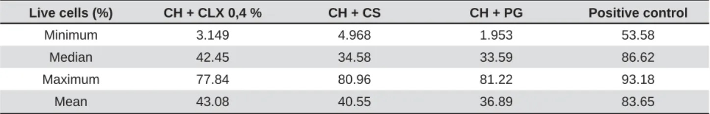
Live cells (%) CH + CLX 0,4 % CH + CS CH + PG Positive control
Minimum 3.149 4.968 1.953 53.58
Median 42.45 34.58 33.59 86.62
Maximum 77.84 80.96 81.22 93.18
Mean 43.08 40.55 36.89 83.65
CH: Calcium hydroxide; CLX: Chlorhexidine; CS: Casearia sylvestris; PG: Propylene glycol
Table 3- Minimum, median, maximum and mean of the percentage of living cells after contact with the experimental substances
5.0 software (GraphPad Software Inc, La Jolla, CA, USA) was used as the analytical tool.
RESULTS
InÀaPPaWory reacWion
The values and statistical comparisons concerning
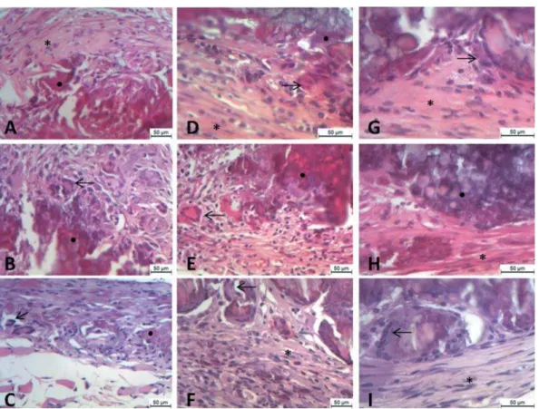
Figure 1- Week one subcutaneous tissue response of the evaluated intracanal dressings (7 days). Chlorhexidine 0.4%+calcium hydroxide (A); Casearia sylvestris Sw+calcium hydroxide (B); Propylene glycol+calcium hydroxide (C). )LEURFHOOXODUWLVVXHPDFURSKDJHLQ¿OWUDWLRQDUURZVDQGPDWHULDOUHPQDQWVƔFDQEHREVHUYHG$WGD\VDGHFUHDVH LQWKHLQÀDPPDWRU\LQ¿OWUDWHDUURZVDQGDGLVFUHWHLQFUHDVHLQ¿EURFHOOXODUWLVVXHLQFRPSDULVRQWRWKHGD\SHULRG were observed. Chlorhexidine 0.4%+calcium hydroxide (D); Casearia sylvestris Sw+calcium hydroxide (E); Propylene glycol+calcium hydroxide (F). After 30 days, organized subcutaneous tissue is evident. Chlorhexidine 0.4%+calcium hydroxide (G) presenting giant cells (arrow). Fibrocellular tissue (*) is present in Casearia sylvestris Sw+calcium hydroxide (H) and propylene glycol+calcium hydroxide (I)
probably due to the alkaline action of the calcium hydroxide. Overall, the lowest values were found at the 30 day period for all pastes tested. In the 7 day period there were no statistically signi cant differences (P>0.05) among the tested pastes (P>0.05), however, during the periods of 14 to 30 days, statistically signi cant differences were found (P<0.05). A representative picture of the in ammatory reaction is shown in Figure 1.
Microbiological analysis
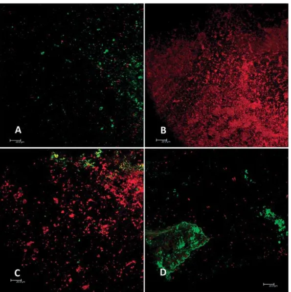
Table 3 shows the minimum, median, maximum and mean of the percentage of living cells after contact with the different calcium hydroxide pastes. All the pastes presented a signi cantly (P<0.05) lower percentage of live cells than the control. There were no statistically signi cant differences (P>0.05) between the medications. Figure 2 represents images obtained from the bio lms after treatment with the different pastes.
DISCUSSION
The objective of this study was to compare the effects of the addition of chlorhexidine or the extract from Casear ia sy lv est r is Sw in propylene glycol to calcium hydroxide on the biocompatibility and antimicrobial activity of the pastes. In endodontic therapy, chlorhexidine is used in concentrations of 0.2% to 2.0%29. Chlorhexidine has an antimicrobial
action in low concentrations (0.006%), and its effectiveness decreases in line with the reduction in concentration. This shows that there is a consensus among the choice of chlorhexidine concentrations used both as an irrigant solution or intracanal medication1. The ndings for the in ammatory
reaction of all groups tested, including the control group at 7 days, was more intense than at 14 and 30 days, as reported by a previous study30.
Figure 2- Representative confocal pictures after treatment with chlorhexidine 0.4%+calcium hydroxide (A); Casearia
sylvestris Sw+calcium hydroxide (B); Propylene glycol+calcium hydroxide (C); Positive control (D). Dead cells are observed as red and live cells are observed as green
the others, which is contrary to another study26. The
differences in the results can be attributed to the fact that the previous study26 used a pure extract,
and in our study the association with the calcium hydroxide paste could have reduced the cytotoxic effect. The characteristics of the in ammatory in ltrate during this period were also de ned by the presence of giant cells and macrophages, however, the beginning of some tissue organization was detected in the three groups.
At the 30 day period, there was a decrease in the intensity of the in ammatory response in all the groups. The 0.4% chlorhexidine group reaction remained high compared to the other groups, establishing a statistically signi cant difference between groups 1 and 3. Such differentiation can be linked to the severity of the tissue injury by the chlorhexidine substance12. The increase in the
percentage of chlorhexidine mixed with calcium hydroxide was noted; proportionally increasing the intensity caused an in ammatory reaction.
Concentrations greater than 0.5%, chlorhexidine can produce tissue necrosis and the in ammatory process is more intense in slowing the healing process12,23,27. For the Casear ia sylvest r is extract
group, a reduction of the in ammatory in ltrate occurred, probably due to its anti-in ammatory potential originating from its essential oils22 and, in
addition, the extract from this plant does not have a genotoxic effect17.
With regards to the antimicrobial analysis of the paste, the Casear ia sy lv est r is Sw extract group showed the lowest performance against E. faecalis compared to the other pastes used in the experiment, suggesting that this vehicle would not be adequate for the preparation of calcium hydroxide, especially to provoke an antimicrobial action, although it had a satisfactory result in the in ammatory response.
vehicle reduces its antimicrobial ef ciency, probably due to a reduction of the ability of the hydroxyl ion’s diffusion of calcium hydroxide in this vehicle. A possible reason is that pastes treated with propylene glycol promote a slower release of hydroxyl ions30.
It should be stressed that Ent erococcus faecalis is a microorganism resistant to pHs up to the magnitude of 11.010,18, which would explain the smaller
antimicrobial action of the pastes with propylene glycol that favor a lower pH level6.
CONCLUSION
Antimicrobial components such as chlorhexdine and Casearia sylvest ris did not improve antimicrobial activity against E. faecalis in comparison to the calcium hydroxide + propylene glycol medication. In addition to this, the incorporation of chlorhexidine in the calcium hydroxide paste gave the highest in ammatory response.
REFERENCES
1- Agerbaek N, Melsen B, Rolla G. Application of chlorhexidine by oral irrigation systems. Scand J Dent Res. 1975;83:284-7. 2- Barbin LE, Saquy PC, Guedes DF, Sousa-Neto MD, Estrela C, Pécora J. Determination of para-chloroaniline and reactive oxygen species in chlorhexidine and chlorhexidine associated with calcium hydroxide. J Endod. 2008;34:1508-14.
3- Basrani B, Ghanem A, Tjäderhane L. Physical and chemical properties of chlorhexidine and calcium hydroxide-containing medications. J Endod. 2004;30:413-7.
4- Bystrom A, Claesson R, Sundqvist G. The antibacterial effect of camphorated paramonochlorophenol, camphorated phenol and calcium hydroxide in the treatment of infected root canals. Endod Dent Traumatol. 1985;1:170-5.
5- Chávez de Paz LE. Image analysis software based on color segmentation for characterization of viability and physiological activity of bio lms. Appl Environ Microbiol. 2009;75:1734-9. 6- Duarte MA, Midena RZ, Zeferino MA, Vivan RR, Weckwerth PH, Santos F, et al. Evaluation of pH and calcium ion release of calcium hydroxide pastes containing different substances. J Endod. 2009;35:1274-7.
7- Esteves I, Souza I, Rodrigues M, Cardoso LG, Santos LS, Sertie JA, et al. Gastric antiulcer and anti-in ammatory activities of the essential oil from Casear ia sylvest r is Sw. J Ethnopharmacol.
2005;101:191-6.
8- Estrela C, Sydney GB, Bammann L, Felippe Júnior O. Mechanism of action of calcium and hydroxyl ions of calcium hydroxide on tissue and bacteria. Braz Dent J. 1995;6:85-90.
9- Estrela C, Sydney GB, Pesce HF, Felippe Júnior O. Dentinal diffusion of hydroxyl ions of various calcium hydroxide pastes. Braz Dent J. 1995;6:5-9.
10- Evans M, Davies JK, Sundqvist G, Figdor D. Mechanisms involved in the resistance of Ent er ococcus faecalis to calcium
hydroxide. Int Endod J. 2002;35:221-8.
11- Evans MD, Baumgartner JC, Khemaleelakul SU, Xia T. Ef cacy of calcium hydroxide: chlorhexidine paste as an intracanal medication in bovine dentin. J Endod. 2003;29:338-9.
12- Faria G, Celes MR, De Rossi A, Silva LA, Silva JS, Rossi MA. Evaluation of chlorhexidine toxicity injected in the paw of mice and added to cultured l929 broblasts. J Endod. 2007;33:715-22. 13- Gentil M, Pereira JV, Sousa YT, Pietro R, Neto MD, Vansan LP, et al. I n vit r o evaluation of the antibacterial activity of Ar ct ium lappa as a phytotherapeutic agent used in intracanal dressings.
Phytother Res. 2006;20:184-6.
14- Grassi TF, Camargo EA, Salvadori DM, Marques ME, Ribeiro DA. DNA damage in multiple organs after exposure to chlorhexidine in Wistar rats. Int J Hyg Environ Health. 2007;210:163-7.
15- Guerreiro-Tanomaru JM, Chula DG, Pontes Lima RK, Berbert FL, Tanomaru-Filho M. Release and diffusion of hydroxyl ion from calcium hydroxide-based medicaments. Dent Traumatol. 2012;28:320-3.
16- Haapasalo M, Qian W, Portenier I, Waltimo T. Effects of dentin on the antimicrobial properties of endodontic medicaments. J Endod. 2007;33:917-25.
17- Maistro EL, Carvalho JC, Mantovani MS. Evaluation of the genotoxic potential of the Casear ia sylvest r is extract on HTC and
V79 cells by the comet assay. Toxicol In Vitro. 2004;18:337-42. 18- McHugh CP, Zhang P, Michalek S, Eleazer PD. pH required to kill Ent er ococcus faecalisin vit r o. J Endod. 2004;30:218-9.
19- Minotti PG, Ordinola-Zapata R, Midena RZ, Marciano MA, Cavenago BC, Bramante CM, et al. Rat subcutaneous tissue response to calcium silicate containing different arsenic concentrations. J Appl Oral Sci. 2015;23:42-8
20- Mizuno M, Banzai Y. Calcium ion release from calcium hydroxide stimulated bronectin gene expression in dental pulp cells and the differentiation of dental pulp cells to mineralized tissue forming cells by bronectin. Int Endod J. 2008;41:933-8. 21- Pereira MS, Rossi MA, Cardoso CR, Silva JS, Bezerra da Silva LA, Kuga MC, et al. Cellular and molecular tissue response to triple antibiotic intracanal dressing. J Endod. 2014;40:499-504. 22- Schneider NF, Moura NF, Colpo T, Flach A. Composição química e atividade antimicrobiana do óleo volátil de Casear ia sylvest r is
Swart. Rev Bras Farm. 2006;87:112-4.
23- Silva RA, Assed S, Nelson-Filho P, Silva LA, Consolaro A. Subcutaneous tissue response of isogenic mice to calcium hydroxide-based pastes with chlorhexidine. Braz Dent J. 2009;20:99-106.
24- Tanomaru JM, Leonardo MR, Tanomaru Filho M, Bonetti Filho I, Silva LA. Effect of different irrigation solutions and calcium hydroxide on bacterial LPS. Int Endod J. 2003;36:733-9. 25- Teske M, Trentini AM. Comp ndio de toterapia. 3 ed. Colombo: Herbarium Laboratório Botânico; 1997.
26- Vianna ME, Gomes BP, Berber VB, Zaia AA, Ferraz CC, Souza-Filho FJ. I n vit r o evaluation of the antimicrobial activity of
chlorhexidine and sodium hypochlorite. Oral Surg Oral Med Oral Pathol Oral Radiol Endod. 2004;97:79-84.
27- Waris G, Ahsan H. Reactive oxygen species: role in the development of cancer and various chronic conditions. J Carcinog. 2006;5:14.
28- Zamany A, Safavi K, Spångberg LS. The effect of chlorhexidine as an endodontic disinfectant. Oral Surg Oral Med Oral Pathol Oral Radiol Endod. 2003;96:578-81.