Arq Neuropsiquiatr 2008;66(2-A):254-255
254
Isolated oculomotor Nerve ParesIs as
the PreseNtINg sIgN of multIPle sclerosIs
Pedro Beleza
1, Álvaro Machado
1, João Soares-Fernandes
2, Gisela Carneiro
1,
Maria José Jordão
1, Fátima Almeida
1, José Figueiredo
1ParesIa Isolada do Nervo oculomotor comum: uma forma de aPreseNtação INvulgar de esclerose múltIPla
São Marcos Hospital, Braga, Portugal: 1MD, Department of Neurology; 2MD, Department of Neuroradiology.
Received 26 September 2007, received in inal form 13 December 2007. Accepted 14 February 2008.
Dr. Pedro Beleza – Serviço de Neurologia / Hospital São Marcos - Largo Carlos Amarante, Apartado 2242 - 4701-965 Braga - Portugal. E-mail: [email protected]
In multiple sclerosis (MS), ocular motor disturbances such as nystagmus or internuclear ophthalmoplegia are frequent and their pathophysiological processes are rel-atively well known. On the contrary, other rare and not so well studied manifestations such as isolated ocular motor nerve palsy may be observed and can represent a diagnos-tic challenge for the clinician as in the case we report.
case
A 35-year-old woman was admitted in the emergency de-partment complaining of double vision on right gaze of sudden onset lasting for three days. Additionally from the beginning of clinical onset she referred constant right periocular pain not related with ocular movements, which spontaneously reverted within 24 hours. She had no recent infectious or traumatic events and her personal and familiar medical history was otherwise un-remarkable. On neurological examination showed a left isolated incomplete III cranial nerve (CN) palsy characterized by eyelid ptosis, limitation on adduction and supraversion of left eye, pare-sis of the four extraocular muscles conirmed by Hess screen and no pupillary dysfunction, either on light or accommodation. The remaining neurological examination, namely visual acuity, fun-doscopy, other cranial nerves and long tracts systems were nor-mal. Also, orbital murmurs, proptosis, quemosis or conjunctival hyperemia were not found. She was apyretic and normotensive. All the analytic work-up was within normal limits. It included he-mogram, C-reactive protein, sedimentation rate, glycemia, lip-id proile, thyrolip-id function, infectious serologies (lyme disease, syphilis), immunologic study (antinuclear antibodies, anticardio-lipin antibodies, anti-neutrophilic cytoplasmic antibodies, an-ti-double stranded antibodies, angiotensin-converting enzyme) and CSF cytochemistry studies. The MRI and the convention-al angiography performed at 24 hours and at the ifth day after admission respectively were normal. A complete and spontane-ous reversion of the III CN palsy occurred within three weeks.
Regarding the clinical follow-up, one year and half after-wards she presented another deicit episode, now consisting of left eye blurred vision which had partially recovered without medication. Further, one month later she had numbness of the
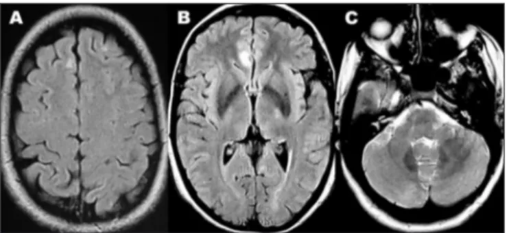
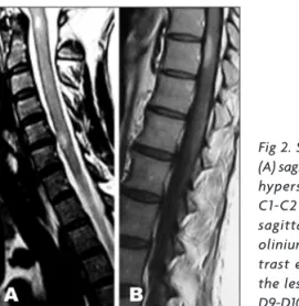
left lower limb. At that time, examination revealed low visual acuity in the left eye (1/10) with normal fundoscopy, bilateral pyramidal syndrome without motor deicits and a left D10 posthesic level. The MRI showed multiple small T2 and Flair hy-perintense lesions located in the subcortical white matter on the high frontal convexity, interhemispheric cortex and left mid-dle cerebellar peduncle, without disruption of the bloodbrain barrier (Fig 1). Left optic nerve was found to be thick although did not show signal abnormalities or gadolinium enhancement. The spinal cord MRI disclosed multiple intraspinal cord lesions in the transitions of C1-C2, C5-C6 and D9-D10 showing T2 hyper-intensity and gadolinium enhancement (Fig 2). Oligoclonal bands were present in the CSF although not in the serum.
Thus, the patient had clinical criteria of time and space dis-semination, corresponding to the optic nevritis and sensitive at-tack, even not considering the cranial nerve episode. The diag-nosis of MS was additionally corroborated by the presence of Barkhof criteria for space dissemination1 and the inding of
oligo-clonal bands only in the CSF. Thus, she was provided beta-1b inter-feron and untill the present time had no other attack (12 months of follow-up) although keeps a sequelar visual deicit (EDSS=2).
dIscussIoN
We describe a case of a 35-year-old woman with an unremarkable medical history who presented an
Arq Neuropsiquiatr 2008;66(2-A)
255
Multiple sclerosis: oculomotor nerve paresis Beleza et al.
lated external III CN ophthalmoparesis associated with ipsilateral periocular pain. Regarding the localization of the presumed lesion, semiology suggested a fascicular or peripheral involvement, however neuroimage was not informative. In fact, features suggesting a nuclear palsy as bilateral eyelid ptosis or contralateral right rectus pal-sy2 were not found. All the etiologic investigation per-formed at that time was normal. So, we could exclude or at least consider as highly improbable the diagnosis of aneurysm, diabetic or vasculitic stroke of the nerve, mesencephalic stroke, tumor and infectious (borrelia, syphilis, tuberculosis) or sarcoidosis meningitis. Also, she had no history suggestive of ophthalmoplegic mi-graine or trauma. Some authors have underlined that minor head trauma can cause an isolated III CN palsy in the absence of abnormal findings on brain MRI3 which was not the case of our patient. MS criteria4 was de-veloped later, so retrospectively the III CN nerve palsy was interpreted as a clinically isolated syndrome (CIS). Isolated CN palsy is a rare inding in MS, occurring in 1.6 % of patients and involving in decreasing order of frequency the CN VI, VIII, VII, III and IV5. This hierarchy of frequency is due to the anatomic variability seen among the differ-ent CN. Namely, the length of the intra-axial traject (less length in CN III, IV and VIII), the density of the fascicular nerves (CN III and VIII have diffuse fascicules)5 and the amount of myelin exposed in the fascicular traject (less in CN IV)6. The largest published series demonstrated that the isolated CN palsy was always associated with brain-stem involvement, either as a lesion seen on MRI (1.5 T) or as a dysfunction disclosed in electrophisiologic tests5.
This study and others7 proved that brainstem audi-tory evoked potentials and blink relex abnormalities are highly sensitive on detection of brainstem demyelinating lesions some of which are not amenable to MRI detection. So, in the light of this, we suggest that the most probable
localization involved in the ophthalmoparesia of the pa-tient currently reported is fascicular mesencephalic. This cannot however be conirmed as long as neurophisiologic tests were not performed by lack of any clinical indication at that time. The MRI performed with a 1.5 T ield could justify the underdetection of a small brainstem lesion in-asmuch as studies demonstrated that high ield MRI such as 3 T provides a signiicantly higher detection rate of in-lammatory brain lesions8. In addition, literature reports only one MS patient with isolated III CN palsy caused by peripheral nervous system involvement who differently from our patient had pupilar involvement and enhance-ment of the cisternal portion of the CN III on MRI9. To the best of our knowledge only two other patients were reported in English-language literature showing an isolat-ed CN III paresis as the presenting sign of MS10,11. As both observations were performed more than 15 years ago, we aim with this case report to provide an updated insight on this rare manifestation of MS.
In conclusion, a CIS indicating a pathological process involving the CN may constitute a hard diagnostic chal-lenge for the clinician. This hypothesis should be consid-ered in a young woman presenting with an isolated III CN palsy even with a normal MRI if all other other possible diagnosis were excluded. From an accurate identiication of the irst MS attack depends a timely initiation of the disease-modifying treatment.
refereNces
1. Barkhof F, Filippi M, Miller DH, et al. Comparison of MRI criteria at irst presentation to predict conversion to clinically deinite multiple sclerosis. Brain 1997;120(Par II):2059-2069.
2. Brazis PW. Localization of lesions of the oculomotor nerve: recent con -cepts. Mayo Clin Proc 1991;66:1029-1035.
3. Levy RL, Geist CE, Miller NR. Isolated oculomotor palsy following mi -nor head trauma. Neurology 2005;65:169.
4. McDonald WI, Compston A, Edan G, et al. Recommended diagnostic criteria for multiple sclerosis: guidelines from the International panel on the diagnosis of multiple sclerosis. Ann Neurol 2001;50:121-127. 5. Thomke F, Lensch E, Ringel K, Hopf HC. Isolated cranial nerve palsies
in multiple sclerosis. J Neurol Neurosurg Psychiatry 1997;63:682-685.
6. Jacobson DM, Moster ML, Eggenberger ER, Galetta SL, Liu GT. Isolat
-ed trochlear nerve palsy in patients with multiple sclerosis. Neurology 1999;53:877-879.
7. Uncini A. Electrophysiological and magnetic resonance imaging cor
-relates of brainstem demyelinating lesions. Electromyogr Clin Neuro
-physiol 1990;30:233-238.
8. Wattjes MP, Lutterbey GG, Harzheim M, et al. Higher sensitivity in the detection of inlammatory brain lesions in patients with clinically iso -lated syndromes suggestive of multiple sclerosis using high ield MRI: an intraindividual comparison of 1.5 T with 3.0 T. Eur Radiol 2006;16: 2067-2073.
9. Bhatti MT, Schmalfuss IM, Williams LS, Quisling RG. Peripheral third
cranial nerve enhancement in multiple sclerosis. AJNR Am J Neurora
-diol 2003;24:1390-1395.
10. Newman NJ, Lessell S. Isolated pupil-sparing third-nerve palsy as the presenting sign of multiple sclerosis. Arch Neurol 1990;47:817-818. 11. Galer BS, Lipton RB, Weinstein S, Bello L, Solomon S. Apoplectic head
-ache and oculomotor nerve palsy: an unusual presentation of multiple sclerosis. Neurology 1990;40:1465-1466.