Management of Fournier’s gangrene: experience of a university
Management of Fournier’s gangrene: experience of a university
Management of Fournier’s gangrene: experience of a university
Management of Fournier’s gangrene: experience of a university
Management of Fournier’s gangrene: experience of a university
hospital of Curitiba
hospital of Curitiba
hospital of Curitiba
hospital of Curitiba
hospital of Curitiba
Manejo da gangrena de Fournier: experiência de um hospital universitário
Manejo da gangrena de Fournier: experiência de um hospital universitário
Manejo da gangrena de Fournier: experiência de um hospital universitário
Manejo da gangrena de Fournier: experiência de um hospital universitário
Manejo da gangrena de Fournier: experiência de um hospital universitário
de Curitiba
de Curitiba
de Curitiba
de Curitiba
de Curitiba
ADRIANO ANTONIO MEHL1; DORIVAM CELSO NOGUEIRA FILHO ACBC-PR2; LUCAS MARQUES MANTOVANI2; MICHELE MAMPRIM GRIPPA2;
RALF BERGER2; DENISE KRAUSS3; DENISE RIBAS4
A B S T R A C T A B S T R A C T A B S T R A C T A B S T R A C T A B S T R A C T
Objective: To analyze the results obtained in the Department of General Surgery, Cajuru University Hospital - PUCPR, with the treatment of Fournier’s gangrene. Methods: We reviewed the charts of 40 patients diagnosed with Fournier’s gangrene admitted to the Cajuru University Hospital from November 1999 to April 2006, analyzing gender, age, predisposing factors, etiology, lesion location, laboratory tests , surgical procedures, antibiotic use and hyperbaric oxygen therapy. Results: The most common etiology was the anorectal origin. The most prevalent etiological agent was E. coli. The predominant predisposing factor was diabetes mellitus. The majority of patients were male. The location and extent of injury was usually in the perineum. All underwent surgical debridement, 17 with associated colostomy and two with combined cystostomy. All patients received antibiotics, the most used being metronidazole and gentamicin. Twenty-six patients underwent hyperbaric therapy. The overall mortality was 20%. Conclusion: Fournier’s syndrome, despite all the advances in treatment today, continues to show high mortality rates. Early recognition of infection associated with invasive and aggressive treatment are essential for attempting to reduce these prognostic indices.
Key words: Key words:Key words:
Key words:Key words: Fournier’s gangrene. Hyperbaric Oxygenation/utilization. Therapy.
Work done at the General Surgery Service and at the Hyperbaric Oxygen Therapy Service, Hospital Universitário Cajuru – PUCPR – Curitiba, Paraná, Brazil.
1. Head, Hyperbaric Oxygen Therapy Service, Hospital Universitário Cajuru – PUCPR - Curitiba, Paraná, Brazil; 2. Former Resident, General Surgery Service, Hospital Universitário Cajuru - Curitiba, Paraná, Brazil; 3. Resident, Otorhinolaryngology, HSCM – Paraná, Brazil; 4. Resident, Gynecology and Obstetrics, HSCM – Paraná, Brazil.
INTRODUCTION
INTRODUCTION
INTRODUCTION
INTRODUCTION
INTRODUCTION
J
ean Alfred Fournier, a French virologist (venerologist), described, in 1883, five cases of gangrene of the scrotum in young healthy patients without an apparent cause1,2. Fournier’s Syndrome or Gangrene is a rare conditioncharacterized by acute onset and progression to fulminant sepsis with high levels of morbidity and mortality.
Fournier’s Syndrome (FS) was classified as primary when a cause was not identified and secondary when causing factors were discovered3. The disease is not
exclusive to men, as there have been cases of vulvar necrosis4-6.
Contemporary series indicate that FS tends to affect patients between the third and sixth decade of life, with predisposing comorbidities, and in most cases, a present etiologic factor7,8.
Despite the controversy in the description of the syndrome, it is characterized by a polymicrobial infection (aerobic and anaerobic bacteria) with an identifiable cause
in 95% of cases, beginning in the genital or perineal regions9. It is characterized by an obliterative endarteritis,
followed by ischemia and thrombosis of subcutaneous vessels, resulting in necrosis of the skin and adjacent subcutaneous tissue2,10, even before evidence of erythema,
crepitus and blistering formation.
Microbial factors can activate the coagulation cascade directly or indirectly by inducing production of proinflammatory cytokines and subsequent tissue factor expression in endothelial cells and monocytes, which activate the clotting cascade11, evolving to thrombosis
of blood vessels, the main characteristic of this syndrome.
Predisposing factors include: diabetes mellitus, local trauma, urine leakage, perirectal or perineal surgery9,
extention of a periuretral/anal infection12,13, anorectal
abscess, genitourinary infection14,15, alcoholism,
immunosuppression and renal or hepatic disease16-18.
Diagnosis is based on clinical signs and physical examination. Radiological methods can help show the existence of gas if there is not crepitus, but false negatives may happen19 22. Laboratory tests are nonspecific, in most
cases showing anemia, leukocytosis, thrombocytopenia, hyperglycemia, hyponatremia, hypokalemia, azotemia and hypoalbuminemia.
The mean time of diagnosis is six days with the conventional methods23 and 21 hours with the mere
identification of necrotizing fasciitis in frozen section biopsy specimens24.
The treatment is based mainly on surgical management, ranging from simple drainage to radical d e b r i d e m e n t w i t h o r w i t h o u t f e c a l o r u r i n a r y diversion2,7,25-27, followed or not by flaps rotation28, use
o f b r o a d s p e c t r u m a n t i b i o t i c s a n d s u p p o r t measures 2,29,30. There are also adjuvant measures such
as hyperbaric chamber to prevent the extension of necrosis, reduce systemic signs of infection and enhance ischemic tissue survival31.
In 1995, Laor and colleagues described an index of severity of Fournier’s Syndrome (FSI - Founier Severity Index) for predicting the outcome of patients with this condition32. Chawla et al. reported that the index indicated
the probability of survival based on clinical and laboratory variables that characterize the severity of the disease and serve to compare patients33.
Despite all the current pathophysiological knowledge, FS remains a disease of high mortality2,7,34 and
improvements in antimicrobial therapy and medical care did not affect its natural history35.
This study aims to analyze the results obtained in our services with the treatment of Fournier’s Gangrene.
METHODS
METHODS
METHODS
METHODS
METHODS
We conducted a retrospective study of medical records of patients treated at University Hospital Cajuru -PUC/PR with diagnosis of Fournier’s Syndrome in the period between November 1998 and April 2006. The diagnosis was established by clinical history and physical examination. The variables were gender, age, location, predisposing factors, etiology, lesion location, laboratory tests and imaging, surgical procedures, antibiotics, use (or not) of an hyperbaric therapy protocol and length of hospi-tal stay.
RESULTS
RESULTS
RESULTS
RESULTS
RESULTS
We evaluated 40 patients with FS in a total of 31 men (77%) and nine women (23%). The mean age was 47.2 years, ranging from 18 to 78.
Twenty-three patients were from the city of Curitiba (capital of Paraná State) and the remaining 17
from other cities in the State. Length of stay ranged from 1 to 62 days with an average of 15.9.
The most common causal factor was the anorectal source, observed in 11 patients (27.5%), followed by an unknown cause in five patients (Table 1). We had two patients who developed Fournier after an intramuscular (IM) injection of potassium diclofenac into the buttock and one case after a diagnostic lumbar puncture (Guillain-Barre’s Syndrome).
The patients were mostly male, totaling 31 (77%). The main predisposing factor was diabetes mellitus, being present in nine patients (22.5%), four type I diabetes and five type II (Table 2). Among the patients who died (eight patients), two were diabetic and one of them had associated hypertension.
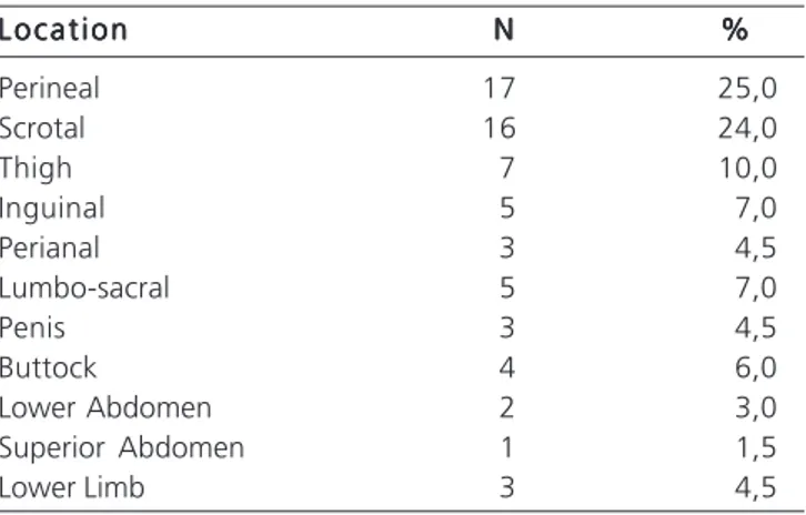
As for the location and extent of the injury, it was observed that it was confined to the perineal area in 17 patients (25.0%), affecting the scrotum in 16 (24.0%) individuals. The gangrene extended to the thigh (10.0%), groin and lumbar (7.0%) (Table 3). The lesions of patients who died were extended to the lower limb, gluteus, lower hemi-abdomen and lumbo-sacral region.
Table 1 Table 1Table 1 Table 1
Table 1 - Causal Factors of Fournier’s gangrene.
E t i o l o g y E t i o l o g yE t i o l o g y E t i o l o g y
E t i o l o g y NNNNN %%%%%
Anorrectal 11 27,5
Urogenital 10 25,0
Traumatic 5 12,5
Gynecologic Surgery 1 2,5
Vascular Surgery 1 2,5
Nursery manipulation 1 2,5
IM Injection 2 5,0
Strangulated Hernia 1 2,5
Peripheral Vascular Disease 1 2,5
Lumbar Puncture 1 2,5
Pressure Ulcer 1 2,5
Unknown 5 12,5
Table 2 Table 2Table 2 Table 2
Table 2 – FS predisposing factors.
Predisposing Factors Predisposing FactorsPredisposing Factors Predisposing Factors
Predisposing Factors NNNNN
Type I Diabetes Mellitus 4
Type II Diabetes Mellitus 5
Arterial Hypertension 7
Paraplegia 3
Tetraplegia Smoking 12
Alcoholism 1
Advanced Age 1
Anti-inflammatory Use 1
Obesity 1
Regarding the exams performed on admission, complete blood count showed the presence of a leukocytosis (average: 15,221/mm3) associated with an increase in bands (average: 14.2%). Among the patients who died leukocytosis and bands were higher (18,364/mm3 and 14.4% respectively) when compared with the survival group (Table 4). The degree of anemia was not severe in both groups. Platelets were increased in six patients and decreased in four.
The treatment of all patients consisted of surgical and medical, the latter with antibiotics alone or associated with hyperbaric therapy.
The antibiotic schemes used were: monotherapy in two patients, one treated with ciprofloxacin for 13 days and the other with cefazolin for three days, both evolving favorably; dual therapy in eight patients (20.0%), five of which with metronidazole and ceftriaxone. Three patients with this scheme died with an average of 2.6 days of hos-pital stay. Therapy with more than two antibiotics was used in most patients (30 – 75.0%).
Metronidazole was the most used antibiotic (23.0%) followed by gentamicin (14.0%), ceftriaxone (11.0%), cefazolin and ampicillin (10.0% each).
Culture results were obtained in 27 patients, and the most prevalent etiological agent was Escherichia coli (26.0%) followed by Pseudomonas aeruginosa, Acinetobacter calcoaceticus-baumannii and Staphylococcus aureus (11.0% each) (Table 5).
All patients underwent radical surgical debridement, ranging from 1 to 7 procedures, with an average of 1.5. Debridement consisted of excision of all necrotic tissue, cleansing with saline and drainage. If there was impairment of anal sphincters or the possibility of contamination of the wound with fecal material, a colostomy was performed. Indeed, 17 patients were submitted to colostomy, of which 12 were lateral and five were terminal. Four patients with a colostomy died. Two patients required cystostomy because of extensive involvement of the external genitalia (both were not submitted to hyperbaric therapy). Plastic surgery procedures were subsequently performed in 11 patients (27.5%). The skin graft was the most common procedure, being done in 10 patients (90.0%). We had a case that required reconstruction of the penis, scrotum, perineum and buttock, and another for which that was necessary to correct a bila-teral degloving of the scrotum.
Hyperbaric therapy was performed in 26 patients (65.0%), the number of cycles ranging from 10 to 30 according to the needs of each case, with an average of 12.5 cycles. The remaining patients were not submitted to hyperbaric oxygen therapy because of early death, delayed indication or contraindication to the procedure. Each session lasted 120 minutes (two hours) at a pressure of absolute atmosphere (ATA) that ranged from 2.0 to 2.8 ATA with a mean of 2.31 ATA. Patients who died (n=3) averaged 2.6 ATA per session against
2.28 ATA for those who survived. Thirteen patients (50%) completed all cycles of hyperbaric chamber established at baseline with an average of 16.2 sessions ranging from 6 to 30. At the request of the attending physician, 10 patients were discharged before the end of the established protocol because of clinical improvement, resulting in an average of 10.1 sessions ranging from 6 to 18. Of the three deaths, there was an average of five sessions ranging from 1 to 13 (Table 6).
The need for hospitalization in an Intensive Care Unit (ICU) was observed in eight patients (20.0%), with
Table 3 Table 3 Table 3 Table 3
Table 3 - Location and extent of injury.
L o c a t i o n L o c a t i o n L o c a t i o n L o c a t i o n
L o c a t i o n NNNNN %%%%%
Perineal 17 25,0
Scrotal 16 24,0
Thigh 7 10,0
Inguinal 5 7,0
Perianal 3 4,5
Lumbo-sacral 5 7,0
Penis 3 4,5
Buttock 4 6,0
Lower Abdomen 2 3,0
Superior Abdomen 1 1,5
Lower Limb 3 4,5
Table 5 Table 5 Table 5 Table 5
Table 5 - Microorganisms isolated in cultures
C u l t u r e C u l t u r e C u l t u r e C u l t u r e
C u l t u r e NNNNN
E.coli 7
P. aeruginosa 3
Acinetobacter sp 3
S.aureus 3
Serratia sp- 2
S. coagulase - 1
Proteus vulgaris 1
Morganella 1
E. cloacae 1
K. pneumoniae 1
No growth 4
Table 4 Table 4 Table 4 Table 4
Table 4 - Results of CBC performed at admission.
E x a m s E x a m s E x a m s E x a m s
E x a m s A l lA l lA l lA l lA l l D e a dD e a dD e a dD e a dD e a d A l i v eA l i v eA l i v eA l i v eA l i v e
Globular Volume (%) 36,1 36,6 33,3
Hemoglobin (g/dl) 10,7 12,4 9,9
Leucocytes (mm3) 15.221 18.364 9.356
five of them needing mechanical ventilation. Of the eight patients who died five were admitted to an ICU. The cau-ses of death in these patients were: severe electrolyte disorders (hypokalemia and hypomagnesemia), sepsis, acute respiratory distress syndrome (ARDS) and multiple organs and systems failure (MODS).
Overall mortality was 20% (eight patients), 35.7% (5 of 14 patients) from the group submitted to only surgical-clinical treatment and 11.5% (3 of 26 patients) in the group with associated hyperbaric oxygen therapy.
DISCUSSION
DISCUSSION
DISCUSSION
DISCUSSION
DISCUSSION
Jean Alfred Fournier was the first to describe this infection in 1883, characterizing it as idiopathic, rapidly progressive and affecting young men1. While much progress
has occurred since the original description of Fournier’s Syndrome, this debilitating disease is still responsible for high morbidity and mortality2. Currently, it is far from being
considered as idiopathic and, in contrast to the original description, affects women as well as men4-6.
Many studies now available on this syndrome are aimed at identifying predisposing factors for its development7,8. Diabetes mellitus has been found as a
common comorbidity, being present in 40-60% of patients who develop FS2. Chronic alcoholism is present in 25 to
50% of cases2. Other comorbidities that may be associated
include male gender, advanced age, immunosuppression, corticosteroid therapy, kidney and liver failure, hypertension, obesity and other less common conditions7,8.
FS is generally considered polymicrobial, involving aerobic and anaerobic organisms9. The basic
pathophysiological event is thrombosis of small vessels, known as obliterative endarteritis10. The bacteria that usually
contribute to this endarteritis are from the Bacteroides species, known to produce heparinases, collagenase and hyaluronidase, and also to inhibit phagocytosis2. Aerobic
species are known to cause platelet aggregation and alter complement fixation2. Most studies have reported E. coli,
Proteus, Klebsiella, Bacteroides, Streptococcus and Staphylococcus as the organisms most commonly isolated2. The microbiological results of our study are
simi-lar to the literature, with E. Coli being the most prevalent organism (26%) (Table 5).
Today, the causative agent of FS is identifiable in almost 100% of cases2. According to the literature,
anorectal infection is present in most cases, although several other factors such as local trauma, urine leakage, perirectal or perineal surgery, extension of periuretral/anal infection12,13 and genitourinary tract infection are also
common14-16. In our study, the causal factors was mostly
of anorectal source (27.5%) agreeing with the literature data (Table 1).
The clinical presentation of patients with FS is variable3. Signs and symptoms are pain, erythema, swelling
of the perineal region, crepitus, serous drainage, fever, shivering and progression to shock3. It is important to
recognize the infection during the early stages, when the patient still has minimal cutaneous manifestations, remembering that these cutaneous manifestations are actually the “tip of the iceberg”, as the infection spreads through the fascial plans3. Pain, swelling and redness in
the affected sites were present in all patients involved in our study.
Laboratory findings usually include anemia, leukocytosis (except in immunocompromised patients), thrombocytopenia, hyperglycemia, hyponatremia, hypokalemia, azotemia and hipoalbuminemia3. We found
an increase in leukocytosis and bands (left shift) in all patients analyzed. We found no marked anemia in any of them (Table 4).
The most important aspect in the management of patients with FS is the high index of suspicion and early diagnosis2. Treatment should be promptly initiated
with intensive resuscitation to stabilize the patient and correction of possible electrolyte disturbances, intravenous antibiotics for gram-positive skin, gram-negative enteric tract and genito-urinary system, as well as anaerobic bacteria, i.e., one should start a broad spectrum antibiotic regimen25 27,30. Surgical treatment consists of extensive
debridement of necrotic and injured tissues until healthy one is found2,27. The separation of the skin from
subcutaneous tissue with a hemostat is a strategy to delineate the actual extent of necrosis, the debridement ending at the level where the planes are not easily separated36. The gangrene progresses at the speed of 2.5
cm2 per hour27,37. Debridement should be repeated if
necessary36. After control of the active infection,
enzymatic debridement with topical lyophilized collagenase can be performed twice daily in patients with large cutaneous defects till definite reconstruction36. Lyophilized collagenase is an enzyme
that digests and debrides necrotic tissues36. According to
Burge, necrosis involving only the scrotum can be treated with minimal debridement, allowing more radical procedures for the cases in which the entire perineum is involved37.
Recommendations for a urinary diversion vary widely in the literature36. If there is urine leakage, periurethral
phlegmon or inability to bladder catheterization, suprapubic catheterization should be held. Atakan et al performed suprapubic catheterization in all patients in their study to avoid uretral catheterization36.
Table 6 Table 6Table 6 Table 6
Table 6 - Hyperbaric Oxygen Therapy and its variables in both groups.
V a r i a b l e V a r i a b l eV a r i a b l e V a r i a b l e
V a r i a b l e Alive (n=23)Alive (n=23)Alive (n=23)Alive (n=23)Alive (n=23) Dead (n=3)Dead (n=3)Dead (n=3)Dead (n=3)Dead (n=3)
Pressure ( ATA) 2,28 2,6
A derivation of the intestinal tract can be accomplished through a colostomy or Hartmann’s operation (proximal colostomy and distal stump closure) in the cases of anal sphincter dysfunction or persistent contamination of the wound with feces38.
Typically the bladder and rectum are spared by FS due to their non-perineal blood supply36. The anatomic
and vascular separation of the surrounding fascia usually spares the testicles36. Gas apparent in X-ray exams is
produced by bacteria within the testis and patients with this condition should be warned that they will almost certainly need orchiectomy36.
In our study, 75% of the patients analyzed underwent triple antibiotic regimen, and metronidazole (23%), ceftriaxone (11%) and gentamicin (14%) were the most commonly used antibiotics. All patients underwent radical surgery treatment. Colostomy was performed in 17 patients (42.5%). Two patients required cystostomy because of extensive involvement of the external genitalia.
Twenty-six patients (65%) underwent hyperbaric oxygen therapy (HBOT). HBOT was first used to treat conditions not related to diving in 1956, when Boerema, a Dutch cardiovascular surgeon, began to use it intraoperatively39. Boerema, along with other surgeons,
found that larger vessels could be clamped for periods significantly longer and that certain cardiac repairs could be made if the operation was performed in a pressurized environment39. With the advent of extracorporeal
membrane oxygenation, better techniques to bypass and deep hypothermia, the need for the operation in a chamber ended39. By the late ’60s, many surgeons have left this
research field and many pressurized rooms were closed39.
The tissues are often in hypoxia, which alters leukocyte function and healing40. On HBOT patients breathe
100% oxygen at high pressures, typically at 2 to 2.5 absolute atmospheres40. HBOT increases tissue oxygen levels and
improves their resilience40.
The altered ability of tissue healing is a major problem in many situations such as infections, diabetes mellitus, peripheral arterial disease, chronic venous insufficiency and post-radiotherapy41. The difficulty of tissue
recovering is most often secondary to inadequate blood supply, which is unable to supply the growing local need for oxygen41.
With HBOT there is the increase in tissue oxygen tension, which enhances collagen synthesis, angiogenesis, epithelialization and resistance to bacteria41.
There are certain contra-indications for HBOT. The untreated pneumothorax is an absolute contraindication to HBOT, as well as therapy with doxorubicin, cisplatin and disulfiram39. Doxorubicin has a
high mortality when used in conjunction with HBOT in animals39. Cisplatin decreases the tensile strength of healing
injuries and disulfiram blocks the production of superoxide dismutase39. This enzyme is protective against the damaging
effects of high partial oxygen pressures39.
All other contraindications are relative, such as upper respiratory infections, which may make it difficult to compensate the pressure difference from the ears and sinuses, low threshold for seizures, which can be improved with anticonvulsants, emphysema with CO2 retention, high fever and congenital spherocytosis, which causes hemolysis39.
Some side effects are seen with HBOT. Barotrauma of the middle ear is the main collateral effect and can be avoided with slow pressurization and the use of decongestants39.
Almost all patients experience a seizure if breathing 100% oxygen continuously for over three hours at three absolute atmospheres39. For this reason, frequent
periods of breathing room air are interposed when oxygen is given at high pressures39. In clinical practice, seizure is
rare39.
Pulmonary toxicity of oxygen becomes apparent after about six hours breathing oxygen at 2 ATA39.
If a patient needs a FiO2 of 40% outside the chamber, particular care should be given to early signs of oxygen pulmonary toxicity39.
Psychological consequences of FS are the result of extreme pain, physical disfigurement and emotional factors like anxiety, fear, worry, anger and hopelessness42. The lack of emotional harmony may delay
the healing process42. Adequate analgesia, optimized
nutrition and social and psychological services support to patients and their families will greatly assist in healing, both physically and emotionally42.
Fournier’s Syndrome, even today, continues with high mortality rates. According to various studies this index is around 0 to 67%2. Laor et al. proposed criteria to try to
define the prognosis of patients with FS27,33. The variables
were: age, hematocrit, serum urea, albumin, alkaline phosphatase and cholesterol on the day of admission, as well as leukocytes, platelets, potassium, bicarbonate, total protein, albumin and LDH in the seventh hospital day27,33. They concluded that the factors at admission that
were associated with poor prognosis were advanced age, low hematocrit, albumin, urea and high levels of alkaline phosphatase2. Renal and liver failures were also associated
with poorer prognosis2.
The overall mortality found in our study was 20%, which corresponds to the rates found in the literature. Patients who had associated hyperbaric chamber with clinical and surgical treatment (n=26) had a mortality rate of 11.5% (n=3) and for those who underwent only medical and surgical treatment (n=14) the index was 37.5%. The leading causes of death were severe electrolyte disturbance (hypokalemia and hypomagnesemia), sepsis, respiratory distress syndrome (ARDS) and multiple organ failure and systems (MODS).
and aggressive treatment is essential for attempting to reduce these prognostic indices. Broad-spectrum antibiotic regimens proved to be more appropriate along with extensive
debridement. Patients who underwent HBOT had a proportionately lower mortality rate when compared to those who did not receive it.
R E S U M O R E S U M O R E S U M O R E S U M O R E S U M O
Objetivo: Objetivo: Objetivo: Objetivo:
Objetivo: Analisar os resultados obtidos no Serviço de Cirurgia Geral do Hospital Universitário Cajuru – PUCPR, com o tratamen-to da gangrena de Fournier. Métratamen-todos: Métodos: Métodos: Métodos: Métodos: Foram revisados os prontuários de 40 pacientes com diagnóstico de Gangrena de Fournier internados no hospital universitário Cajuru de Novembro de 1999 a Abril de 2006, analisando-se as variáveis: sexo, idade, fatores predisponentes, etiologia, localização da lesão, exames laboratoriais, procedimentos cirúrgicos realizados, antibioticoterapia e utilização de câmara hiperbárica. Resultados: Resultados: Resultados: Resultados: Resultados: A etiologia mais comum foi de origem anorretal. O agente etiológico mais prevalente foi a E. coli. O fator predisponente predominantes foi a diabetes mellitus, A maioria dos pacientes eram do sexo masculino. A localização e extensão da lesão mais freqüente foi a perineal. Todos foram submetidos à desbridamento cirúrgico, 17 à colostomia e dois à cistostomia. Todos os pacientes utilizaram antibiótico, sendo os mais usados: metronidazol e gentamicina. Vinte e seis pacientes submeteram-se à terapia hiperbárica. A mortalidade global foi de 20%. Conclusão:Conclusão:Conclusão:Conclusão:Conclusão: A Síndrome de Fournier, apesar de todos os avanços terapêuticos atuais, continua apresentando altos índices de mortalidade. O reconhecimento precoce da infecção associado a tratamento agressivo e invasivo são medidas essenciais para se tentar diminuir esses índices prognósticos.
Descritores: Descritores: Descritores: Descritores:
Descritores: Gangrena de Fournier. Oxigenação hiperbárica/utilização. Terapêutica.
REFERENCES
REFERENCES
REFERENCES
REFERENCES
REFERENCES
1. Fournier AJ. Gangrene foudroyanta de la verge. Semaine Medicale 1883;3:345.
2. Noton k., Johnson LW.,Perry T., Perry KH., Sehon JK., Zibari GB.: Management of Fournier’s Gangrene: An Eleven Year Retrospective Analysis of Early Recognition, Diagnosis and Treatment. Annal Meeting Southeastern Surgical Congress, Nashville, Tennesse. 2002.
3. Eltorai IM, Hart GB, Strauss MB, Montroy R, Juler GL. The role of hyperbaric oxygen in the management of Fournier´s gangrene. Int Surg 1986;71:53-58.
4. Adinolfi MF, Voros DC, Moustoukas NM, Hardin WD, Nichols RL. Severe systemic sepsis resulting from neglected perineal infections. South Med J 1983;76:746-749.
5. Addison WA, Livengood CH III, Hill GB et al. Necrotizing fasciitis of vulvar origin in diabetic patients. Obstet Gynecol 1984;63:473-478.
6. Adams JA, Culkin DJ, Mata JA et al. Fournier´s gangrene in children. Urology 1990;35:439-441.
7. Eke N. Fournier´s gangrene: A review of 1726 cases. Br J Surg 2000;87:718-728.
8. Huber P, Kissack A, Simonton T. Necrotizing soft-tissue infection from rectal abscess. Dis Colon Rectum 1983;26:507-511. 9. Klic A, Aksoy Y, Klic L. Fournier´s gangrene: etiology, treatment
and complications. Ann Plast Surg 2001;47:523-527.
10. Vick R, Carson CC. Fournier´s disease. Urol Clin North Am 1999;26:841-849.
11. Levi M, Ten CH. Dissemined intravascular coagulation. N Engl J Med 1999;341:586-592.
12. Smith GL, Bunker CB, Dinneen MD. Fournier´s gangrene. Br J Urol 1998;81:347-355.
13. Efem SEE. The features and aetiology of Fournier´s gangrene. Postgrad Med J 1994;70:568-571.
14. Patey R, Smith A. Gangrene and Fournierá gangrene. Urol Clin North Am 1992;19:149.
15. Spirnack J, Resnick M, Hampel N. Fournier´s Gangrene. Report of 20 patients. J Urol 1984;131:289.
16. Iorianni P, Oliver GC. Synergistic soft tissue infections of the perineum. Dis Colon Rectum 1992;35:640-644.
17. Williamson M, Thomas A, Webster DJT, Young HL. Management of synergistic bacterial gangrene in severely immunocompromised patients. Dis Colon Rectum 1993;36:862-865.
18. La Ganga, Arata A, Montobbio A, Gianotti G. Clinical evaluation and therapeutic strategy in necrotizing fasciitis or Fournier´s syndrome. Minerva Chir 1995;50:929-932.
19. Bernardo Quiros JMde, Arguelles RY, Portela CM et al. Fournier´s gangrene: computerized tomography findings. Arch Esp Urol 1997;50(3):294-296.
20. Begley MG, Shawker TH, Robertson CN et al. Fournier´s gangre-ne: diagnosis with scrotal US. Radiology 1988;169(2):387-389. 21. Amendola MA, Cassilas J, Joseph R et al. Fournier´s gangrene: CT
findings. Abdom Imaging 1994;19(5):471-474.
22. Okizukz H, Sugimura K, Yoshizako T. Fournier´s gangrene: diagnosis based on MR findings. AJR Am J Roentgenol 1992;158(5):1173-1174.
23. Karpman E, Das S, Takasugi S. Fournier´s gangrene: etiopathology, diagnosis and contemporary management. Contemporary Urology 2000;1:31-43.
24. Stamenkovic I, Lew PD. Early recognition of potentially fatal necrotizing fasciitis. The use of frozen-section biopsy. N Engl J Med 1984;310(26):1689-1693.
25. Adeyokunnu AA. Founier´s syndrome in infants 1983;22:101-103. 26. Carroll PR, Cattolica EV, Turzan CW, McAninch JW. Necrotizing soft-tissue infections of the perineum and genitalia. West J Med 1986;144:174-178.
27. Sáenz Ev., Hernandez-Magro PM., Ovalle MA., Alvarez-Tostado JMV.: Experience in Management of Fournier’s Gangrena. TechColoproctol (2002) 6:5-13.
28. Brenner P, Krause BA, Axmann D, Berger A. Fournier gangrene: therapy with a pedicle rectus abdominis flap. Chirurgic 1995;66:537-540.
29. Bahlmann JCM, Fourie IJvH, Arndt TCH. Fournier´s gangrene: necrotizing fasciitis of the male genitalia. Br J Urol 1983;55:85-88. 30. Difalco G, Guccione C, D´Annibale A et al. Fournier´s gangrene
31. Efem SE, Udoch KT, Iwara CI. The antimicrobial spectrum of honey and its clinical significance. Infection 1992;20:227-229.
32. Laor E, Palmer LS, Tolia BM, reid RE, Witer HI. Outcome prediction in patients with Fournier´s gangrene. J Urol 1995;154:89-92. 33. Chawla SN, Gallop C, Mydlo JH. Fournier´s gangrene: na analysis
of repeated surgical debridement. Eur Urol 2003;43:572-575. 34. Stephens BJ, Lathrop JC, Rice WT, Gruenberg JC. Fournier´s
gan-grene: historic versus contemporary differences in etiology and clinical importance. Am Surg 1993;59:149-154.
35. Yaghan RJ, Al-Jaberi TM, Bani-Hani I. Fournier´s gangrene: changing face of the disease. Dis Colon Rectum 2000;43:1300-13008. 36. Atakan IH, Kaplan M, Kaya E, Aktoz T, Inci O. A Life-Threatening
Infection: Fournier‘s Gangrene. International Urology and Nephrology 2003;34:387-392.
37. Frezza EE, Atlas I. Minimal Debridement in the Treatment of Fournier‘s Gangrene. The American Surgeon 1999;65(11):1031-5.
38. Korkut M, Iços G, Dayangaç M; Akgün E, Yeniay L, Erdogan O, Çal Ç. Outcome Analysis in Patients With Fournier‘s Gangrene. Dis Colon Rectum 2003:649-53.
39. Kindwall EP, Gottlieb LJ, Larson DL. Hiperbaric Oxygen Therapy in Plastis Surgery: A Review Article. Plastic and Reconstructive Surgery, 1991;88(5):898-908.
40. Wang C, Lau J. Hyperbaric Oxygen Therapy in Treatment of Hypoxic Wounds. Hyperbaric Oxigen 2001.
41. Uhl E Sirsjö A, Haapaniemi T, Nilsson G; Nylander G. Hyperbaric Oxygen Improves Wound Healing in Normal and Ischemic Skin Tissue. Plastic and Reconstructive Surgery 1994;93(4):835-41. 42. Schroeder JL Steinke EE. Necrotizing Fasciitis: The Importance of
Early Diagnosis and Debridement. AORN Journal 2005;82(6):1031-40.
Received 09/11/2009
Accepted for publication 09/01/2010 Conflict of interest: none
Source of funding: none
How to cite this article: How to cite this article: How to cite this article: How to cite this article: How to cite this article:
Mehl AA, Nogueira Filho DC, Mantovani LM, Grippa MM, Berger R, Krauss D, Ribas D. Management of Fournier’s gangrene: experience of a university hospital of Curitiba. Rev Col Bras Cir. [periódico na Internet] 2010; 37(6). Disponível em URL: http://www.scielo.br/rcbc