Journal of
Coloproctology
w w w . j c o l . o r g . b r
Original Article
Anorectal melanoma – histopathological and
immunohistochemical features and treatment
夽
Geraldo Magela Gomes da Cruz
a,e, José de Souza Andrade Filho
b,c, Gil Patrus
b,
Sinara Mônica de Oliveira Leite
a,c,∗, Ilson Geraldo da Silva
a,c,d,
Ricardo Guimarães Teixeira
a,c, Áurea Cassia Gualberto Braga
a,c,
Renata Magali Ribeiro Silluzio Ferreira
aaGrupo de Coloproctologia, Faculdade de Ciências Médicas de Minas Gerais, Belo Horizonte, MG, Brazil
bSociedade Brasileira de Patologia (SBP), São Paulo, SP, Brazil
cFaculdade de Ciências Médicas de Minas Gerais, Belo Horizonte, MG, Brazil
dService of Male Coloproctology, Santa Casa de Belo Horizonte, Belo Horizonte, MG, Brazil
eService of Female Coloproctology, Santa Casa de Belo Horizonte, Belo Horizonte, MG, Brazil
a r t i c l e
i n f o
Article history:
Received 27 October 2013 Accepted 4 February 2014 Available online 13 April 2014
Keywords:
Anorectal melanoma Anorectal cancer Histopathology Immunohistochemistry Surgical treatment
a b s t r a c t
Anorectal melanomas should be characterized by location (anal, rectal and anorectal), color, size, shape and mobility and microscopically, by melanocyte subtypes, grade of melanin pigmentation, junctional changes in the squamous epithelium, atypical mitotic index, cel-lular atypia, inflammatory infiltrate, vascular and perineural invasion, sentinel lymph node, and anorectal parietal penetration. Anorectal melanomas must be staged by American Joint Committee on Cancer (AJCC) and/or TNM Classification of Malignant Tumours (TNM) crite-ria. As melanocytes can present with several shapes, sometimes the differential diagnosis with other tumors in this region may be difficult. Because of this, immunohistochemistry is mandatory to attain a precise diagnosis. This study is a report of 14 patients with anorectal melanoma, in whom histological examinations were remade and immunohistochemistry was performed with several markers for melanocytes and for other tumor cells of the anorec-tal region, properly establishing the diagnosis. The most rational surgery is the extended local resection, when the disease is restricted to the area or the abdominoperineal resection to advanced lesions. Regardless of the technique used, the results are always poor. The authors deny any efficacy of current radio and/or chemotherapy as part of treatment of anorectal melanoma. Target-therapy for metastatic disease has been considered a good strategy, but the results are still inconclusive.
© 2014 Sociedade Brasileira de Coloproctologia. Published by Elsevier Editora Ltda. All rights reserved.
夽
Study carried out at Coloproctology Group of Santa Casa de Belo Horizonte (GCP-SCBH), Post-Graduation and Research Program of Santa Casa de Belo Horizonte (PPGP-SCBH) and Faculdade de Ciências Médicas de Minas Gerais (FCMMG).
∗ Corresponding author.
E-mail: [email protected] (S.M.d.O. Leite). http://dx.doi.org/10.1016/j.jcol.2014.02.006
96
j coloproctol.2 0 1 4;3 4(2):95–103Melanoma anorretal – diagnósticos histopatológico e imunohistoquímico
e tratamento
Palavras-chave: Melanoma anorretal Câncer anorretal Histopatologia Imunohistoquímica Tratamento cirúrgico
r e s u m o
Os melanomas anorretais (ARM) devem ser caracterizados pela localizac¸ão (anal, retal e anorretal), colorac¸ão, dimensão, forma e mobilidade. Microscopicamente, por tipos de melanócitos, graduac¸ão da pigmentac¸ão melânica, alterac¸ões juncionais sob o epitélio escamoso, índice mitótico atípico, atipias celulares e citoplasmáticas, infiltrado infla-matório, invasões vascular e perineural, linfonodo sentinela e penetrac¸ão parietal. Devem ser estadiados pelos critérios American Joint Committee on Cancer (AJCC) e/ou TNM Classi-fication of Malignant Tumours (TNM). Como as células do ARM são variáveis, isto torna difícil o diagnóstico diferencial com outros tumores da região anorretal. Assim, faz-se necessária a realizac¸ão de IHC. Apresentamos uma série de 14 pacientes, nos quais foram refeitos exames histológicos e realizados IHCs com vários marcadores, firmando corretamente o diagnóstico. Os trabalhos mostram que a cirurgia mais racional é a excisão local alargada (ELA) em casos de doenc¸a localizada e ressecc¸ão abdominoperineal do reto (APR) para lesões avanc¸adas. Independente da técnica, a sobrevida de cinco anos é inferior a 35%; a sobrevida média não ultrapassa 26 meses; o tempo livre de doenc¸a é inferior a 10 meses; e a sobre-vida global não ultrapassa é de 32 meses. Não há correlac¸ão entre melhora dos resultados com qualquer tratamento adjuvante. As terapias-alvo para doenc¸a metastática comec¸am a apresentar resultados animadores, ainda inconclusivos.
© 2014 Sociedade Brasileira de Coloproctologia. Publicado por Elsevier Editora Ltda. Todos os direitos reservados.
Introduction
The anorectal melanoma (ARM) is a tumor that originates in melanocytes – cells that produce melanin – and which devel-ops in the anal canal. The first description of ARM in the literature dates from 1897, by Moore.1ARMs are rare. The most
common sites of incidence of melanomas are the skin (91.2%), followed by eyes (5.2%) and the anorectal region (less than 1%).2 ARMs occur more often between the sixth and eighth
decades of life3and are more frequent in women.4The
etiol-ogy of ARMs is associated to exposure of the skin to ultraviolet rays, which explains its rarity in the anorectal region, usually not exposed.5
The symptoms are common to other tumors of the anorec-tal region: elimination of mucus and blood through the anal canal, anal pain, feeling of rectal fullness or incomplete evac-uation, externalization of tumor and changes in bowel habits. The proctologic examination allows detection of the tumor, but it may be misdiagnosed as other diseases of the anal canal: thrombosed hemorrhoids or other tumor lesions, especially if the lesion is not pigmented. Examination of the inguinal regions should be performed to search for metastatic nodes. A biopsy is mandatory to attain proper diagnosis.5
Histological (hematoxylin–eosin) examination character-izes the lesion regarding cell type, degree of melanin pigmentation and mitotic index. Melanocytes can be found in four different forms: epithelioid, lymphoma-like, spindle-cell and pleomorphic, which complicates the differential diag-nosis of some diseases such as Paget, Bowen, lymphomas, undifferentiated carcinomas, sarcomas and gastrointestinal stromal tumor (GIST). Thus, especially in amelanic ARMs (but
also in melanocytic ones), immunohistochemistry should be performed – the study of protein expressions of melanocytes.6
The possible surgical procedures are: local excision, which may be extended, and abdominoperineal resection with or without inguinal lymphadenectomy. Several authors advo-cate extended local excision as the preferred procedure, as the prognosis is poor and similar, regardless of the surgi-cal approach (whether economic or radisurgi-cal).3,7,8However, in
advanced cases, or as rescue surgery after extended exci-sion and recurrence, abdominoperineal resection can be performed.9
Adjunctive treatments show that low efficacy and melanomas are radio-resistant. Thus, radiation therapy is indicated only in special cases, as a palliative measure. Sev-eral chemotherapeutic agents have been tried without any substantiated conclusions about their benefit.10 The
possi-bility of studying specific mutations in ARMs has shown that melanomas are heterogeneous regarding their tumor biology.11 Target molecules can be identified in some
sub-groups, allowing more specific treatment with better response. One of these subgroups includes patients with mutations in the BRAFV600E gene. BRAF inhibitors induce tumor regression in up to 70% of patients with metastatic disease.12
Another subgroup includes patients with melanomas with KIT gene aberrations, who can benefit from c-KIT blockers: imatinib, dasatinib, sunitinib and sorafenib. There are several ongoing phase II case reports, with promising results.13
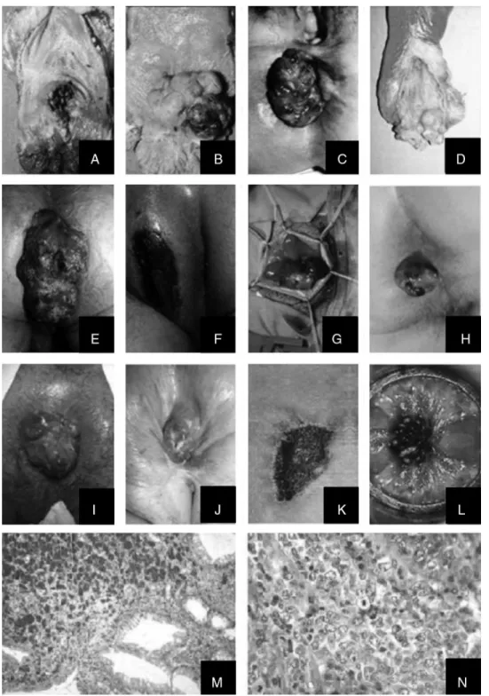
Fig. 1 – Illustrations of the 14 patients with ARMs: four surgical specimens of abdominoperineal resection (patients A, B, C and D), five patients submitted to local resection (patients E*, F, G, I, L), one with protective stoma (patient E*), one to extended local resection (patient H), one to relief colostomy (patient K), two to hemorrhoidectomy with the incidental finding of melanoma (patients M and N), and one who did not undergo surgery (patient J).
Methods
This is a historical series, given the rarity of the lesion, where the organization and method of the main investiga-tor were essential for the completion of the work. It was
only possible to carry it out because the necessary mate-rial was stored and cataloged, allowing new tests to be performed.
98
j coloproctol.2 0 1 4;3 4(2):95–103Table 1 – Main IHC markers used in the diagnosis of ARM and several other anorectal diseases, making the differential diagnosis between them.
ARM Paget Bowen Lymphoma UCA GIST LMS
ARM markers
S-100 protein S
HBM-45 S
Melan-A/MART-1 S
Vimetime S S
C117 (c-kit) S/N S/N S
PDGFRA S S
TU markers no ARM
CEA S
CK high MW (34BE12, AE1) S
CK low MW (35BE12, AE2, AE3) S
CD34 (QBEN-10) S
CD45 (PTPRC) S
CD68
Chromogranin A (CHGA) S
Synaptophysin (SYP) S
Desmin (DES) S S
Caldesmon (CALD-1) S
DOG-1 S
GIST, Gastrointestinal Stromal Tumor; UCA, undifferentiated carcinoma; TU, Tumor.
of patients, type of surgery, TNM classification and sur-vival/mortality data were collected from the sample.
The patients were submitted to the four basic types of surgery: local resection, extended local resection, colostomy derivation (alone or associated with local resection) or abdominoperineal resection of the rectum (APR).
Histopathological analysis was performed with H&E stain-ing (hematoxylin–eosin) in 5-mm slides. Cell type (epithelioid, spindle cell, lymphoma-like and pleomorphic); degree of melanin pigmentation (severe, moderate and focal, or absent); junctional activity under the squamous epithelium; melanocytic involvement of the anorectal junction; presence of abundant and eosinophilic cytoplasm; type of nuclei (round and vesicular); prominent eosinophilic nucleoli; presence of inflammatory infiltrate; perineural and vascular invasion; parietal invasion and mitotic index per microscopic field were characterized. New slides were made from the paraffin blocks of eight patients. In the remaining patients, the slides were reviewed (four) or only considered (two). All analyses were performed by the same pathologist.
Immunohistochemistry (IHC) was performed in 12 patients. In four cases IHC was performed only for ARM markers. In eight cases IHC was performed for ARM markers and other non-ARM anorectal tumors. The immunohisto-chemical study was not possible in only two patients. All examinations were performed by the same pathologist. Immunohistochemistry allows confirmation of melanoma and the differential diagnosis with other anorectal tumors. Table 1 shows the major markers studied.
All the materials used (clinical records, histopathology and immunohistochemistry reports, slides-12 patients and paraffin-9 patients); the free and informed consent forms signed by the four patients still alive and approval by the Research Ethics Committee of Santa Casa de Belo Horizonte
approving the project are in the possession of the main inves-tigator.
Results
Table 2 shows the sample descriptive data: age, gender, ASA classification of patients, type of surgery, TNM classification and survival/mortality. Age ranged from 44 to 81, with a mean age of 64.7 years. There were nine (64.3%) females and only two melanodermic patients (14.2%). The time of anorectal symp-toms varied between three and 12 months (85.7%) with a mean of 7.7 months. Only eight patients (57.1%) perceived the pres-ence of an anal tumor, but 92.2% complained of bleeding in stools and/or clothing. Most patients complained of symp-toms during evacuation (92.9%): anal pain (57.1%), sensation of incomplete evacuation (14.2%), tumor prolapse during evac-uation (21.4%). Fecal incontinence occurred in four (28.6%). Six (42.8%) had clinical symptoms of anemia and five patients (35.7%) had overall poor health status.
Comorbidities
According to the ASA – American Society of Anesthesiology – classification, six patients were ASA I, three were ASA II and five were ASA III. The comorbidities were hypertension in eight patients, diabetes mellitus in three, congestive heart failure in two, chronic obstructive pulmonary disease in one, lower-limb varicose disease in two, anemia in four and two patients were cachectic.
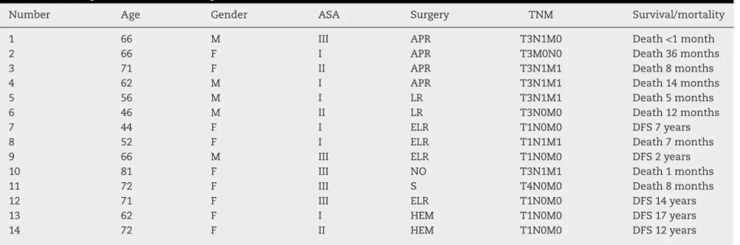
Table 2 – Sample characteristics – patients numbered from 1 to 14.
Number Age Gender ASA Surgery TNM Survival/mortality
1 66 M III APR T3N1M0 Death <1 month
2 66 F I APR T3M0N0 Death 36 months
3 71 F II APR T3N1M1 Death 8 months
4 62 M I APR T3N1M1 Death 14 months
5 56 M I LR T3N1M1 Death 5 months
6 46 M II LR T3N0M0 Death 12 months
7 44 F I ELR T1N0M0 DFS 7 years
8 52 F I ELR T1N1M1 Death 7 months
9 66 M III ELR T1N0M0 DFS 2 years
10 81 F III NO T3N1M1 Death 1 months
11 72 F III S T4N0M0 Death 8 months
12 71 F III ELR T1N0M0 DFS 14 years
13 62 F I HEM T1N0M0 DFS 17 years
14 72 F II HEM T1N0M0 DFS 12 years
M, male; F, female; ASA, American Society of Anesthesiology, classification; APR, abdominoperineal resection; LR, local resection; ELR, extended local resection; NO, not operated; S, palliative stoma; HEM, hemorrhoidectomy; TNM, tumor staging system; DFS, disease-free survival.
Proctologic examination and colonoscopy
The tumor was visible on inspection in eight (57.1%) patients. In 11 patients the tumor was palpable and the pectineal line was affected by the tumor in 13 (92.9%) cases. In 10 patients the tumor was anorectal, in three cases it was located in hemor-rhoidal papilla and in one case the tumor was entirely rectal. The mean size was 3.7 cm. Nine tumors were dark, one was light in color (amelanic) and two had dark and light mixed areas. In nine patients (64.3%) the tumor was fixed and in five mobile, of which one was pediculated and two found in hemorrhoidal disease. In these two patients the diagnosis was made after histological analysis of the hemorrhoidectomy specimen. The tumors were ulcerated in one case, vegetating in 10 cases and flat in one case, in addition to the two located in hemorrhoids.
Staging
With the exception of the two patients whose diagnosis was attained in hemorrhoidectomy specimens, all others under-went staging according to the tests available at the time of diagnosis. Imaging studies were most often performed.
ARM staging characteristics
1. Tumor location: in 10 cases (71.4%) the tumor was anorec-tal; in three cases (21.4%) it was located in hemorrhoidal papilla, of which two were casual findings during hemor-rhoidectomy, and in one case (7.1%) the tumor was rectal. 2. Anorectal wall thickening: present in five patients (35.7%),
absent in seven (50.0%) and HD in two cases (14.2%). 3. Lymph nodes: para-iliac lymph nodes were observed in six
patients (42.9%); inguinal chain lymph node in five patients (35.7%); paravertebral chain lymph nodes in four patients (28.6%), concomitantly. Invasion of the vagina and bladder was observed in one patient (7.1%).
4. Metastases: liver metastases were diagnosed in five patients (35.7%); lung metastases in four (28.6%), no bone metastases; subcutaneous metastases spread throughout
the body in two (14.2%). At the second visit, due to disease recurrence, pelvic-perineal recurrences, bone metastases and overall metastases occurred.
Surgeries performed, chemotherapy and radiotherapy, staging (TNM) of tumors, survival, disease-free interval and mortality
1. Surgeries performed: four patients underwent APR (28.6%), three underwent ELR with curative intent (21.4%), three underwent LR without curative intent (21.4%), two did not have the tumors resected (14.2%), and two underwent surgery for HD with HP examination showing ARM post-operatively (14.2%). Two were reoperated due to recurrent disease (14.2%). One was operated after the first surgery and underwent left lobe hepatectomy for single metastasis in the left lobe of the liver (7.1%).
2. Staging (TNM classification): S (stage)-I (To-T2, No/Nx, Mo), three patients; S-II (T3-T5, No/Nx, Mo), two patients; S-III (To-T5, N1, Mo), one patient; and S-IV (To-T5, No/Nx, M1), six patients.
3. Chemotherapy/radiotherapy: only one patient received CT and none received RT.
4. Survival, disease-free interval and mortality: of the 14 patients, nine died (64.3%) and five remain alive and disease-free (35.7%). For the nine patients who died, sur-vival ranged from one to 36 months, with a mean sursur-vival of 8.4 months. Disease-free survival ranged from 2 to 17 years in the 5 patients who are still alive (Table 2).
Histopathological analysis
1. Cell subgroups: pleomorphic cells were identified in two cases; all the remaining 12 patients (85.7%) had epithelioid cells concomitantly with spindle cells and “lymphoma-like” cells were not observed in any cases.
100
j coloproctol.2 0 1 4;3 4(2):95–103Table 3 – IHC test results of 14 cases of ARM.
Immunohistochemistry analysis (IHC)
Antigens associated to the melanoma Partial antigens Antigens not associated to the
melanoma–associated to other anorectal tumors
S-100 HMB-45 MART-1 Vimetina (V9) c-kit CEA AE-1 AE-2 ACT DES h-CAL SYN QBEN-10 CD10
NEP CD45 PTPRC
1 PD PD PD PD PF N N N N N N N N – N
2 PD PD PD P/N N N – – – – – – – – –
3 PD PD PD – –N N N N – – – – – – –
4 PD PF PD PD N N N N N N N N N – –
5 PD PD PD PD N N N N – – – – – – –
6 – – – – – – – – – – – – – –
7 PD PD PD PD N N N N – – – – – – –
8 – – – – – – – – – – – – – –
9 PD PD PD – N N N N – – – – – N –
10 PD PD PD – PF N N N – – – – – N –
11 – – – – – – – – – – – – – –
12 PD PD PD PD PF N N N – – – N – – N
13 – – – – – – – – – – – – – –
14 – – – – – – – – – – – – –
PD, Positive/Diffuse; PF, Positive/Focal; N, Negative; –, not performed.
3. Junctional activity under the squamous epithelium: it was not found in one case and it was not reported in five cases; in the remaining eight patients, junctional activity under the squamous epithelium was described (57.1%).
4. Elevated and Atypical Mitotic Index: except for one patient, all the other 13 patients had elevated and atypical mitotic index described (92.9%).
5. Perineural and Vascular Invasion: it was not found in three patients, not reported in three patients and it was found in eight patients, being only perineural in one patient, vascu-lar in two patients and perineural and vascuvascu-lar in the other five patients.
6. Parietal Invasion: could not be studied in five patients (three unresectable tumors and two inoperable ones). In the remaining nine patients: tumor invasion beyond the musculature in four patients; mucosal invasion only in two patients, up to the lamina propria in two and up to the submucosa even in one case.
7. Chain lymph nodes: studied in four cases in which radi-cal surgery was performed. Fourteen ganglia were found in two, all without metastases, and two had 18 ganglia, three with malignancy and 14 ganglia, 12 with metastases. 8. Other lymph nodes: ganglia found by imaging tests could
not be studied.
Immunohistochemical findings
Immunohistochemical (IHC) analyses were not performed in five of the 14 patients: in the first three because they were very old patients and IHC was not available, and in the last two due to the scarcity of material (hemorrhoidectomy). IHC analysis was performed in all other nine patients with S-100 protein and melanoma-associated antigens (HMB45 and Melan-A or MART-1). Vimentin (V9) was tested in six patients. The c-kit, CEA and cytokeratins (AE-1 and AE-2) were tested in the nine patients studied with S-100 protein and melanoma-associated
antigens. Actin, desmin, caldesmon-h and CD34 or QBEN-10 were tested in two patients; synaptophysin in three patients, CD10 or NEP was tested in two patients, and CD45 or PTPRC was tested in two patients (Table 3).
The tumor cells stained by markers and counterstained with Giemsa, showed strong positivity for Vimentin – V9 (100%) and for melanoma-associated antigens – S-100 protein (100%), HMB45 (100%) and MART-1 or Melan-A (100%). The c-kit showed focal positivity in three cases (33%), and CD-68 and Iron were positive in one case in which they were tested. They showed negativity for high molecular weight cytokeratins (AE1 and 34BE12); they were negative for low molecular weight cytokeratin (35BE12 and AE2); negativity for CEA, smooth mus-cle actin, Desmin, h-caldesmon, synaptophysin, CD34 (QBEN -10), CD-10 (NEP) and CD45 (PTPRC) in cases in which they were tested (Table 3 and Fig. 2).
Discussion
Anorectal melanomas (ARMs) are rare tumors. In a review of cancer cases in nine states, until 1993, Weinstock4found
an incidence of 1.7 ARM cases per million inhabitants. In other studies, the incidence ranged from 1 to 2% per million inhabitants.14,15
In this study we cannot assess the real impact of ARMs. Of the series of 14 cases, six were treated by the main investiga-tor, within a universe of 40,000 anorectal examinations (0.15% incidence) and among 973 cases of colorectal cancers (0.6% of them).
The highest incidence of ARM is between the sixth and eighth decades of life.3In a review of 126 cases of ARMs treated
from 1973 to 2001, Podnosy et al. reported a higher prevalence in women (61%), with a mean age of 69.2 years.16Weinstock4
Fig. 2 – IHC of the ARM of patient I (9) (counterstaining with Giemsa). (A) S-100 protein: positive result (enucleate
cytoplasmic staining, with evident brownish color), (B) Melan-A: positive result (brownish cytoplasmic staining), (C) HMB-45 positive result (brownish cytoplasmic staining with granular pattern, corresponding to melanosomes), (D) CK-AE1/AE3: negative result (the melanin pigment, originally brownish, acquires a greenish hue); (E) CD10: negative result (no brownish staining), (F) c-kit: negative result (absence of brownish staining).
among women (nine). Of the fourteen patients, twelve were white.
Slingluff, Collmer and Seigler, in a review of 24 ARM cases, identified rectal bleeding as the most common symptom, fol-lowed by anal pain, visible or palpable mass, pruritus ani, tenesmus, prolapse and change in bowel habits.17The
symp-toms of patients in this series coincide with this report, in addition to overall poor health status in five patients (35.7%).
Proctologic examination in this series showed that the tumor was visible (57.1%) or palpable (92.9%) in almost all cases. The mean size was 3.7 cm and in 12 patients the tumor was dark in color. These data show that the diagno-sis is easily suspected, although almost always delayed. In 13 patients the pectineal line was affected, which is consis-tent with the literature.18 Tumor staging showed stage I in
21.4% of patients (two incidentalomas found during hem-orrhoidectomy) and stage IV (42.9%) in most symptomatic patients. Lymph node metastasis was found in six patients and distant metastases in five (liver, lung and disseminated subcutaneous). These findings are variable in the literature,3,18
but corroborate the fact that the disease is severe at the diagnosis.
Chute et al.6 evaluated 17 cases of primary ARM with
special reference to histopathology and IHC. The morpho-logical characteristics evaluated microscopically included cell morphology, melanin pigmentation, junctional change and mitotic rate. Morphological subtypes of ARM were: epithe-lioid (12 cases), spindle-cell (seven cases), lymphoma-like (10 cases) and pleomorphic (six cases). Melanin pigmentation was present in nine cases; junctional change was present in six cases and mitotic rate was three or more per high-frequency microscopy field in eight cases.
These findings coincide with those of this series: as cell subgroups, in 12 patients (85.7%) the ARMs had epithelioid cells concomitantly with spindle cells; 10 patients (71.4%) had diffuse and intense melanin hyperpigmentation; junc-tional activity under the squamous epithelium was present in eight patients (57.1%) and the high and atypical mitotic index occurred in 13 of the 14 patients (92.9%). However, there is no correlation between the morphological characteristics described and prediction of survival.
As the differential diagnosis between amelanic ARM with epithelioid cells and other anorectal diseases (Paget’s disease, lymphoma, undifferentiated carcinoma and GIST) can be dif-ficult when considering only the histopathological criteria, IHC has become a very important resource to establish the correct differential diagnosis. Thus, after this test became available (after 1985), almost all studies mention perform-ing IHC for the diagnosis of ARM and differential diagnosis with other diseases. Almost all test the ARM panel of mark-ers (S-100 protein, Vimentin, Melan-A, HMB-45) and include specific markers such as cytokeratins (Paget’s disease), CD45 (lymphoma), chromogranin and synaptophysin (undifferen-tiated carcinoma), CD34 (GIST) and Desmin and caldesmon (sarcoma).19
In this series there was a strong positivity for Vimentin (100%), S-100 protein (100%), HMB45 (100%) and MART-1 or Melan-A (100%), consistent with the literature.19c-Kit showed
focal positivity in 33% and CD-68 and iron showed focal pos-itivity in the only case in which they were tested. As for the markers tested for differential diagnosis, they were all negative (cytokeratins, CEA, smooth muscle actin, Desmin, h-caldesmon, synaptophysin, CD34, CD10 and CD45).
None of these markers were predictive of survival.6
102
j coloproctol.2 0 1 4;3 4(2):95–103(Proliferating Cell Nuclear Antigen), which were associated with advantage in survival – patients with high Ki67 and low PCNA scores. These data have not been validated by subse-quent studies.20
As for surgery, there is a consensus that the disease is severe, with poor results and there is no difference in survival of patients undergoing local excision or extended surgery, such as abdominoperineal resection of the rectum. Consid-ering the risks of major surgery and the inconvenience of a definitive stoma, with significant impact on quality of life, the choice of most authors is the extended local excision, reserv-ing APR for advanced cases or rescue in case of recurrence after local resection.9,21–23
Four of the 14 patients in this series underwent APR due to invasive ARMs, as ELR was not feasible. Three patients underwent ELR with curative intent (21.4%), as they had local-ized ARMs. Three patients underwent ELR without curative intent (21.4%), as APR was not possible due to patient over-all status. Two patients did not have their tumors resected (14.2%), as they were not fit to undergo the surgical procedure and were considered inoperable. And two patients underwent surgery for hemorrhoidal disease, with the histopathologi-cal analysis disclosing the presence of ARM postoperatively (14.2%).
Of the 14 patients studied, nine died and five remain alive and disease-free. The mean survival time was 8.4 months. In the literature, the overall survival varies among authors, but there is consensus that it depends on the surgical technique used (extended local resection or APR).9,22–23
Only one case series mentions the incidental finding of ARM in an eventual anal surgery. In eight of the 50 patients with ARM, in the report of Thibault et al., ARM was found incidentally during anal surgery (16.0%), and five of them were reoperated, two with APR and three with ELR, with no tumor trace in the surgical specimen at the microscopy.
The findings of these authors are similar to findings in this series of 14 ARMs: two were ARM cases incidentally found during hemorrhoidectomy (14.2%). However, none of them required reoperation, and both are alive – one 17 years after surgery and the other 12 years after surgery. Therefore, it can be concluded that these small tumors were cured with local resection only.
Radiotherapy and chemotherapy are considered ineffec-tive for treatment of ARM. Some authors who have used RXT reported that they did not observe any advantage.3,24In
this study, only one patient received chemotherapy and none received RXT. The use of targeted therapies against metastatic melanoma was considered frustrating by Satzger et al.11
How-ever, knowledge of the different processes of oncogenesis of melanomas can lead to more precise therapies. One of the sub-groups of ARM, with mutations in the BRAF gene, respond to the action of BRAF inhibitors (PLX4032 and RAF265), leading to regression of disease in up to 70% of patients with metastatic melanoma with BRAF V600E mutation.25
Another subgroup of ARM, with KIT gene mutation, is sen-sitive to c-KIT (imatinib) blocker action.25 Other blockers of
c-KIT have been studied in ARMs, however, in a very small number of patients, which prevents drawing an immediate conclusion.
Conclusion
ARMs are tumors with high malignant potential, and their rar-ity makes it difficult to establish diagnostic and therapeutic procedures with statistically significant results. Histopatho-logy does not always confirm the diagnosis and IHC is essential for attaining a definitive diagnosis.
The most rational surgical approach is the ELR in cases of localized ARMs and APR for advanced cases of the disease. Regardless of the surgical technique used, the overall survival is very low.
There is no outcome improvement with CXR and CT. The use of targeted therapies is starting to show encouraging results for some subtypes (with mutations in BRAF and/or KIT genes).
Conflicts of interest
The authors declare no conflicts of interest.
Acknowledgements
To the assistants of the Coloproctology Unit of Santa Casa de Belo Horizonte, during whose meetings the cases showed here were discussed and strategies were drawn, with several of them having participated in some of the surgeries.
r e f e r e n c e s
1. Moore WD. Recurrent melanosis of the rectum after previous removal from the verge of the anus in a man aged sixty-five. Lancet. 1897;1:290–4.
2. Chang AE, Karnell LH, Menck HR, The American College of Surgeons Commission on Cancer and the American Cancer Society. The National Cancer Data Base report on cutaneous and noncutaneous melanoma: a summary of 84,836 cases from the past decade. Cancer. 1998;83:1664–78.
3. Thibault C, Sagar P, Nivatvongs S, et al. Anorectal melanoma – an incurable disease? Dis Colon Rectum. 1997;40:661–8. 4. Weinstock MA. Epidemiology and prognosis of anorectal
melanoma. Gastroenterology. 1993;104:174–8.
5. Abbas JS, Karakousis CP, Holyoke ED. Anorectal melanoma: clinical features, recurrence and patient survival. Int Surg. 1980;65:423–6.
6. Chute DJ, Cousar JB, Mills SE. Anorectal malignant melanoma – morphologic and immunohistochemical features. Am J Clin Pathol. 2006;126:93–100.
7. Droesch JT, Flum DR, Mann GN. Wide local excision or abdominoperineal resection as the initial treatment for anorectal melanoma? Am J Surg. 2005;189:446–9.
8. Ward MW, Romano G, Nicholls RJ. The surgical treatment of anorectal malignant melanoma. Br J Surg. 1986;73:68–9. 9. Brady MS, Kavolius JP, Quan SH. Anorectal melanoma. A
64-year experience at Memorial Sloan-Kettering Cancer Center. Dis Colon Rectum. 1995;38:146–51.
10. Moss RW. The Moss reports for cancer patients. A sample from the Moss reports: treatment of anorectal melanoma. J Surg Oncol. 1995;58:118.
12. Hersey P, Bastholt L, Chiarion-Sileni V, et al. Small molecules and targeted therapies in distant metastatic disease. Ann Oncol. 2009;20 Suppl. 6:vi35–40.
13. Batus M, Waheed S, Ruby C, et al. Optimal management of metastatic melanoma: current strategies and future directions. Am J Clin Dermatol. 2013;14:179–94,
http://dx.doi.org/10.1007/s40257-013-0025-9. PMID: 23677693 [PubMed – in process].
14. Goldman S, Glimelius B, Pahlman L. Anorectal malignant melanoma in Sweden: report of 49 patients. Dis Colon Rectum. 1990;33:874–7.
15. Rossetti C, Koukouras D, Eboli M, et al. Primary anorectal melanomas: an institutional experience. J Exp Clin Cancer Res. 1997;16:81–5.
16. Podnosy YD, Tsai NC, Smith D, et al. Factors affecting survival in patients with anal melanoma. Am Surg. 2006;72:917–20. 17. Slingluff C, Collmer R, Seigler H. Anorectal melanoma:
clinical characteristics and results of surgical management in twenty-four patients. Surgery. 1990;107:1–9.
18. Konstadoulakis MM, Ricaniadis N, Walsh D. Malignant melanoma of the anorectal region. J Surg Oncol. 1995;58:118–20.
19. Canales JBL, Blesa JMG. Amelanotic anorectal malignant melanoma: case report with immunohistochemical study and literature review. Case Rep Oncol. 2009;2: 30–5.
20. Ben-Izhak O, Bar-Chana M, Sussman L, et al. Ki67 antigen and PCNA proliferation markers predict survival in anorectal malignant melanoma. Histopathology. 2002;41:519–25.
21. Das G, Gupta S, Shukla PJ, et al. Anorectal melanoma: a large clinicopathologic study from India. Int Surg. 2003;88:21–4.
22. Chiu YS, Unni KK, Beart RW. Malignant melanoma of the anorectum. Dis Colon Rectum. 1980;23:122–4.
23. Che X, Zhao DB, Wu YK, et al. Anorectal malignant melanomas: retrospective experience with surgical management. World J Gastroenterol. 2011;17:534–9. 24. Moozar KL, Wong CS, Couture J. Anorectal malignant
melanoma: treatment with surgery or radiation therapy, or both. Can J Surg. 2003;46:345–9.