Original article
osteoporosis: is there a difference?
Controle postural de idosas com e sem osteoporose: há diferenças?
Thomaz Nogueira Burke
I, Fabio Jorge Renovato França
I, Sarah Rúbia Ferreira de Meneses
II, Viviam Inhasz Cardoso
II,
Rosa Maria Rodrigues Pereira
III, Camille Figueredo Danilevicius
IV, Amélia Pasqual Marques
VPhysical Therapy and Electromyography Laboratory, School of Medicine, Universidade de São Paulo (USP), São Paulo, Brazil
IMSc student. Department of Physical Therapy, Speech and Occupational Therapy, Universidade de São Paulo (USP), São Paulo, Brazil. IIStudent, Department of Physical Therapy, Speech Therapy and Occupational Therapy, Universidade de São Paulo (USP), São Paulo, Brazil. IIIMD, PhD. Associate professor, Division of Rheumatology, School of Medicine, Universidade de São Paulo (USP), São Paulo, Brazil. IVMD. Rheumatologist, Division of Rheumatology, School of Medicine, Universidade de São Paulo (USP), São Paulo, Brazil.
ABSTRACT
CONTEXT AND OBJECTIVE: Little is known about postural control among elderly individuals with osteoporosis and its relationship with falls. It has been suggested that elderly women with kyphosis and osteoporosis are at greater risk of falling. The aim of this study was to evaluate posture and postural
control among elderly women with and without osteoporosis.
DESIGN AND SETTING: Cross-sectional study conducted at the Physical Therapy and Electromyography Laboratory, School of Medicine, Universidade de São Paulo (USP).
METHODS: Sixty-six elderly women were selected from the bone metabolism disorders clinic, Division of Rheumatology, USP, and were divided into two groups: osteoporosis and controls, according to their bone mineral density (BMD). Postural control was assessed using the Limits of Stability (LOS) test and the Modiied Clinical Test of Sensory Interaction and Balance (CTSIBm) and posture, using photometry.
RESULTS: The elderly women with osteoporosis swayed at higher velocity on a stable surface with opened eyes (0.30 versus 0.20 degrees/second; P = 0.038). In both groups, the center of pressure (COP) was at 30% in the LOS, but with different placements: 156° in the osteoporosis group and
178° in the controls (P = 0.045). Osteoporosis patients fell more than controls did (1.0 versus 0.0; P = 0.036).
CONCLUSIONS: The postural control in elderly women with osteoporosis differed from that of the controls, with higher sway velocity and maximum displacement of COP. Despite postural abnormalities such as hyperkyphosis and forward head, the COP position was posteriorized.
RESUMO
CONTEXTO E OBJETIVO: Pouco se sabe sobre o controle postural de idosos com osteoporose e sua relação com as quedas. Foi sugerido que idosas cifóticas com osteoporose têm maior risco de quedas. Esta pesquisa teve como objetivo avaliar o controle postural e a postura em idosas com e sem osteoporose.
TIPO DE ESTUDO E LOCAL: Estudo transversal realizado no Laboratório de Avaliação Fisioterapêutica e Eletromiograia da Faculdade de Medicina da Universidade de São Paulo (USP).
MÉTODOS: Sessenta e seis mulheres idosas foram selecionadas da Clínica de Doenças Osteometabólicas da Divisão de Reumatologia da Universidade de São Paulo e divididas em dois grupos: osteoporose e controle, de acordo com a densidade mineral óssea (DMO). Foi avaliado o
controle postural pelos testes Limite de Estabilidade (LOS) e Modiied Clinical Test of Sensory Interaction and Balance (CTSIBm) e a postura pela fotometria.
RESULTADOS: As idosas com osteoporose oscilaram com maior velocidade em superfície irme com olhos abertos (0,30 x 0,20 graus/segundo, P = 0,038). O COP (centro de pressão) de ambos os grupos encontrava-se a 30% do LOS, porém com posicionamentos distintos: 156° no grupo
osteoporose e 178° no grupo controle (P = 0,045). As osteoporóticas caíram com maior frequência em comparação aos controles (1,0 x 0,0, P = 0,036).
CONCLUSÃO: O controle postural de idosas com osteoporose diferiu dos controles, com maior velocidade de oscilação e máximo deslocamento do COP, e que apesar da presença de alterações posturais como hipercifose e anteriorização de cabeça, o COP se encontrou posteriorizado. Key words:
Aged. Osteoporosis.
Postural balance. Posture.
Kyphosis.
Palavras-chave: Idoso.
Osteoporose. Equilíbrio postural.
Postura. Cifose.
INTRODUCTION
Osteoporosis is a common disorder characterized by reduced bone mass and by deterioration of the microarchitecture of the bone tissues, thereby leading to increased bone fragility.1 It afects around 55% of
the population over the age of 50 years in the United States.2
Postural control is the inherent ability to maintain the center of mass on a supporting base, between stability limits. hese limits are the operational areas up to which the center of mass can be displaced without the need to change the supporting base.3 hus, balance
limits.4 In order to avoid falling, the center of body mass must be kept
within the supporting base or, even better, within the stability limits. It has been suggested that elderly people present reduced ability to control their posture, which may predispose them to increased risk of falling.5 According to Jonson,6 age-related deterioration of balance or
postural control has a negative impact on the ability to safely carry out day-to-day activities.
Among the likely causes of postural instability among the elderly, changes in the relationship between sensory information and motor ac-tion are of importance. he elderly have greater diiculty in interpret-ing sensory information and prioritizinterpret-ing it accordinterpret-ing to its relevance, and in selecting the proper response in order to maintain their balance in speciic positions.7
Little is known about postural control among elderly individuals with osteoporosis and its relationship with falls. Lynn8 suggested that
elderly women with kyphosis and osteoporosis were at greater risk of falling. his author suggested that the changes to the body caused by osteoporosis would displace the center of pressure (COP) closer to the limit of stability, thereby making it easier to lose balance, with conse-quent falls. However, Lynn’s study only assessed six women with osteo-porosis and ive controls, and the participants difered in age substan-tially (ranging from 52 to 85 years). Furthermore, although that study suggested that thoracic hyperkyphosis and forward head position gave rise to falls, these parameters were not objectively and quantitatively as-sessed.
Studying osteoporosis among the elderly is particularly important, since this group is at greater risk of developing fractures and comorbidi-ties associated with falls. It has been estimated that for every decrease of one standard deviation of bone mineral density (BMD) in the head of the femur, there is a proportional 2.6-fold increase in the risk of frac-tures in the hip.9
OBJECTIVE
Since few studies have measured postural control among elderly in-dividuals with osteoporosis,8,10 and these studies did not measure the
COP quantitatively, our aim here was to conduct a study to investi-gate postural control and posture among elderly women with and with-out osteoporosis. We hypothesized that women with osteoporosis would present diminished postural control and an anteriorly shifted COP, caused by postural abnormalities such as hyperkyphosis and forward head, in relation to elderly individuals without osteoporosis.
METHODS
Sample characteristics
Our sample consisted of 66 women with ages ranging from 66 to 81 years. hey were recruited from the bone metabolism diseases outpa-tient clinic of the Division of Rheumatology, Universidade de São Pau-lo (USP). he participants were divided into two groups, according to their BMD: the osteoporosis group (n = 46) presented BMD that was at least 2.5 standard deviations (SD) lower than the standard values for young adults, in relation to the lumbar spine, femoral neck and total femur regions.11 he control group (n = 20) presented BMD that was
above -2.5 SD in relation to the same areas.
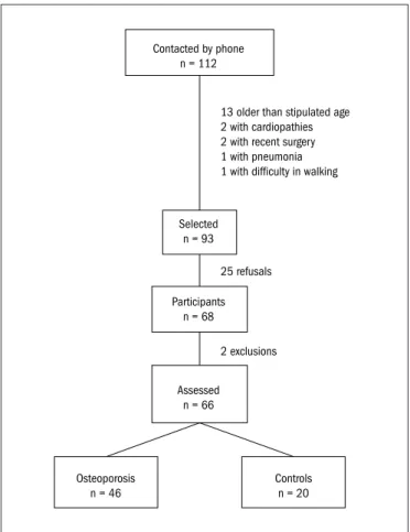
Patients were excluded if they presented signiicant visual impair-ment; inability to walk more than 10 meters without assistance; neu-rological or musculoskeletal diseases (e.g. Parkinson’s disease, stroke or neurodegenerative disorders); or amputations and prostheses for the arms or legs. Figure 1 displays the low of our study. he participants signed an informed consent form. he study and consent forms were approved by the Ethics Committee of Hospital das Clínicas (HC) of the USP School of Medicine.
Measurements
History of falls
A blinded investigator (i.e. blinded to group status) applied a questionnaire in order to obtain information on age, weight, height and history of falls over the past year. Falls were deined as non-inten-tional contact of hands, arms, chest or hips with the loor, after los-ing balance.
Postural control
Postural control was assessed using the Modiied Clinical Test of Sensory Interaction and Balance (CTSIBm) and the 100% Limits of Stability (LOS) test. he CTSIBm measures static equilibrium under four sensory conditions: stable surface and opened eyes (SS-OE); stable
Contacted by phone n = 112
13 older than stipulated age 2 with cardiopathies 2 with recent surgery 1 with pneumonia 1 with dificulty in walking
Selected n = 93
25 refusals
Participants n = 68
2 exclusions
Assessed n = 66
Osteoporosis n = 46
Controls n = 20
surface and closed eyes (SS-CE); unstable surface and opened eyes (US-OE); and unstable surface and closed eyes (US-CE).
In order to pick up the COP sway velocity (degrees per second), we used a force platform (model: NeuroCom Balance Master). he COP indicated the location of the vector resulting from the reaction force applied on the ground, as measured by the force platform. his vec-tor is the same as (but opposite in direction to) the weighted mean of all the forces acting on the force platform, such as weight and the in-ternal forces (from the muscles and joints) that are transmitted to the ground.12 Each experiment was repeated three times, for 10 seconds; we
have presented the means from the experiments. While performing the CTSIBm, we also measured the position of the COP in relation to the center of the ellipse in the LOS test. he results have been presented as percentages of LOS and in degrees.
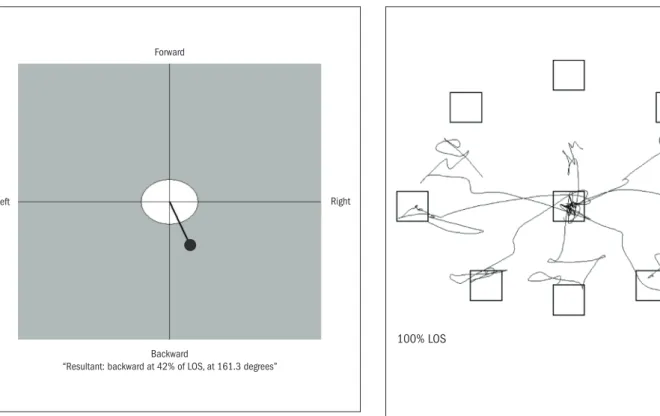
During the 100% LOS test, the participants had to reach out to touch eight diferent targets that were distributed symmetrically around a central point that represented the maximum distance (theoretical LOS) through which the subjects would be able to shift their COP without losing balance (Figure 2), and without moving their feet ( Fig-ure 3). We deined the variable of maximum excursion as the greatest distance reached out towards the targets by the center of gravity at any point during the attempts. his was expressed as a percentage of the the-oretical LOS.13 We decided to merge all shifts in the anterior-posterior
and side-to-side directions.
For the CTSIBm and 100% LOS tests, the participants remained in the orthostatic position, with arms extended along the sides of the body, without wearing shoes. hey were placed in one of the three standard positions recommended by the manufacturer.
Posture
Posture was assessed by means of photometry, which consisted of capturing images using a digital camera for subsequent analysis of ana-tomical points that had previously been marked out. We used a specii-cally designed postural assessment software: Software de Avaliação Pos-tural (SAPO), available at www.sapo.incubadora.fapesp.br.14
Anatomical points were marked on the skin using markers of 15 mm in diameter. Photographs were taken with the individuals mini-mally dressed, such that it was possible to view the following anatomical points: tragus of the ear, 7th cervical vertebra, 12th thoracic vertebra and
midpoint of the acromion. From analyzing the points and their rela-tionships, we made measurements relating to the conditions of forward head and thoracic kyphosis.
he head position was determined in terms of the angle between the vertical line passing through the midpoint of the acromion and the line drawn between the midpoint of the acromion and the tragus.15 Positive
values signiied that the tragus was advanced in relation to the acromion and, therefore, a forward head position; negative values indicated retrac-tion of the head.
he measurements of thoracic kyphosis were based on the kyphosis index, as assessed using the lexicurve method proposed by Takahashi and Atsumi,16 with the modiications suggested by Teixeira and
Carval-ho.17 he results were presented in degrees. his method presents high
intraclass correlation (ICC = 0.906), in comparison with kyphosis mea-surements using Cobb’s angle.17 horacic hyperkyphosis was diagnosed
when the angles were greater than 50º, as described by Wilner.18
In order to be photographed, the participants remained in the or-thostatic position, with their feet in the position recommended by the
Forward
Backward
“Resultant: backward at 42% of LOS, at 161.3 degrees”
Right Left
Figure 2. Example of positioning the center of pressure (COP) in relation to the center of the ellipse of the limits of stability.
100% LOS
Figure 3. Example of the 100% limits of stability test.
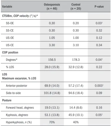
Table 2. Postural control among individuals with osteoporosis and in controls
Variable Osteoporosis
(n = 46)
Control
(n = 20) P-value
CTSIBm,COP velocity (°/s)*
SS-OE 0.30 0.20 0.03†
SS-CE 0.30 0.30 0.32
US-OE 1.05 1.00 0.12
US-CE 3.30 3.10 0.34
COP position
Degrees* 156.5 178.3 0.04†
% LOS 28.0 (15.9) 32.9 (12.8) 0.22
LOS
Maximum excursion, % LOS
Anterior-posterior 69.9 (14.0) 57.2 (17.4) 0.003†
Side-to-side 101.8 (14.8) 94.6 (18.4) 0.09
Posture
Forward head, degrees 19.0 (13.1) 14.4 (8.6) 0.16 Kyphosis, degrees 53.1 (13.8) 45.9 (10.1) 0.05†
Hyperkyphosis, n (%) 70% 40%
*Median values; SS-OE = stable surface and opened eyes; †Statistically signiicant difference between the
groups (P < 0.05); SS-CE = stable surface and closed eyes; US-OE = unstable surface and opened eyes; US-CE = unstable surface and closed eyes.
Statistical analyses
he data were compared using the SigmaStata 3.5 software. he sample size was determined by taking the power to be 80% and the standard deviation and expected diference in means to be 20%, with α
= 0.05. hus, a minimum number of 17 subjects per group was deter-mined. We used the t test for parametric variables such as age, weight, body mass index (BMI), BMD (total femur), COP position (%LOS) and maximum COP excursion COP. he results were presented as means and standard deviations. For non-parametric variables, such as BMD (lumbar spine), COP location (degrees), falls and COP velocity, we used the Mann-Whitney test, and medians were presented. he sig-niicance level was established as 5% (α = 0.05), with 95% conidence intervals.
RESULTS
he anthropometric and demographic characteristics of the 66 in-dividuals are described in Table 1. he participants were non-institu-tionalized elderly women (age range: 66-80 years), who were able to walk independently. he two groups were similar in terms of age. he individuals with osteoporosis presented signiicantly lower weight, and height, in comparison with the controls. he individuals in the osteopo-rosis group reported signiicantly more falls than did those in the con-trol group (P = 0.036).
In the osteoporosis group, 70% of the individuals had thoracic hy-perkyphosis, versus 40% among the controls. Table 2 displays the values for postural control among the individuals with and without osteopo-rosis. he individuals with osteoporosis presented higher sway velocities under all four conditions tested by the CTSIBm. However, these dif-ferences only reached signiicance in the tests on a stable surface with opened eyes (P = 0.038). In the LOS test, the individuals with osteo-porosis presented signiicantly higher amplitudes of displacement than observed among the controls, when asked to reach their LOS. he dif-ference was signiicant when the displacement was in the anterior-pos-terior direction, but not in the side-to-side direction. he COP was placed at the same percentage as the LOS, but with diferent angula-tion (P = 0.045).
DISCUSSION
he aim of this study was to assess posture and postural control among women with and without osteoporosis. he elderly women with osteoporosis presented higher sway velocity and higher numbers of falls, in comparison with the controls. When asked to shift their COP closer to their stability limits, the women with osteoporosis had higher sway amplitudes in the anterior-posterior direction, in comparison with the controls. Furthermore, among the women with osteoporosis, the COP was displaced posteriorly and in the right lateral direction; among the controls, the COP was displaced posteriorly. Both groups showed for-ward head and thoracic hyperkyphosis: the proportions were diferent (osteoporosis group = 70%; controls = 40%), but this diference was not signiicant.
manufacturer of the equipment. To facilitate this procedure, we manu-factured a rug with the same dimensions and marks as on the force plat-form. he camera was positioned at a distance of two meters from the participants, and at an elevation of one meter above ground level, in the horizontal plane, i.e. parallel to the plane that was to be observed.19 A
ribbon guide was placed on the individual, in the same plane. All the images were taken with the camera, tripod, ribbon guide and patient in the same positions. We used a Sony Cybershot P-92 digital camera, and images were transferred to the postural analysis software (SAPO) to make the assessments of interest.
Table 1. Anthropometric and demographic characteristics of individuals with osteoporosis and controls
Variable Osteoporosis
(n = 46)
Control
(n = 20) P-value
Age, years 73.0 (4.2) 71.9 (3.2) 0.262 Weight, kg 57.7 (8.6) 68.4 (7.3) 0.000 Height, m 1.50 (0.06) 1.55 (0.06) 0.003 BMI, kg/m2 25.6 (3.4) 28.3 (2.4) 0.002
BMD, T-score*
Lumbar spine -3.5 -1.0 0.000 Total femur -2.1 (0.6) -0.4 (0.8) 0.000 Falls/patient†, n 1.0 0.0 0.036
*BMD = bone mineral density, as compared with individuals with maximum bone density; †preceding year;.
According to Hageman et al.,20 postural sway in the vertical
posi-tion seems to increase with age, and this is prompted when the support base is modiied (through decreasing its size or changing its surface to a foam surface), the body coniguration is changed (standing on one foot) or the visual input is changed. his happens because postural control depends on harmony between the visual, vestibular, proprioceptive and musculoskeletal systems.21
We found that the sway velocity among the women with osteopo-rosis was 16.7% higher than among the controls. Our indings are in agreement with those of Liu-Ambrose et al.22, who found balance scores
that were 11% lower among women with osteoporosis. Considering the four diferent conditions of our test, the greatest diference was seen in relation to the stable surface and opened eyes, in which the women with osteoporosis swayed at a velocity that was 50% greater than shown by the controls. Under the remaining conditions (stable surface with closed eyes and unstable surface with opened or closed eyes), the women with osteoporosis also swayed at higher velocity, but the diferences were not signiicant.
It is important to emphasize that greater diferences between the groups were seen when the eyes were opened. his may relect a “ceiling efect”, caused by the additional diiculties when performing the tasks with closed eyes (reduced discriminatory properties of the test). In situ-ations of increased diiculty (e.g. eyes closed or unstable surfaces), the diferences decreased and both groups swayed at a higher velocity.
Our data suggest that the individuals with osteoporosis displaced the COP with a higher amplitude in the anterior-posterior direction, in comparison with the controls. his inding was unexpected, since vol-untarily shifting the COP towards the stability limit depends on lower-limb muscle strength, along with the trust that individuals have in their own ability to move. Studies have suggested that individuals with osteo-porosis present reduced strength and greater fear of falling.3,23,24
Speciically focusing on our indings, it may be that the lower weight (8.1 kg lower) and BMI (3.3 kg/m2 lower) relative to the
con-trols, which is characteristic of individuals with osteoporosis,25,26 may
have had an inluence. Weight may have negatively inluenced the abil-ity to displace the COP. his behavior was less evident in the side-to-side direction. Era et al.27 described an association between low BMI
and deicient balance among elderly individuals, thus supporting our indings.
Melzer et al.5 suggested that decreased postural control may be a risk
factor for falls among the elderly, and that the impairment is probably caused by conlicting sensory-motor inputs. hrough such conlicts, el-derly people would have greater diiculty in identifying the most rel-evant sensory information, and in developing the proper postural reac-tion to maintain their balance in the desired posireac-tion.7
Both groups presented forward head posture and increased thoracic kyphosis, which are typical of the elderly. According to Lynn et al.,8 a
kyphotic posture displaces the center of gravity towards the anterior sta-bility limit, thereby requiring increased efort in order to maintain bal-ance, even after minor changes. In this regard, our study yielded contra-dictory indings, since both groups presented the COP displaced poste-riorly and to the right, at 30% of the LOS. Among the individuals with
osteoporosis, it was displaced to the right (156º), while among the con-trols, it was displaced posteriorly (178º). It may be that displacement of body structures caused simply by a kyphotic posture and by a forward head posture are insuicient to disrupt balance, and to shift the COP towards the stability limit.
he only two postural parameters that we assessed were forward head and thoracic kyphosis. However, compensatory displacements of other structures, such as hip antepulsion and trunk extension, may have inluenced the inal position of the COP, since it is determined not only by the sum of the segmental weights but also by their spatial positions.10
Our indings support the concept that posture should not be analyzed segmentally, but in an overall manner. It is important to observe the re-lationships between all segments in the body, since compensatory dis-placements inluence the COP and, therefore, body equilibrium. Ac-cordingly, future larger studies with overall assessment of posture are need in order to conirm or refute our indings.
CONCLUSIONS
Our study suggests that postural control among individuals with osteoporosis is diferent from postural control among the general elder-ly population. Individuals with osteoporosis are more likeelder-ly to present higher sway velocities and greater maximum shift of the COP. Despite postural abnormalities such as forward head and kyphosis, the COP is located posteriorly. Better understanding of the determinants of the COP among elderly individuals with osteoporosis, as well as of its rel-evance in relation to causing falls, is of importance in order to develop preventive strategies with the aims of improving quality of life and re-ducing comorbidities among the elderly.
REFERENCES
1. Versluis RG, Papapoulos SE, de Bock GH, et al. Clinical risk factors as predictors of postme-nopausal osteoporosis in general practice. Br J Gen Pract. 2001;51(471):806-10. 2. Kuczyński M, Ostrowska B. Understanding falls in osteoporosis: the viscoelastic modeling
perspective. Gait Posture. 2006;23(1):51-8
3. Alexander NB. Postural control in older adults. J Am Geriatr Soc. 1994;42(1):93-108. 4. Konrad HR, Girardi M, Helfert R. Balance and aging. Laryngoscope. 1999;109(9):
1454-60.
5. Melzer I, Benjuya N, Kaplanski J. Postural stability in the elderly: a comparison between fallers and non-fallers. Age Ageing. 2004;33(6):602-7.
6. Jonson E. Effects of healthy aging on balance: a quantitative analysis of clinical tests [the-sis]. Stockholm: Karolinska Institutet; 2006. Available from: http://diss.kib.ki.se/2006/91-7140-633-6/thesis.pdf. Accessed in 2010 (Jun 18).
7. Freitas Júnior P, Barela JA. Alterações no funcionamento do sistema de controle postural de idosos. Uso da informação visual. Rev Port Cien Desp. 2006;6(1):94-105.
8. Lynn SG, Sinaki M, Westerlind KC. Balance characteristics of persons with osteoporosis. Arch Phys Med Rehabil. 1997;78(3):273-7.
9. Cummings SR, Black DM, Nevitt MC, et al. Bone density at various sites for predic-tion of hip fractures. The Study of Osteoporotic Fractures Research Group. Lancet. 1993;341(8837):72-5.
10. Wendlova J. Determination of the centre of gravity in the methodology of kinesiotherapy for osteoporotic patients. Bratisl Lek Listy. 2008;109(5):231-7.
11. World Health Organization. Assessment of osteoporotic fracture risk and its role in screening for menopausal osteoporosis. Geneva: WHO Technical Report Series; 1994.
12. Winter DA. Biomechanics and motor control of human movement. 2nd ed. USA: John Wiley
13. Boulgarides LK, McGinty SM, Willett JA, Barnes CW. Use of clinical and impaired-based tests to predict falls by community-dwelling older adults. Phys Ther. 2003;83(4):328-39. 14. Ferreira EAG. Postura e controle postural: desenvolvimento e aplicação de método
quanti-tativo de avaliação postural [Posture and postural control: development and application of a quantitative method for postural evaluation]. [thesis]. São Paulo: Faculdade de Medicina da Universidade de São Paulo; 2005.
15. Kendall FP, McCreary EK, Provance PG. Postura: alinhamento e equilíbrio muscular. In: Ken-dall FP, McCreary EK, Provance PG, editores. Músculos provas e funções. 4a ed. São Paulo:
Manole; 1995. p. 69-118.
16. Takahashi E, Atsumi H. Age differences in thoracic form as indicated by thoracic index. Hum Biol. 1955;27(2):65-74.
17. Teixeira FA, Carvalho GA. Coniabilidade e validade das medidas da cifose torácica através do método lexicurva [Reliability and validity of thoracic kyphosis measurements using le-xicurve method]. Rev Bras Fisioter. 2007;11(3):199-204.
18. Willner S. Spinal pantograph - a non-invasive technique for describing kyphosis and lordosis in the thoraco-lumbar spine. Acta Orthop Scand. 1981;52(5):525-9.
19. Munhoz WC, Marques AP, Siqueira JT. Evaluation of body posture in individuals with internal temporomandibular joint derangement. Cranio. 2005;23(4):269-77.
20. Hageman PA, Leibowitz JM, Blanke D. Age and gender effects on postural control measures. Arch Phys Med Rehabil. 1995;76(10):961-5.
21. Berg KO, Maki BE, Williams JI, Holliday PJ, Wood-Dauphinee SL. Clinical and labora-tory measures of postural balance in an elderly population. Arch Phys Med Rehabil. 1992;73(11):1073-80.
22. Liu-Ambrose T, Eng JJ, Khan KM, Carter ND, McKay HA. Older women with osteoporosis have increased postural sway and weaker quadriceps strength than counterparts with nor-mal bone mass: overlooked determinants of fracture risk? J Gerontol A Biol Sci Med Sci. 2003;58(9):M862-6.
23. Granito RN. Efeitos do envelhecimento e da osteoporose na cifose torácica, na propriocep-ção e no torque dos músculos do tronco [dissertation]. São Carlos: Universidade Federal de São Carlos; 2005.
24. Cook DJ, Guyatt GH, Adachi JD, et al. Quality of life issues in women with vertebral fractures due to osteoporosis. Arthritis Rheum. 1993;36(6):750-6.
25. Marone MM, Gouveia CH, Lewin S, et al. Inluence of body composition on the bone mass of postmenopausal women. Sao Paulo Med J. 1997;115(6):1580-8.
26. Ramalho AC, Lazaretti-Castro M, Hauache O, et al. Osteoporotic fractures of proximal femur: clinical and epidemiological features in a population of the city of São Paulo. Sao Paulo Med J. 2001;119(2):48-53.
27. Era P, Schroll M, Ytting H, et al. Postural balance and its sensory-motor correlates in 75-year-old men and women: a cross-national comparative study. J Gerontol A Biol Sci Med Sci. 1996;51(2):M53-63.
Sources of funding: This work was supported by Fundação de Amparo à Pesquisa do Estado de São Paulo (Fapesp); grant number 2007/01611-2
Conlict of interest: None
Date of irst submission: December 11, 2009 Last received: March 16, 2010
Accepted: June 23, 2010
Address for correspondence: Amélia Pasqual Marques
Foito - Departamento de Fisioterapia, Fonoaudiologia e Terapia Ocupacional da Faculdade de Medicina da Universidade de São Paulo
Rua Cipotânea, 51
Cidade Universitária — São Paulo — Brasil CEP 05360-160