Plasma clearance and biodistribution
of oxidatively modified
99m
Tc-ß-VLDL
in rabbits
1Departamento de Análises Clínicas e Toxicológicas,
Faculdade de Ciências Farmacêuticas, Universidade de São Paulo, 05508-900 São Paulo, SP, Brasil
2Instituto do Coração (INCOR), Faculdade de Medicina, Universidade de São Paulo, 05403-000 São Paulo, SP, Brasil E.L. Silva1,
J.C. Meneghetti2, I.J.C. Coelho2 and D.S.P. Abdalla1
Abstract
The biodistribution and removal from plasma (measured as fractional clearance rate, FCR, per hour) of native and oxidatively modified 99mtechnetium-labeled ß-very low density lipoprotein (99mTc-ß-VLDL) were investigated in hypercholesterolemic (HC) and control (C) three-month old New Zealand rabbits. The intracellular accumulation of ß-VLDL labeled with 99mTc was studied in vitro in THP-1 cells and monocyte-derived macrophages isolated from rabbits. After intrave-nous injection into C rabbits, copper-oxidized ß-VLDL (99m Tc-ox-ß-VLDL) was cleared from the circulation faster (0.362 ± 0.070/h) than native ß-VLDL (99mTc-nat-ß-VLDL, 0.241 ± 0.070/h). In contrast, the FCR of 99mTc-ox-ß-VLDL in HC rabbits was lower (0.100 ± 0.048/h) than that of 99mTc-nat-ß-VLDL (0.163 ± 0.043/h). The hepatic uptake of radiolabeled lipoproteins was lower in HC rabbits (0.114 ± 0.071% injected dose/g tissue for 99mTc-nat-ß-VLDL and 0.116 ± 0.057% injected dose/g tissue for 99mTc-ox-ß-VLDL) than in C rabbits (0.301 ± 0.113% injected dose/g tissue for 99mTc-nat-ß-VLDL and 0.305 ± 0.149% injected dose/g tissue for 99mTc-ox-ß-VLDL). The uptake of 99mTc-nat-ß-VLDL and 99mTc-ox-ß-VLDL by atherosclerotic aorta lesions isolated from HC rabbits (99mTc-nat-ß-VLDL: 0.033 ± 0.012% injected dose/g tissue and 99mTc-ox-ß-VLDL: 0.039 ± 0.017% in-jected dose/g tissue) was higher in comparison to that of non-athero-sclerotic aortas from C rabbits (99mTc-nat-ß-VLDL: 0.023 ± 0.010% injected dose/g tissue and 99mTc-ox-ß-VLDL: 0.019 ± 0.010% in-jected dose/g tissue). However, 99mTc-nat-ß-VLDL and 99m Tc-ox-ß-VLDL were taken up by atherosclerotic lesions at similar rates. In vitro studies showed that both monocyte-derived macrophages isolated from rabbits and THP-1 macrophages significantly internalized more 99mTc-ox-ß-VLDL than 99mTc-nat-ß-VLDL. These results indicate that in cholesterol-fed rabbits 99mTc-ox-ß-VLDL is slowly cleared from plasma and accumulates in atherosclerotic lesions. However, although the extent of in vitro uptake of 99mTc-ox-ß-VLDL by macrophages was high, the in vivo accumulation of this radiolabeled lipoprotein by atherosclerotic lesions did not differ from that of 99mTc-nat-ß-VLDL. Correspondence
D.S.P. Abdalla Faculdade de Ciências Farmacêuticas
Universidade de São Paulo Caixa Postal 66083 05389-970 São Paulo, SP Brasil
Fax: 55 (011) 813-2197
Research supported by FAPESP (No. 92/4406-3) to D.S.P. Abdalla. E.L. Silva is the recipient of a post-graduate fellowship from CAPES.
Received May 27, 1996 Accepted April 7, 1997
Key words
•ß-Very low density lipoprotein
•Copper-mediated oxidation •Biodistribution
•Fractional clearance rate •Atherosclerosis
Introduction
ß-Very low density lipoprotein (ß-VLDL) is an atherogenic lipoprotein which induces the transformation of macrophages into foam cells and accumulates in plasma of hyper-cholesterolemic (HC) rabbits and in patients with type-III hyperlipoproteinemia (1). The oxidative modification of this lipoprotein may be induced by reactive oxygen species released by diverse cell types and increases the atherogenicity of this lipoprotein (2-6) in the presence of transition metals such as copper and iron. These oxidant species may induce changes in low density lipoprotein (LDL) and ß-VLDL (4,7) and they also act as mediators in the atherosclerotic process and other vascular diseases (8).
In hypercholesterolemia, the uptake of lipoproteins by the LDL receptor is reduced due to a down-regulation (9). However, na-tive and oxidized ß-VLDL can be taken up by cells by other receptors whose expression is not regulated by cell sterol content (10). Therefore, the uptake of ß-VLDL by these receptors may be increased even during hy-percholesterolemia, favoring the formation of foam cells. Increasing evidence demon-strates the presence of oxidatively modified lipoproteins in blood plasma from patients with heart disease and in cholesterol-fed rabbits (11-13). However, the pathophysi-ological role of oxidized lipoproteins pres-ent in blood plasma concerning the athero-genic process is not fully understood. There-fore, the objective of the present study was to compare the plasma clearance rate and the biodistribution of native and oxidized-ß-VLDL in cholesterol-fed (HC) and control rabbits (C) using 99mtechnetium (99m
Tc)-ra-diolabeled lipoproteins (14).
Material and Methods
Cholesterol, benzamidine, phenylmeth-ylsulfonyl fluoride (PMSF), aprotinin, bu-tylated hydroxytoluene (BHT) and EDTA
were purchased from Sigma Chemical Co. (St. Louis, MO). Sephadex G25 was from Pharmacia (Uppsala, Sweden). The enzy-matic reagents for the cholesterol and tri-glyceride determinations were kindly do-nated by CELM (São Paulo, Brazil). All other chemicals were of the purest analytical grade available. Rompun® was kindly
do-nated by Bayer Co. (São Paulo, Brazil).
Animals
Male New Zealand white rabbits weigh-ing 2.5-3.5 kg were kept in an animal room on a 12-h light/12-h dark cycle, with free access to water and to the indicated diet. The 1% cholesterol diet was prepared by dissolv-ing cholesterol (Sigma) in diethylether and then mixing it with Purina Rabbit Labora-tory Chow (Purina, Campinas, SP, Brazil). The control diet consisted of Purina Rabbit Laboratory Chow without added cholesterol. The plasma cholesterol and triacylglycerols of control rabbits (C) and cholesterol-fed rabbits (HC) were determined by enzymatic methods.
ß-VLDL isolation and copper-mediated oxidation
Blood from HC rabbits (4-8 weeks of a cholesterol-rich diet) was collected into tubes containing EDTA (1 mg/ml) and centrifuged at 1000 g for 10 min at 4oC to isolate plasma.
Immediately after isolation, 2 mM benzami-dine, 1 mM PMSF, 1 µg/ml aprotinin and 20 µM BHT were added to plasma to inhibit proteolytic degradation of apolipoproteins and lipid peroxidation. ß-VLDL (density <1.019 g/ml) was isolated from plasma by sequential ultracentrifugation as described previously (15). ß-VLDL was dialyzed against 6 liters of 150 mM NaCl, 1 mM EDTA, and 3 mM NaN3 in Tris buffer, pH
protein/ml) was incubated with 200 µM cop-per sulfate for 18 h at 37oC in a shaking water
bath.
Lipoprotein labeling with 99mTc
Native and copper-oxidized ß-VLDL were labeled with 99mTc according to Lees et
al. (16). Briefly, ß-VLDL (4-8 mg protein) was mixed with 40-60 mCi 99m
Tc-pertechne-tate (TcO4-) and 10 mg sodium dithionite,
which was dissolved just before use in 0.5 M glycine buffer, pH 9.8, and the mixture was incubated for 30 min. 99mTc-ß-VLDL was
separated from free 99mTc by Sephadex G 25
chromatography. The purified 99m
Tc-ß-VLDL was dialyzed against 150 mM NaCl, 1 mM EDTA, and 3 mM NaN3 in Tris buffer,
pH 7.4, at 4oC for 2 h, sterilized through a
0.22-µm filter to eliminate aggregates and immediately used in the experiments. Label-ing efficiency was 75 to 85% for native VLDL and 83-93% for copper-oxidized ß-VLDL. Upon precipitation with 20% (w/v) trichloroacetic acid (TCA), an average of 86% and 80% of the radioactivity of native and oxidized ß-VLDL, respectively, was re-covered in the precipitate.
Biodistribution and plasma clearance studies
Overnight fasted rabbits were injected with native (99mTc-nat-ß-VLDL) or oxidized
(99mTc-ox-ß-VLDL) 99mTc-ß-VLDL in a
bo-lus (1-3 ml, iv) corresponding to 0.5-1.5 mg protein (5-10 mCi) into the marginal ear vein. Blood samples were drawn from the central ear artery into heparinized tubes 5, 15, 30, 60, 120, 160 and 240 min after injection. Plasma was isolated by centrifuga-tion at 1000 g for 10 min and radioactivity was measured with a gamma-counter (Logic, Abbott Corp., Chicago, IL). The fractional clearance rate (FCR), defined as the fraction of the intravascular pool cleared per hour, was calculated from biexponential curves obtained from the radioactivity found in
plasma after injection fitted by the least square procedure. The percentage of plasma radio-activity was calculated considering the value obtained at 5 min as 100% and the FCR of the 99mTc-lipoproteins from the
intravascu-lar compartment was estimated according to Matthews (17). After 6 h, the rabbits were sacrificed by exsanguination under ketamine/ Rompun® anesthesia and the organs were
removed. After rinsing the tissues with 150 mM NaCl, radioactivity per gram of wet tissue was counted. The radioactivity, ex-pressed as percentage of the injected dose, was calculated for the liver, kidney, heart, ileo-psoas muscle, adrenal gland, lung, spleen, bone marrow and aorta.
Imaging studies
The biodistributions of native 99m
Tc-ß-VLDL and copper-oxidized 99mTc-ß-VLDL
were imaged with a gamma camera (model ON 100, Ohio Nuclear Inc., Solon, OH) equipped with a high resolution collimator and stored in 128 x 128 matrices in a com-puter system. Planar images were acquired for the anterior view of the full body 6 h after the injection of 99mTc-labeled lipoproteins.
Autoradiography
Cholesterol-fed and control rabbits were sacrificed 6 h after injection of 99m
Tc-la-beled lipoproteins and the aortas were re-moved, cleaned and washed with saline. The arteries were then opened, covered with plas-tic wrap, placed on ray film (Kodak X-OMAT XRP-5) and stored in a cassette for 48 h to perform autoradiography.
Rabbit monocyte-derived macrophages
ml) were spread onto culture dishes in RPMI-1640 medium supplemented with 100 U/ml penicillin and 100 µg/ml streptomycin. Af-ter 1-h incubation at 37oC in a humidified
incubator in a 5% CO2 atmosphere, the
con-taminant lymphocytes and nonadherent cells were removed by gentle washing with PBS. To differentiate monocytes from macrophag-es, the adherent cells were cultured in fresh RPMI-1640 medium containing 10% fetal calf serum and antibiotics for 7 days. The medium was changed twice weekly. The adherent macrophages were then incubated with the medium described above supple-mented with 1 mg/ml rabbit lipoprotein-de-ficient serum (LPDS) for 24 h at 37oC in a
5% CO2 atmosphere. After incubation the
cells were used for ß-VLDL uptake and deg-radation measurements.
THP-1 macrophages
Human THP-1 monocytic leukemia cells were maintained in RPMI-1640 containing 10% fetal calf serum, 100 U/ml penicillin and 100 µg/ml streptomycin at 37oC in a 5%
CO2 atmosphere. Cells cultured in the
pres-ence of 40 nM phorbol myristoyl acetate (PMA) were seeded onto multiwell dishes at a cell density of 1 x 10⁶ cells/ml and
incu-bated for 3 days. Induction of macrophage differentiation in THP-1 cells was character-ized by increased adherence to culture dishes and typical changes in cell morphology. Thereafter, the cells were incubated for 24 h in medium containing 1 mg protein/ml LPDS and used for ß-VLDL uptake and degrada-tion measurements.
Uptake of 99mTc-ß-VLDL by macrophages
After preincubation in RPMI-1640 con-taining LPDS for 24 h, the THP-1 phages or rabbit monocyte-derived macro-phages were incubated with fresh medium containing increasing concentrations of na-tive or copper-oxidized 99mTc-ß-VLDL for 4
h at 37oC in a 5% CO
2 atmosphere. The cells
were then washed twice for 10 min with PBS containing 0.2% bovine serum albumin (BSA), pH 7.4, and once with PBS without BSA. The incorporation of 99mTc-ß-VLDL
into the cells was determined after solubiliz-ing the cells in 0.1 N NaOH and measursolubiliz-ing the radioactivity with a gamma-counter. Aliquots of digested cells were assayed for protein content (19). Cell degradation of
99mTc-ß-VLDL was determined by
measur-ing the 20% TCA-soluble radioactivity with a gamma-counter. Blank values were ob-tained with parallel cell-free incubations with
99mTc-β-VLDL and subtracted to calculate
total cell-mediated lipoprotein degradation.
Other assays
The concentration of thiobarbituric acid-reactive substances (TBARS) was measured as described elsewhere (20). Hydroperox-ides of cholesteryl esters, triacylglycerols and phospholipids were monitored by HPLC as described elsewhere (21,22). Formation of carbonyls was monitored according to Levine et al. (23) and thiol (-SH) content was determined according to Elman (24). Elec-trophoretic mobility of native and oxidized lipoproteins was evaluated in 1% agarose gel.
Statistical analysis
Data are reported as mean ± SD. All comparisons were made using ANOVA and the Scheffé test. Statistical significance was defined as P<0.05.
Results
Biochemical analysis of native and oxidized ß-VLDL
Incubation of ß-VLDL (1 mg/ml) with 200 µM copper sulfate for 18 h at 37oC
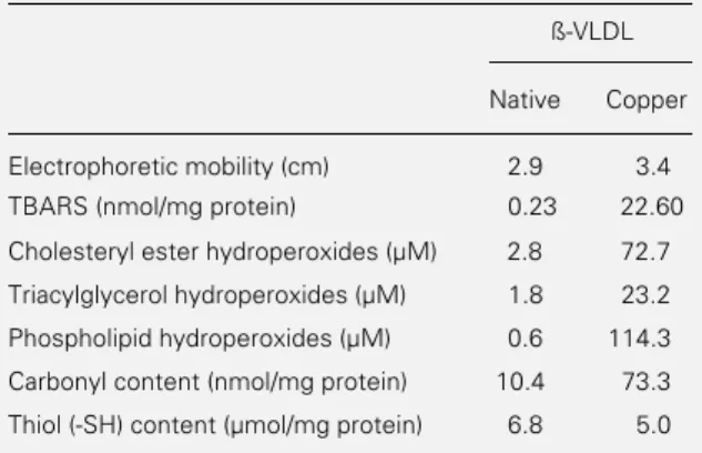
mobil-ity on agarose gel as compared to native ß-VLDL (3.4 cm vs 2.9 cm, respectively) (Table 1). Higher amounts of TBARS and lipid hydroperoxides were found in ß-VLDL after copper-dependent oxidation and the most significant increase was observed in phos-pholipid hydroperoxides (Table 1). The in-cubation of ß-VLDL with copper sulfate decreased the amount of apolipoprotein thiol (-SH) by 26% (Table 1). In addition, there was a significant increase of carbonyl con-tent in oxidized ß-VLDL (Table 1).
Concentration of lipids in blood plasma
Two months after starting cholesterol feeding, the total cholesterol concentration in blood plasma increased approximately 40-fold in comparison to the basal choles-terol content of HC rabbits (1182.4 ± 172.0 mg/dl vs 31.9 ± 9.5 mg/dl, respectively, N = 20, P<0.001). Triacylglycerol concentration also increased on account of the cholesterol-rich diet (57.5 ± 21.8 mg/dl for basal vs
260.3 ± 72.3 mg/dl after 60 days, P<0.01). In HC rabbits, more than 85% of blood plasma cholesterol was recovered in the lipoprotein fraction with a density less than 1.019 g/ml.
Fractional clearance rate
After injection of the native and copper-oxidized 99mTc-labeled ß-VLDL into C and
HC rabbits, the radioactivity in plasma was measured and the FCR (per hour) was calcu-lated. Plasma radioactivity was also deter-mined after 20% TCA precipitation. The FCR values calculated after TCA precipita-tion were similar to those obtained with total plasma (data not shown). In addition, the radioactivity was measured in the lipopro-tein fraction with a density <1.019 g/ml after ultracentrifugation of plasma samples ob-tained from 2 rabbits of each group (C and HC) 5 min and 6 h after injection of the
99mTc-labeled lipoproteins. Approximately
85% radioactivity was observed in the
lipo-protein fraction with a density <1.019 g/ml 6 h after injection of 99mTc-nat-ß-VLDL into C
and HC rabbits. In contrast, when animals of both groups were injected with 99m
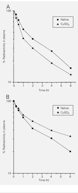
Tc-ox-ß-VLDL, 95-98% of the radioactivity was pres-ent in the lipoprotein fraction with a density <1.019 g/ml. In both C and HC groups the plasma clearance of 99mTc-nat-ß-VLDL and 99mTc-ox-ß-VLDL reflects a biexponential
decay mode (Figure 1). Decay curves showed that plasma clearance of 99mTc-ox-ß-VLDL
was faster than plasma clearance of 99m
Tc-nat-ß-VLDL in C rabbits (P<0.05). How-ever, both 99mTc-labeled lipoproteins were
removed from HC rabbit plasma at a slower rate than from C rabbits (P<0.05). More-over, 99mTc-ox-ß-VLDL remained in HC
rab-bit plasma for a longer period of time in comparison to 99mTc-nat-ß-VLDL. FCR data
are shown in Table 2.
Biodistribution studies
Biodistribution data for injected native and oxidized 99mTc-ß-VLDL were obtained
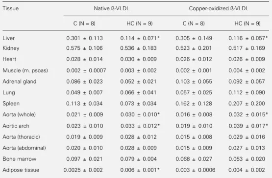
for male New Zealand white control rabbits fasted overnight (N = 8) and cholesterol-fed rabbits (N = 9) 6 h after injection. Table 3 shows the percent of the injected dose of
Table 1 - Biochemical determinations of native and copper-oxidized ß-VLDL.
ß-VLDL (1 mg/ml) was incubated with 400 µM peroxynitrite at 37oC for 18 h. Results are the mean of duplicates from two
determinations. TBARS = Thiobarbituric acid-reactive sub-stances.
ß-VLDL
Native Copper
Electrophoretic mobility (cm) 2.9 3.4 TBARS (nmol/mg protein) 0.23 22.60
Cholesteryl ester hydroperoxides (µM) 2.8 72.7
Triacylglycerol hydroperoxides (µM) 1.8 23.2
Phospholipid hydroperoxides (µM) 0.6 114.3
Carbonyl content (nmol/mg protein) 10.4 73.3
99mTc-lipoproteins per gram tissue. No
dif-ference in 99mTc-ox-ß-VLDL or 99m
Tc-nat-ß-VLDL uptake was observed among the tis-sues studied (liver, kidney, spleen, adrenal glands, aorta, heart, muscle and adipose tis-sues, lung and bone marrow) in C rabbits.
In HC rabbits, both 99mTc-nat-ß-VLDL
and 99mTc-ox-ß-VLDL were taken up to a
lesser extent by the liver than in C rabbits. In contrast, the aorta and other tissues showed a similar accumulation of 99mTc-nat-ß-VLDL
and 99mTc-ox-ß-VLDL in both C and HC
rabbits (Table 3). This suggests that internal-ization of these lipoproteins by these tissues may be mediated by receptors that are not down-regulated by cell cholesterol content. A 1.5- and 2-fold higher uptake of 99m
Tc-nat-ß-VLDL and 99mTc-ox-ß-VLDL,
respec-tively, was noted in the whole aorta and in the aortic arch containing atherosclerotic le-sions when compared to normal aorta. How-ever, there was no significant difference be-tween 99mTc-ox-ß-VLDL and 99m
Tc-nat-ß-VLDL accumulation in the atherosclerotic aorta.
The cholesterol-rich diet increased the weight of liver, adrenals and spleen but did not affect the weight of the aorta (data not shown). Moreover, the uptake of both 99m
Tc-ox-ß-VLDL and 99mTc-nat-ß-VLDL
calcu-lated as % of injected dose per whole organ (Table 4) was similar to the uptake calcu-lated as % of injected dose per gram tissue. The effect of tissue weight enhancement due to the cholesterol-rich diet was observed in the spleen where an increased 99m
Tc-nat-ß-VLDL uptake was found in HC rabbits. Since the presence of atherosclerotic lesions did not affect the weight of the aorta, the 99m
Tc-labeled lipoprotein uptake by the whole aorta was similar to that obtained by % of injected dose per gram tissue.
Imaging studies
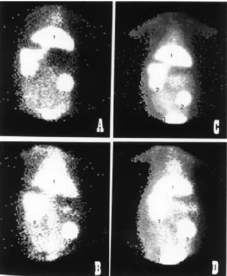
The gamma camera images of the biodis-tribution of 99mTc-labeled lipoproteins in one
Figure 1 - Plasma decay curves of native 99mTc-ß-VLDL and
cop-per-oxidized 99mTc-ß-VLDL in
control (A) and hypercholester-olemic rabbits (B). Data are re-ported as means for 8-9 rabbits.
% Radioactivity in plasma
10
0 1 2 3 4 5 6
Time (h)
A
Native CuSO4
% Radioactivity in plasma
10
0 1 2 3 4 5 6
Time (h)
B
100
Native CuSO4
Table 2 - Removal of native and copper-modified
99mTc-labeled ß-VLDL from plasma of cholesterol-fed
and control rabbits.
Data are reported as means ± SD of results obtained from (N) rabbits. *P<0.05 compared to the respective control rabbits; **P<0.05 compared to native ß-VLDL from control rabbits (ANOVA and Scheffé test).
Fractional clearance rate/h
99mTc-ß-VLDL Cholesterol-fed Control
Native 0.163 ± 0.043* 0.241 ± 0.020
(N = 9) (N = 8)
Copper-modified 0.100 ± 0.048* 0.362 ± 0.070**
(N = 8) (N = 8)
Table 3 - Biodistribution of 99mTc-ß-VLDL in rabbits.
Data are reported as means ± SD of results obtained from control (C) and hypercholesterolemic (HC) rabbits and are expressed as percentage of the injected radioactivity dose/g tissue. *P<0.05 compared to the respective C group (ANOVA and Scheffé test).
Tissue Native ß-VLDL Copper-oxidized ß-VLDL
C (N = 8) HC (N = 9) C (N = 8) HC (N = 9)
Liver 0.301 ± 0.113 0.114 ± 0.071* 0.305 ± 0.149 0.116 ± 0.057*
Kidney 0.575 ± 0.106 0.536 ± 0.183 0.523 ± 0.201 0.517 ± 0.169
Heart 0.028 ± 0.014 0.030 ± 0.009 0.026 ± 0.012 0.026 ± 0.009
Muscle (m. psoas) 0.002 ± 0.0007 0.003 ± 0.002 0.002 ± 0.001 0.004 ± 0.002
Adrenal gland 0.086 ± 0.023 0.052 ± 0.021 0.103 ± 0.055 0.092 ± 0.057
Lung 0.049 ± 0.007 0.066 ± 0.041 0.057 ± 0.025 0.112 ± 0.090
Spleen 0.113 ± 0.034 0.073 ± 0.034 0.162 ± 0.128 0.207 ± 0.200
Aorta (whole) 0.021 ± 0.009 0.030 ± 0.010* 0.016 ± 0.008 0.032 ± 0.015*
Aortic arch 0.023 ± 0.010 0.033 ± 0.012* 0.019 ± 0.010 0.039 ± 0.017*
Aorta (thoracic) 0.019 ± 0.009 0.028 ± 0.012 0.015 ± 0.008 0.029 ± 0.016
Aorta (abdominal) 0.020 ± 0.010 0.028 ± 0.009 0.015 ± 0.009 0.027 ± 0.013
Bone marrow 0.097 ± 0.021 0.079 ± 0.004 0.068 ± 0.027 0.053 ± 0.020
Adipose tissue 0.0025 ± 0.002 0.006 ± 0.001* 0.003 ± 0.0006 0.004 ± 0.002
Table 4 - Biodistribution of 99mTc-ß-VLDL in rabbits (% injected dose/organ).
Data are reported as means ± SD of results obtained from (N) rabbits. C = Control rabbits; HC = hypercholes-terolemic rabbits. *P<0.05 compared to the respective control group; **P<0.05 compared to native ß-VLDL of the HC group (ANOVA and Scheffé test).
Tissue Native ß-VLDL Copper-oxidized ß-VLDL
C (N = 8) HC (N = 9) C (N = 8) HC (N = 9)
Liver 25.50 ± 3.97 15.35 ± 1.38* 28.25 ± 4.47 8.31 ± 2.40*,**
Kidney 6.11 ± 1.19 5.82 ± 2.11 5.85 ± 1.66 4.83 ± 1.32
Heart 0.123 ± 0.057 0.121 ± 0.056 0.104 ± 0.052 0.113 ± 0.046
Adrenal 0.039 ± 0.022 0.051 ± 0.033 0.046 ± 0.032 0.073 ± 0.031
Spleen 0.085 ± 0.040 0.157 ± 0.030* 0.111 ± 0.072 0.312 ± 0.215
Aorta 0.012 ± 0.004 0.019 ± 0.006 0.010 ± 0.005 0.021 ± 0.010*
control rabbit and one cholesterol-fed rabbit obtained 6 h after injection are shown in Figure 2. The liver was the predominant uptake site for 99mTc-nat-ß-VLDL (Figure
2A) and 99mTc-ox-ß-VLDL (Figure 2C) in
the control rabbit. The uptake of all 99m
Tc-labeled lipoproteins by the liver was higher in the control rabbit than in the cholesterol-fed rabbit. In the latter, there was a high uptake of both 99mTc-nat-ß-VLDL and 99m
ographs showing the same pattern were ob-served for 99mTc-nat-ß-VLDL.
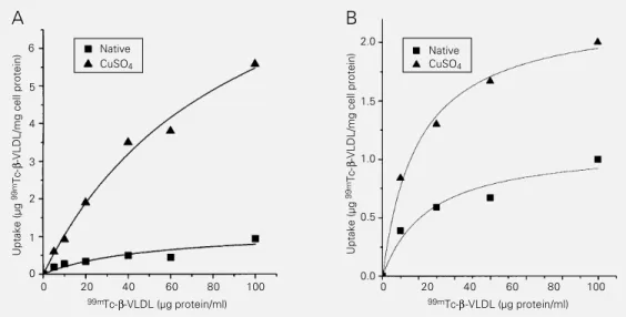
Uptake of 99mTc-ß-VLDL by macrophages
To determine whether 99mTc-labeling
af-fects the binding of lipoproteins by cell mem-brane receptors, as well as to validate our in vivo findings, the uptake of 99mTc-labeled
lipoproteins was tested in THP-1 phages and rabbit monocyte-derived macro-phages. For uptake rate measurements these cells were initially preincubated with LPDS and further incubated with increasing a-mounts of 99mTc-nat-ß-VLDL and 99m
Tc-ox-ß-VLDL (Figure 4). Data obtained after 4-h incubation showed that both THP-1 phages and rabbit monocyte-derived macro-phages internalized 99mTc-ox-ß-VLDL to a
greater extent than 99mTc-nat-ß-VLDL. 99m
Tc-ox-ß-VLDL uptake was approximately 5-fold higher in THP-1 macrophages (Figure 4A) and 2-fold higher in monocyte-derived macrophages (Figure 4B) than 99m
Tc-nat-ß-VLDL uptake.
Discussion
Hypercholesterolemia is an important risk factor for the development of the atheroscle-rotic process, particularly in terms of the cholesterol transported in the LDL and in ß-VLDL of rabbits fed a cholesterol-rich diet.
In vitro studies have shown that oxidative modification of ß-VLDL can contribute to atherogenesis (25). Oxidized ß-VLDL in-creases foam cell formation through binding and internalization into macrophages via macrophage scavenger receptors (25). The presence of oxidatively modified lipopro-teins in the plasma of animals and athero-sclerotic patients has been demonstrated (11-13). However, the pathophysiological role of these circulating oxidized lipoproteins re-mains a matter of speculation. The oxidized lipoproteins present in blood plasma may participate in atherogenesis by different
Figure 2 - Biodistribution of 99mTc-labeled ß-VLDL 6 h after tracer injection. A, 99m
Tc-nat-ß-VLDL in a control rabbit; B, 99mTc-nat-ß-VLDL in a cholesterol-fed rabbit; C, copper-oxidized 99mTc-ß-VLDL in a control rabbit; D, copper-oxidized 99mTc-ß-VLDL in a cholesterol-fed
rabbit. 1, Liver; 2, kidney; 3, urinary bladder.
Accumulation of 99mTc-labeled ß-VLDL by the atherosclerotic lesion
Figure 3 shows the uptake of 99m
Tc-ox-ß-VLDL by rabbit aorta. For comparison, we analyzed the aortas of one control (Figure 3A) and one cholesterol-fed rabbit (Figure 3B). The hypercholesterolemic rabbit aorta accumulated 99mTc-ox-ß-VLDL mainly in the
aortic arch where numerous atherosclerotic lesions occurred (Figure 3B). In control rab-bits, the accumulation of 99mTc-ox-ß-VLDL
Autoradi-Figure 3 - Autoradiography for 99mTc-ox-ß-VLDL uptake
in the aorta. A, Aorta from a control rabbit; B, aorta from a hypercholesterolemic rabbit. The arrow indicates the accumulation of copper-oxidized 99mTc-ß-VLDL in the
atherosclerotic lesions of the aortic arch from choles-terol-fed rabbits.
Uptake (µg
99m
Tc-β
-VLDL/mg cell protein)
0
0 20 40 60 80 100
Native CuSO4
Native CuSO4 6
5
4
3
2
1
99mTc-β-VLDL (µg protein/ml)
Uptake (µg
99m
Tc-β
-VLDL/mg cell protein)
0.0
0 20 40 60 80 100
0.5
99mTc-β-VLDL (µg protein/ml) 1.0
1.5 2.0
A B
Figure 4 - Uptake of 99m
Tc-nat-ß-VLDL and 99mTc-ox-ß-VLDL
by THP-1 macrophages (A) and rabbit monocyte-derived mac-rophages (B). Uptake was de-termined after incubating the cells with medium containing increasing concentrations of labeled lipoproteins for 4 h at 37oC. The internalization of
the 99mTc-labeled ß-VLDL into
cells was determined after solubilizing cells in 0.1 N NaOH and measuring the ra-dioactivity with a gamma-counter. Each point is the mean of triplicate determinat i o n s m a d e i n 2 3 e x p e r i -ments.
mechanisms. The present study shows data of biodistribution, fractional clearance rate and atherosclerotic lesion uptake of oxidized
99mTc-ß-VLDL in cholesterol-fed rabbits.
This radiotracer was chosen because 99m
Tc-labeled lipoproteins can be used for nonin-vasive quantitative biodistribution studies by scintigraphic imaging and for quantita-tively determining the uptake and degrada-tion of lipoproteins (14).
The biodistribution and fractional clear-ance rate differ between native and oxidized ß-VLDL in cholesterol-fed and control rab-bits. A relevant and new finding observed here was the significant reduction of plasma
99mTc-ox-ß-VLDL removal in
hypercholes-terolemic rabbits (Table 2 and Figure 1). Our data show that the exogenously oxidized ß-VLDL remains in plasma for as long a time as the native one. This contrasts with previ-ous findings showing that exogenprevi-ously oxi-dized lipoproteins are rapidly removed from blood plasma by cells of the monocytic phag-ocyte system (26). This result suggests that in these animals there is a significant pool of oxidatively modified ß-VLDL in blood plasma responsible for the competitive inhi-bition or saturation of common cell recep-tors, leading to a slower removal of 99m
Tc-ox-ß-VLDL from plasma. Indeed, we
cently demonstrated that ß-VLDL isolated from blood of cholesterol-fed rabbits has an increased content of lipid hydroperoxides (13).
Consistent with the low fractional clear-ance rate of 99mTc-ß-VLDL is the decreased
uptake of 99mTc-nat-ß-VLDL and 99m
Tc-ox-ß-VLDL by the liver of hypercholesterolemic rabbits. Down-regulation of B/E receptors in cholesterol-fed rabbits has been previously shown (27-29). Accordingly, our results in-dicate that the binding and internalization of oxidatively modified ß-VLDL by hepatic cells can be partially mediated by B/E or other down-regulated receptors. Native ß-VLDL is recognized by remnant and B/E receptors of liver parenchymal cells from rats and rabbits (30). In contrast, copper-oxidized ß-VLDL is taken up mainly by Kupffer cells of rat liver (31). In vitro studies have shown that acetylated LDL competes only 10-20% with oxidized ß-VLDL for cell association and degradation by both liver endothelial cells and Kupffer cells (31). Thus, distinct receptors responsible for the recog-nition of oxidized ß-VLDL appear to occur in rabbit liver, similarly to mouse peritoneal macrophages which recognize modified li-poproteins by different receptors (32,33).
In contrast to the phenomenon observed in the liver, the other organs from hypercho-lesterolemic rabbits incorporated the native and oxidized ß-VLDL possibly through re-ceptors which are not down-regulated by cell cholesterol content, since both control and cholesterol-fed animals showed a simi-lar accumulation of 99mTc-labeled
lipopro-teins in these organs (Tables 3 and 4). The accumulation of native 99mTc-ß-VLDL by
tissues with low LDL receptor activity, such as spleen and bone marrow, suggests that the uptake of these radiolabeled particles occur-ring in the reticulo-endothelial system is prob-ably mediated by the scavenger receptor, LDL receptor-related protein (LRP) or by phagocytosis.
The high uptake of 99mTc-labeled ß-VLDL
by the kidney observed in our study may be related to the diverse lipoprotein receptors present in the cells of this organ. The pres-ence of apolipoprotein E on the surface of ß-VLDL allows this lipoprotein to be internal-ized by receptors other than the LDL recep-tor such as the LRP, the VLDL receprecep-tor and glycoprotein-330 (Gp-330) (10). In the kid-ney, the epithelial cells of the plasma mem-brane in proximal tubules and glomeruli show a high expression of Gp-330, a recently iden-tified receptor that mediates the endocytosis of ß-VLDL (10). Gp-330 is also present in type-2 pneumocytes and in the epithelium lining the epididymis and yolk sac (10). Ac-cumulation of 99mTc-ß-VLDL in kidneys was
not decreased in cholesterol-fed rabbits, in-dicating that the receptor mediating its inter-nalization is not down-regulated by the ste-rol content of the cells (Tables 3 and 4). Although lipoproteins are not expected to be filtered by the kidneys due to their size, they may bind to Gp-330 present on the glomeru-lar plasma membrane. Another possibility for the appearance of radioactivity in kid-neys is the presence of free 99mtechnetium
(99mTc-pertechnetate) released from labeled
lipoproteins in the body. However, it has been reported that in both animals and hu-mans only 5% to 12% of injected 99mTc
activity was excreted through the urine within a 24-h period following an injection of 99m
Tc-LDL, indicating that 99mTc-labeled
lipopro-tein acts as an intracellular ligand (27). In fact, 99mtechnetium is frequently used as a
radiotracer because it acts as an intracellu-larly trapped ligand providing an accurate measurement of lipoprotein uptake by tis-sues (14).
charged oxidized lipoproteins (35). In fact, the copper-dependent oxidation of ß-VLDL originated particles with increased negative charge (Table 1) due to derivation of ε -amino groups of apolipoprotein lysine resi-dues by aldehydes released from perdized lipids, allowing the recognition of oxi-dized lipoproteins by the scavenger receptor (25,36). Although the atherosclerotic lesions of aorta arteries from cholesterol-fed rab-bits, mainly in the aortic arch, accumulated more 99mTc-ox-ß-VLDL than aortas from
control rabbits (Table 3 and Figure 3), the atherosclerotic lesions of cholesterol-fed rab-bits did not accumulate higher amounts of
99mTc-ox-ß-VLDL than of 99m
Tc-nat-ß-VLDL. Therefore, both 99mTc-labeled
lipo-proteins when present in the blood circula-tion could cross the endothelium to a similar extent. In fact, Simionescu et al. (37) and Vasile et al. (38) showed that in rats and in hypercholesterolemic rabbits, lipid deposi-tion on the vessel wall occurs by transcytosis not mediated by specific receptors.
Previous data have shown (14,26,39) that
99mTc-LDL is intracellularly trapped, thus
representing an adequate radiotracer for non-invasive imaging of LDL metabolism. More-over, 99mTc-LDL is recognized by the high
affinity LDL receptor (39) and is also taken up by the scavenger pathways (14). A pos-sible effect of ß-VLDL 99mTc-labeling on
lipoprotein uptake in our study was ruled out by in vitro cell culture experiments. Our results with THP-1 macrophages and rabbit monocyte-derived macrophages (Figure 4) showed that 99mtechnetium did not affect the
binding or internalization of either native or oxidized ß-VLDL. Accordingly, oxidatively modified ß-VLDL was taken up in high
a-mounts by macrophages which express scav-enger receptors (40). However, our biodis-tribution data indicate that when oxidized ß-VLDL is present in blood plasma its accu-mulation on the artery wall is not significant-ly higher than that of native ß-VLDL. These data agree with those previously reported for
99mTc-labeled oxidized LDL whose uptake
was not different from that of 99mTc-native
LDL by aorta of cholesterol-fed rabbits (41). Besides macrophages, the atherosclerotic lesion contains other cells such as smooth muscle cells that can take up oxidized and non-oxidized ß-VLDL (42). Therefore, the
in vivo removal of both ß-VLDL particles from plasma reflects the total uptake by the different cells present in atheroma. The slow removal of oxidized ß-VLDL from plasma of cholesterol-fed rabbits suggests that this oxidatively modified lipoprotein may also participate in the atherogenic process in other ways, including the induction of increased leukocyte adherence to endothelium and its cytotoxicity to endothelial cells (43). Finally, our in vitro experiments support previous data showing that oxidized ß-VLDL is taken up more than the native lipoprotein by the macrophage scavenger receptors (25). How-ever, this does not correspond to the in vivo
process, where other cells and receptors may also participate in ß-VLDL uptake.
Acknowledgments
The authors wish to acknowledge Bayer Co. (São Paulo, Brazil) for the donation of Rompun® and Paula M. Andrade for
References
1. Mahley RW (1983). Development of ac-celerated atherosclerosis: concepts de-rived from cell biology and animal model studies. Archives of Pathology and Labo-ratory Medicine, 107: 393-399.
2. Witztum JL (1994). The oxidation hypoth-esis of atherosclerosis. Lancet, 344: 793-795.
3. Morel DW, DiCorleto PE & Chilson GM (1984). Endothelial and smooth muscle cells alter low density lipoprotein in vitro
by free radical oxidation. Arteriosclerosis, 4: 357-364.
4. Hiramatsu K, Rosen H, Heinecke JW, Wolfbauer G & Chait A (1987). Superox-ide initiates oxidation of low density lipo-protein by human monocytes. Arterioscle-rosis, 7: 55-60.
5. Abdalla DSP, Campa A & Monteiro HP (1992). Low density lipoprotein oxidation by stimulated neutrophils and ferritin. Ath-erosclerosis, 97: 149-159.
6. Jessup W, Darley-Usmar VM, O’Leary VJ & Bedwell S (1991). 5-Lipoxygenase is not essential in macrophage-mediated oxidation of low-density lipoprotein. Bio-chemical Journal,278: 163-169. 7. Parthasarathy S (1994). Mechanisms of
oxidation of LDL. In: Parthasarathy S (Edi-tor), Modified Lipoproteins in the Patho-genesis of Atherosclerosis. R.G. Landes Company, Austin, 91-119.
8. Halliwell B & Gutteridge JMC (1985). The importance of free radicals and catalytic metal ions in human diseases. Molecular Aspects of Medicine, 8: 89-193. 9. Brown MS & Goldstein JL (1986). A
re-ceptor-mediated pathway for cholesterol homeostasis. Science, 232: 34-47. 10. Moestrup SK (1994). The α2
-macroglobu-lin receptor and epithelial glycoprotein-330: two giant receptors mediating en-docytosis of multiple ligands. Biochimica et Biophysica Acta, 1197: 197-213. 11. Salonen JT, Ylä-Herttuala S, Yamamoto R,
Butler S, Korpela H, Salonen R, Nyyssönen K, Palinski W & Witztum J (1992). Autoantibody against oxidized LDL and progression of carotid atherosclero-sis. Lancet, 339: 883-891.
12. Virella G, Virella I, Leman RB, Pryor MB & Virella MFL (1993). Anti-oxidized low den-sity lipoprotein antibodies in patients with coronary heart disease and normal healthy volunteers. International Journal of Clini-cal and Laboratory Research, 23: 95-101.
13. Silva EL, Moriel P, Chang YH & Abdalla DSP (1995). Plasma antioxidant enzymes and oxidized lipoproteins in hypercholes-terolemic rabbits. Biochemical and Mo-lecular Biology International, 36: 679-687. 14. Leitha T, Hermann M, Hüttinger M, Angelberger P & Dudczack R (1993). Tech-netium-99m labelled LDL as a tracer for quantitative LDL scintigraphy. I. Tracer purification, in vitro and in vivo long-term stability, in vitro validation and biodistri-bution. European Journal of Nuclear Medi-cine, 20: 667-673.
15. Havel RJ, Eder HA & Bradgon HJ (1955). The distribution and chemical composi-tion of ultracentrifugally separated lipo-proteins in human serum. Journal of Clini-cal Investigation, 34: 1345-1353. 16. Lees RS, Garabedian HD & Lees AM
(1985). Technetium-99m low density lipo-protein preparation and biodistribution.
Journal of Nuclear Medicine, 26: 1056-1062.
17. Matthews CME (1957). The theory of tracer experiments with 131I-labeled
plasma proteins. Physics in Medicine and Biology, 2: 36-56.
18. Boyum A (1968). Isolation of mononuclear cells and granulocytes from human blood: isolation of mononuclear cells by one g
centrifugation and of granulocytes by combining centrifugation and sedimenta-tion at 1 g. Scandinavian Journal of Labo-ratory Investigation, 21: 77-81.
19. Lowry OH, Rosebrough NJ, Farr AL & Randall RJ (1951). Protein measurement with the Folin phenol reagent. Journal of Biological Chemistry, 193: 265-269. 20. Winterbourn CC, Gutteridge JMC &
Halliwell B (1985). Doxorubicin-depend-ent lipid peroxidation at low partial pres-sures of O2. Free Radical Biology and
Medicine,1: 43-49.
21. Terao J, Shibata SS & Matsushita S (1988). Selective quantification of arachi-donic acid hydroperoxides and their hy-droxy derivatives in reverse-phase high performance liquid chromatography. Ana-lytical Biochemistry, 169: 415-423. 22. Terao J, Asano I & Matsushita S (1985).
Preparation of hydroperoxy and hydroxy derivatives of rat liver phosphatidylcho-line and phosphatidylethanolamine. Lip-ids, 20: 312-317.
23. Levine RL, Garland D, Oliver CN, Amici S, Climent I, Lenz A-G, Ahn B-W, Shaltel S & Stadtman ER (1990). Determination of car-bonyl content in oxidatively modified pro-teins. Methods in Enzymology, 186: 464-478.
24. Elman GE (1935). Tissue sulphydryl groups. Archives of Biochemistry and Bi-ophysics, 82: 70-79.
25. Parthasarathy S, Quinn MT, Scwenke DC, Carew TE & Steinberg D (1989). Oxidative modification of beta-very low density lipo-protein. Arteriosclerosis, 9: 398-404. 26. Steinbrecher UP, Witztum JJ,
Parthasara-thy S & Steinberg D (1987). Decrease in reactive amino groups during oxidation or endothelial cell modification of LDL. Arte-riosclerosis, 7: 135-143.
27. Vallabhajosula S & Goldsmith S (1990).
99mTc-Low density lipoprotein:
intracellu-larly trapped radiotracer for noninvasive imaging of low density lipoprotein me-tabolism in vivo. Seminars in Nuclear Medicine, 1: 68-79.
28. Asai K, Hayashi T, Funaki C, Kuzuya M, Naito M & Kuzuya F (1991). Comparison of plasma clearance of low density lipo-protein with ß-very low density lipopro-tein or acetoacetylated low density lipo-protein in cholesterol-fed rabbits. Bio-chemistry International, 23: 327-334. 29. Kovanen PT, Brown MS, Basu K,
Bilheimer DW & Goldstein JL (1981). Saturation and suppression of hepatic li-poprotein receptors: a mechanism for the hypercholesterolemia of cholesterol-fed rabbits. Proceedings of the National Acad-emy of Sciences, USA, 78: 1396-1400. 30. Gudmundsen O, Berg T, Roos N &
Nenseter MS (1993). Hepatic uptake of ß-VLDL in cholesterol-fed rabbits. Journal of Lipid Research, 34: 589-600.
31. Rijke YB, Hessels MAJ & Berkel TJC (1992). Recognition sites on rat liver cells for oxidatively modified ß-very low den-sity lipoproteins. Arteriosclerosis and Thrombosis, 12: 41-49.
33. Sparrow CP, Parthasarathy S & Steinberg D (1989). A macrophage receptor that rec-ognized oxidized LDL but not acetylated LDL. Journal of Biological Chemistry, 264: 2599-2604.
34. Ylä-Herttuala S, Palinski W, Rosenfeld ME, Parthasarathy S, Carew TE, Butler S, Witztum JL & Steinberg D (1989). Evi-dence for the presence of oxidatively modified low density lipoprotein in ath-erosclerotic lesions of rabbit and man.
Journal of Clinical Investigation, 84: 1086-1095.
35. Geng Y, Kodama T & Hansson GK (1994). Differential expression of scavenger re-ceptor isoforms during monocyte-macro-phage differentiation and foam cell forma-tion. Arteriosclerosis and Thrombosis, 14: 798-806.
36. Haberland ME, Olch CL & Fogelman AM (1984). Role of lysines in mediating inter-action of modified low density lipopro-teins with the scavenger receptor of hu-man monocyte macrophages. Journal of Biological Chemistry, 259: 11305-11311.
37. Simionescu N, Vasile E, Lupu F, Popescu G & Simionescu M (1986). Prelesional events in atherogenesis. American Jour-nal of Pathology, 123: 109-125.
38. Vasile E, Simionescu M & Simionescu N (1983). Visualization of the binding, en-docytosis, and transcytosis of low-density lipoprotein in the arterial endothelium in situ. Journal of Cell Biology, 96: 1677-1689.
39. Lees AM & Lees RS (1991). 99m
Techne-tium-labeled low density lipoprotein: re-ceptor recognition and intracellular se-questration of radiolabel. Journal of Lipid Research, 32: 1-8.
40. Hara H, Tanishita H, Yokoyama S, Tajima S & Yamamoto A (1987). Induction of acetylated low density lipoprotein recep-tor and suppression of low density lipo-protein receptor on the cells of human monocytic leukemia cell line (THP-1 cell).
Biochemical and Biophysical Research Communications, 146: 802-808.
41. Ali KSM, Vallabhajosula S, Censi C, Lipszyc H, Lee H, Machac J, Violi F & Luliano L (1993). Biodistribution and im-aging of Tc-99m-native and oxidized-LDL.
Journal of Nuclear Medicine, 34: 67P (Ab-stract).
42. Horrigan S, Campbell JH & Campbell GR (1991). Effect of endothelial cells on ß-very low density lipoprotein by endotheli-al cells enhances its metabolism by smooth muscle cells in culture. Arterio-sclerosis and Thrombosis, 11: 279-289. 43. Chisolm GM (1993). Oxidized lipoproteins