BIOMEDICAL SCIENCES
AND
CLINICAL INVESTIGATION
www.bjournal.com.br
www.bjournal.com.br
Campus
Institutional Sponsors
The Brazilian Journal of Medical and Biological Research is partially financed by
Hotsite of proteomics metabolomics developped by:
Braz J Med Biol Res, October 2010, Volume 43(10) 931-941
doi: 10.1590/S0100-879X2010007500091
Evaluation of anti-Wnt/β-catenin signaling agents by pGL4-TOP
transfected stable cells with a luciferase reporter system
Evaluation of anti-Wnt/β-catenin signaling
agents by pGL4-TOP transfected stable
cells with a luciferase reporter system
K.A. Chuang
1, C.H. Lieu
1*, W.J. Tsai
2,3, M.H. Wu
4,
Y.C. Chen
4, J.F. Liao
4, C.C. Wang
5and Y.C. Kuo
6*
1Department of Biotechnology and Laboratory Science in Medicine, School of Biomedical Science
and Engineering, National Yang-Ming University, Taipei, Taiwan, ROC
2National Research Institute of Chinese Medicine, Taipei, Taiwan, ROC 3Institute of Life Science, National Tai-Tung University, Taichung, Taiwan, ROC 4Institute of Pharmacology, National Yang-Ming University, Taipei, Taiwan, ROC 5School of Medicine, Fu-Jen University, Taipei, Taiwan, ROC 6Institute of Life Science, Fu-Jen University, Taipei, Taiwan, ROC
Abstract
Refractory and relapsedleukemiais a major problem during cancer therapy, which is due to the aberrant activation of
Wnt/β-catenin signaling pathway. Activation of this pathway is promoted by wingless (Wnt) proteins and induces co-activator β-Wnt/β-catenin
binding to lymphoid enhancer factor (LEF)/T-cell factor protein (TCF). To provide a convenient system for the screening of
anti-Wnt/β-catenin agents, we designed a bi-functional pGL4-TOP reporter plasmid that contained 3X β-catenin/LEF/TCF binding
sites and a selectable marker. After transfection and hygromycin B selection, HEK 293-TOP and Jurkat-TOP stable clones were established. The luciferase activity in the stable clone was enhanced by the recombinant Wnt-3A (rWnt-3A; 100-400 ng/mL)
and GSK3β inhibitor (2’Z,3’E)-6-bromoindirubin-3’-oxime (BIO; 5 µM) but was inhibited by aspirin (5 mM). Using this reporter
model, we found that norcantharidin (NCTD; 100 µM) reduced 80% of rWnt-3A-induced luciferase activity. Furthermore, 50 µM NCTD inhibited 38% of BIO-induced luciferase activity in Jurkat-TOP stable cells. Employing 3H-thymidine uptake assay and
Western blot analysis, we confirmed that NCTD (50 µM) significantly inhibited proliferation of Jurkat cells by 64%, which are the dominant β-catenin signaling cells and decreased β-catenin protein in a concentration-dependent manner. Thus, we established a stable HEK 293-TOP clone and successfully used it to identify the Wnt/β-catenin signaling inhibitor NCTD.
Key words: Cancer; Plasmids; Reporter assay; Screening; Wnt; β-catenin
Introduction
Correspondence: Y.C. Kuo, LS212, Laboratory of Molecular Pharmacology, Institute of Life Science, Fu-Jen University,
No. 510, Chung Cheng Rd., Hsinchuang, Taipei Hsien 242, Taiwan (ROC). Fax: +886-2-2905-2193; E-mail: [email protected]
*These authors contributed equally to this study.
Received May 21, 2010. Accepted August 19, 2010. Available online September 10, 2010. Published October 18, 2010.
Constitutive activation of the wingless (Wnt)/β-catenin
signaling pathway has been reported to be associated with carcinogenesis and to correlate with both shortened relapse-free survival and overall survival in leukemia patients (1). Mutation of the phosphorylation sites in the NH2-terminal
domain of β-catenin has been observed in T-cell malignan
-cies (2). Increased expression of Wnt ligands or frizzled (Fz)
receptors has been described in acute myeloid leukemia
(3). Autocrine Wnt/β-catenin signaling would also seem to
contribute to leukemic cancer cell proliferation (2,3). These
lines of evidence support a crucial role for Wnt/β-catenin
signaling in the maintenance of leukemia. Although
chemo-therapy and radiochemo-therapy do help long-term patient survival, relapse is still a major cause of death in many kinds of cancer (4). Recently, many studies have indicated that the cancer stem/progenitor cells from patients with leukemia,
colon cancer, or skin cancer inappropriately express
Wnt/β-catenin signaling. These cells possess self-renewal ability like stem cells and are responsible for relapse even under
Imatinib treatment (5-8). Thus, blocking the Wnt/β-catenin
signaling may help reduce the frequency of relapses in cancer patients.
Nonsteroidal anti-inflammatory drugs (NSAIDs) such as
agents to suppress β-catenin activity (9). Unfortunately, NSAIDs also have an inhibitory effect on cyclooxygenase-1
(COX-1) and COX-2 activity and cause gastrointestinal bleeding. Thus, it becomes important to search for other
Wnt/β-catenin signaling inhibitors that do not have severe side effects. Lepourcelet et al. (10) have reported six natural
products, ZTM00090, PKF118-310, PKF118-744, PKF115-584, PKF222-815, and CGP049090, that seem to abolish
β-catenin and T-cell factor (TCF) protein-protein interaction. These compounds significantly decreased cyclin D1 gene expression and inhibited cell proliferation in colon cancer
cells. Employing a transient reporter assay in a cell-based system, one small molecule, ICG-001, was shown to block
the formation of the β-catenin/CBP complex and to arrest
cancer cell proliferation. This molecule also attenuated the
growth of xenograft colon cancers in mice (11). Since the
development of new drugs has been limited, we suggest that
searching for new antagonists of Wnt/β-catenin signaling
would seem to require a high-throughput assay.
The hallmark of the Wnt/β-catenin signaling pathway is β-catenin, which acts as a multiple function protein including
adhesion to the cell membrane and transcription activation in the nucleus (12). In the absence of Wnt proteins, little
β-catenin is present in the cytoplasm; this is due to the fact that β-catenin interacts with glycogen synthase kinase 3β (GSK-3β), which causes its phosphorylation, and the pro -tein is then degraded by the ubiquitin-proteasome system
(13). When Wnt proteins bind to Fz receptors, GSK-3β activity is disrupted and β-catenin escapes degradation. The unphosphorylated β-catenin then translocates into
the nucleus, binds to the lymphoid enhancer factor (LEF)/ TCF transcription factors, and turns on downstream gene
expression including c-myc and cyclin D1, which regulate
cell proliferation and apoptosis (14,15).
In the present study, we cloned the LEF/TCF binding sequences into a pGL4.3 luciferase reporter plasmid, which was part of an antibiotic-resistant plasmid. This new construct was transfected into human embryonic kidney (HEK) 293 and human Jurkat leukemic T cells to establish the HEK 293-TOP and Jurkat-TOP stable reporter cell lines. The activities of these cell lines were indicated by luciferase activity induced by recombinant Wnt-3A (rWnt-3A) protein
and (2’Z,3’E)-6-bromoindirubin-3’-oxime (BIO). We also
used the cell lines for drug screening and discovered the norcantharidin (NCTD) that inhibited the activation of the
Wnt/β-catenin signaling pathway in both HEK 293-TOP and Jurkat-TOP cells and reduced the β-catenin levels and cell
proliferation in Jurkat T cells.
Material and Methods
Material
BIO, 1-methyl-BIO (MeBIO), lithium chloride (LiCl) and N-(2-methyl-4-nitro)-2,4-dichlorosulfonamide (FH535) were purchased from Merck (Germany). NCTD (C8H8O4; MW
168) was synthesized as described previously (16). Mouse rWnt-3A was obtained from R&D (USA). Mouse monoclonal
antibodies raised against human β-catenin (cat. No. sc-7963), β-actin (cat. No. sc-47778) and GSK-3β (cat. No.
610201) were purchasedfrom Santa Cruz (USA) and BD Bioscience (USA). Goat anti-mouse IgG conjugated with
horseradish peroxidase was purchased from Pierce (USA).
Other reagents were obtained from Sigma (USA).
Cell culture
HEK 293 (FIRDI, Taiwan), L cells (FIRDI), and L Wnt-3A cells (ATCC, USA) were cultured in DMEM (Invitrogen, USA). Jurkat cells (FIRDI) were cultured in RPMI-1640 medium (Invitrogen). Both media were supplemented with
10% fetal bovine serum (FBS), 2 mM L-glutamine, 0.1 mM sodium pyruvate, 100 U/mL penicillin, and 100 μg/
mL streptomycin. HEK 293-TOP/FOP and Jurkat-TOP/ FOP cells were regularly maintained in selected medium
containing 500 and 1000 µg/mL hygromycin B, respectively. All cells were incubated at 37°C in a humidified atmosphere under 5% CO2. Wnt-3A and control conditioned media were
prepared according to ATCC recommendation.
Generation of pGL4-TOP/FOP reporter plasmids
Two primers, TOP265-F: 5’GTCAGATCTCTTAATATG CGAAGTGGACC3’ and TOP265-R: 5’CGTGGTACCGT
AACGCCAGGGTTTTCC3’, were designed from the se
-quence data of the TOPFLASH plasmid and used to amplify either the TOP (wild-type 3X LEF/TCF binding sites; 265 bp) or the FOP elements (mutant 3X LEF/TCF binding sites; 255 bp) from TOPFLASH or FOPFLASH (Upstate Biotechnol-ogy, USA) by the polymerase chain reaction (PCR). The
TOP and FOP elements were amplified using the following
program: 94°C for 10 min, followed by 30 cycles of 94°C for
1 min, 54°C for 1 min, 72°C for 30 s, then finally 4°C for 10 min. The amplified products were cloned into the pGL4.3
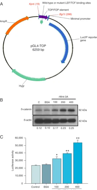
luciferase reporter plasmid (Promega, USA) using the BglII and KpnI cleavage sites and the two successfully created ampicillin-selected plasmids were called pGL4-TOP and pGL4-FOP (Figure 1A).
Establishment of the HEK 293-TOP/FOP and Jurkat-TOP/FOP reporter cell lines
The 0.8 μg pGL4-TOP (or FOP) reporter plasmid was transfected into HEK 293 cells (2 x 10⁵) with LipofectaminTM
2000 (Invitrogen) according to manufacturer instructions. The transfected cells were selected in medium containing
500 μg/mL hygromycin B (MDBio, Taiwan) for 3 weeks to
obtain a stable clone. Medium was replaced with fresh medium every 3 days. At the end of 3 weeks, the surviving cells were diluted in a 96-well plate (Nunclon, Denmark) and a single cell-derived clone was selected and cultured to give a stable cell line in the presence of hygromycin B. These stable clones were called HEK 293-TOP or HEK 293-FOP.
by electroporation using a MicroPorator system (MP-100; Digital Bio Technology, Korea) according to manufacturer instructions. The stable cells were selected in medium
containing 1500 μg/mL hygromycin B.
Luciferase assay
HEK 293-TOP cells (1 x 10⁴) or Jurkat-TOP/FOP (2 x
104) cells were seeded into 96-well plates and incubated
in medium with 10% FBS for one day. The cells were then
stimulated with rWnt-3A (100, 200, 400 ng/mL), L Wnt-3A conditioned medium (Wnt-3A CM), BIO, or LiCl. After
incuba-tion for the indicated times, total cell lysates were extracted with 1X reporter lysis buffer (Promega) and 10 μg total cell
proteins were used to determine luciferase activity by the Luciferase Assay System (Promega) using a Microplate Luminometer (Berthold, Germany).
Proliferation assay
Jurkat cells (2 x 10⁴/well) were cultured in a 96-well
plate for 24 h. Various concentrations of NCTD (12.5 to
50 μM) were added to the cells and the plates were incu
-bated in a 5% CO2-air humidified atmosphere at 37°C for
2 days. Subsequently, tritiated thymidine (1 μCi/well; New
England Nuclear, USA) was added to each well. After 16-h
incubation, the cells were harvested onto glass-fiber filters
with an automatic harvester (Multimash 2000, Dynatech,
UK). The radioactivity of the filters was measured with a
scintillation counter.
Western blot analysis
HEK 293-TOP/FOP cytosolic proteins were examined
by Western blot analysis. Proteins were separated by
10% SDS-PAGE and transferred to an Immobilon-P PVDF membrane (Millipore, USA). Antibodies against β-actin (1:1000), β-catenin (1:1000) and GSK-3β (1:1000) were used for protein detection. The specific reactive proteins
were detected by an enhanced chemiluminescence method that employed rabbit anti-mouse IgG conjugated with
horse-radish peroxidase. The immunoblots were visualized with
the immobilon Western chemiluminescent HRP substrate (Millipore).
RNA extraction and RT-PCR
Total RNA was isolated from Jurkat cells with RNA-BeeTM (Tel-Test, USA) according to manufacturer
instruc-tions. For cDNA synthesis, 1 µg total RNA was added to a 10-µL reaction volume, including reaction buffer, oligo dT,
dNTP and reverse transcriptase and the procedure was used according to manufacturer instructions. The primer sequences
used to detect the GAPDH and β-catenin gene CTNNB1
were as follows: GAPDH: TGAAGGTCGGAGTCAACGGATT TGGT (sense) and CATGTGGGCCATGAGGTCCACCAC (anti-sense); CTNNB1: ACTCTAGGAATGAAGGTGTGGC (sense) and AGTGTGTCAGGCACTTTCTGAG (anti-sense). PCR was carried out using the following procedure:
94°C for 10 min, followed by 30 cycles of 94°C for 1 min,
60°C for 1 min, 72°C for 30 s, and finally 4°C for 10 min. After reaction, the amplified products were run on a 2%
agarose gel.
Statistical analysis
Data are reported as means ± SD of at least three
experiments. The differences between groups were deter -mined by the Student t-test, with the level of significance
set at P < 0.05.
Results
Generation of the HEK 293-TOP/FOP stable cell lines for drug screening
The TOPFLASH plasmid contains three LEF/TCF
binding sites (17). In order to establish a Wnt/β-catenin
signaling-response stable cell line for drug screening, we
amplified the TOP element that contained three LEF/TCF
binding sites from TOPFLASH and inserted them into the pGL4.3 reporter plasmid that contains a hygromycin B phosphotransferase coding gene (Hygr) and a luciferase reporter gene. This new constructed plasmid was named pGL4-TOP (Figure 1A). The integrity of the three LEF/TCF
sequences was confirmed by DNA sequence analysis. No
mutations of binding sequences were detected (data not shown). In addition, the pGL4-FOP, which contained three mutated LEF/TCF binding sites derived from the FOPFLASH plasmid, was constructed as a negative control. These new bi-functional reporter constructs pGL4-TOP and pGL4-FOP can be used to create stable clones using hygromycin B selection without co-transfection with a plasmid containing a selectable marker. Therefore, the pGL4-TOP/FOP reporter
plasmids were transfected into HEK 293 cells and expres -sion of the Hygr gene protected the cells, HEK 293-TOP and HEK 293-FOP, from antibiotic treatment.
Luciferase activity of HEK 293-TOP cells was activated
by Wnt-3A proteins and a specific inhibitor of GSK-3β. It is well known that Wnt proteins activate the
down-stream signaling by stabilization of β-catenin and enhance target gene expression in the nucleus. We evaluated basal and inducible levels of β-catenin signaling in HEK 293-TOP by measuring protein expression of β-catenin and
by measuring the transcriptional activity of the reporter. HEK 293-TOP stable cells were cultured in the presence of 100, 200, and 400 ng/mL rWnt-3A for 24 h. The cell
proteins were then extracted from the cells and subjected
to Western blotting and a luciferase assay. As shown in
Figure 1B, β-actin was used as a loading control and rWnt-3A stimulation increased the levels of β-catenin protein in
a dose-dependent manner. Comparison with the control
group (medium only), the vehicle (0.1% BSA) did not affect β-catenin protein expression. When stimulated with 100,
293-TOP significantly increased by 1.3-fold (P < 0.05),
1.5-fold (P < 0.01), and 2.1-1.5-fold (P < 0.01), respectively (Figure 1C). Furthermore, there was a 2.5-fold increase (P < 0.001)
of luciferase activity upon stimulation with 50% (v/v) Wnt-3A
CM in HEK 293-TOP cells. HEK 293-FOP cells were not affected (Figure 2A). These results indicate that Wnt-3A
specifically increased the amount of β-catenin protein and
induced luciferase activity in HEK 293-TOP stable cells.
GSK-3β, a negative regulator of the Wnt/β-catenin
signaling, is involved in the TCF-mediated transcriptional pathway and its inhibitor has been used as a
pharmaco-logical agent to activate this pathway (18). To confirm the specificity of Wnt/β-catenin signaling induction, a specific inhibitor of GSK-3β, BIO (5 μM), was incubated with HEK
293-TOP cells in the presence of rWnt-3A (400 ng/mL) for 24 h and luciferase activity was then determined. As shown
in Figure 2B, neither the vehicle for rWnt-3A (0.1% BSA) nor the vehicle for BIO (0.05% DMSO) affected luciferase
activity in HEK 293-TOP cells. However, both rWnt-3A and BIO activated the luciferase reporter by 2.6-fold (P < 0.001) and 4.9-fold (P < 0.001), respectively. MeBIO is an analog
of BIO that does not inhibit GSK-3β activity. MeBIO (5 μM)
alone did not affect the luciferase activity. When HEK 293-TOP cells were co-treated with rWnt-3A and MeBIO, MeBIO did not change the stimulatory effect of rWnt-3A on luciferase activity. However, an additive effect was observed in the cells treated with BIO and rWnt-3A (P < 0.01). As shown in
Figure 2D, while BIO did not affect GSK-3β protein expres
-sion, it increased the levels of β-catenin in HEK 293-TOP
cells in a dose-dependent manner. On the other hand, we
also used another GSK-3β inhibitor, LiCl, to confirm the
HEK 293-TOP response. As shown in Figure 2C, LiCl (20 mM) also activated the luciferase reporter by 10.9- and 4.9-fold in the presence or absence of rWnt-3A, respectively.
These results confirmed that HEK 293-TOP cells faithfully responded to the effects of rWnt-3A and the GSK-3β inhibi
-tors during canonical Wnt/β-catenin signaling.
Aspirin and NCTD inhibit rWnt-3A-induced luciferase activity in HEK 293-TOP cells
According to the report of Cho et al. (19), aspirin is able to decrease Wnt-3A CM-induced luciferase activity in HEK 293 cells. To determine whether HEK 293-TOP could be used as a drug screening platform, HEK 293-TOP cells were incubated with aspirin in the presence of rWnt-3A (400 ng/mL) and luciferase activity was analyzed. As shown in Figure 3A, rWnt-3A induced a 4.6-fold increase
in luciferase activity (P < 0.01), while the vehicle (0.1%
BSA) did not affect activity. In contrast, treatment with 1.25, 2.5, and 5 mM (P < 0.05) aspirin decreased the luciferase activity of the rWnt-3A-induced cells in a dose-dependent manner. Therefore, we predicted that the HEK 293-TOP
cell line could be applied to screen potential Wnt/β-catenin
signaling blockers.
Thus, we collected several synthetic compounds or
Figure 1. Map of the pGL4-TOP/FOP reporter plasmids and
ef-fects of rWnt-3A on β-catenin protein levels and luciferase ac -tivities in HEK 293-TOP stable cells. A 265-bp fragment
contain-ing the 3X LEF/TCF elements (19-286 bp) was amplified from
TOPFLASH and FOPFLASH by PCR and inserted into the pGL4.3 reporter plasmid. The pGL4-TOP/FOP plasmid map was drawn with the Vector NTI software. LEF = lymphoid enhancer factor; TCF = T-cell factor protein; AmpR = beta-lactamase cod-ing region; Hygr = hygromycin B codcod-ing region. HEK 293-TOP
cells (1 x 10⁴) were seeded into a 96-well plate for 24 h and then
treated with medium (C), BSA (0.1%) or the indicated concentra -tions of rWnt-3A (100, 200, 400 ng/mL) for another 24 h. Total cell proteins were collected and subjected to Western blotting (B)
and the luciferase assay (C). The ratios of β-catenin to β-actin
natural products for screening. We analyzed the effects of
these compounds (100 µM) on luciferase activity in HEK
293-TOP cells induced by Wnt-3A CM and calculated their inhibitory activities. As shown in Figure 3B, one of these
compounds, compound 12 (NCTD), had an 80% inhibitory
activity on luciferase activity in HEK 293-TOP stimulated
with Wnt-3A CM. The specific inhibitory effect of NCTD on the Wnt/β-catenin signaling was determined with HEK
293-TOP and HEK 293-FOP cells treated with Wnt-3A CM (Figure 3C). After stimulation with Wnt-3A CM, luciferase activity was induced in HEK 293-TOP cells but not in HEK
293-FOP cells. The vehicle (0.05% DMSO) did not affect
the luciferase activity of either HEK TOP or HEK 293-FOP cells. Furthermore, NCTD had little or no effect on luciferase activity in HEK 293-FOP cells. By contrast, NCTD
significantly impaired luciferase activity in HEK 293-TOP
cells in a concentration-dependent manner. As shown in Figure 3D, rWnt-3A was also used to stimulate the cells and
to confirm the inhibitory activity of NCTD. When compared
to the BSA-treated group, rWnt-3A induced luciferase ac-tivity by 3.9-fold (P < 0.01) and NCTD inhibited luciferase activity in a concentration-dependent manner (P < 0.001).
These results indicate that potential Wnt/β-catenin signal -ing inhibitors such as NCTD could be detected by the HEK 293-TOP reporter system.
Luciferase activity of Jurkat-TOP cells was activated by Wnt-3A proteins and BIO inhibitor
It has been reported that Jurkat cells express high levels of Wnt/β-catenin signaling and play an important role in Jurkat cell proliferation (20). To examine whether activa
-tion of Wnt/β-catenin signaling in a leukemic cancer cell
line was blocked by NCTD, we established the Jurkat-TOP and Jurkat-FOP stable cells by pGL4-TOP and pGL4-FOP transfection, respectively. The stable Jurkat-TOP/FOP cells were stimulated with 100, 200, and 400 ng/mL rWnt-3A for 24 h. As shown in Figure 4A, rWnt-3A stimulation
in-creased the levels of β-catenin protein in both Jurkat-TOP and Jurkat-FOP stable cells. The vehicle (0.1% BSA) did not affect β-catenin protein expression in either stable cell
lines. Furthermore, rWnt-3A (100, 200, and 400 ng/mL)
significantly induced luciferase activities in Jurkat-TOP by
1.3-fold (P < 0.05), 1.6-fold (P < 0.01), and 1.8-fold (P< 0.01), respectively. In contrast, the rWnt-3A-mediated
in-Figure 2. Effects of Wnt-3A proteins on luciferase activities in HEK 293-TOP/FOP stable cells treated or not with BIO or LiCl. A, HEK
293-TOP/FOP cells (1 x 10⁴) were seeded into a 96-well plate for 24 h and then treated with L cell conditioned medium (CM; control)
or 50% (v/v) L Wnt-3A CM for another 24 h. Total cell proteins were collected and subjected to the luciferase assay. ***P < 0.001 vs
HEK 293-TOP treated with L cell CM (Student t-test). B and C, HEK 293-TOP cells (1 x 10⁴) were seeded into a 96-well plate for 24
h. The cells were stimulated or not with rWnt-3A (400 ng/mL) in the presence or absence of BIO (5 μM) or MeBIO (5 μM) or LiCl (20 mM) for 24 h. Total cell proteins were extracted and the luciferase activities were determined. Each bar is the mean ± SD of three independent experiments. **P < 0.01, ***P < 0.001 vs BSA (0.1%) group; ##P < 0.01, ###P < 0.001 vs DMSO (0.1%) or control (medium)
group; ++P < 0.01 vs BIO or LiCl group (Student t-test). D, HEK 293-TOP cells (1 x 10⁴) were seeded into a 96-well plate for 24 h and
stimulated with BIO (2.5 and 5 μM) for another 24 h. Finally, total cell proteins were extracted and subjected to Western blotting. The ratios of β-catenin or GSK-3β to β-actin proteins are shown in the bottom panel. BIO = (2’Z,3’E)-6-bromoindirubin-3’-oxime. Lane M =
crease in LEF/TCF luciferase activities had no significant effect on Jurkat-FOP cells. We also observed if GSK-3β
inhibitor was able to activate luciferase activities in Jurkat-TOP/FOP cells. As shown in Figure 4B, while the vehicle
(DMSO) did not affect β-catenin protein expression, BIO markedly increased the levels of β-catenin protein in both
Jurkat-TOP and Jurkat-FOP stable cells at 12 h post-activation. For the reporter assay, the relative luciferase activities of Jurkat-TOP cells were 20-fold higher than those of Jurkat-FOP cells. After BIO treatment, the transcriptional activity of luciferase in Jurkat-TOP cells was increased in a dose-dependent manner (2.2-, 3.1- and 4.0-fold; P < 0.001). However, these results were not due to the decrease of
GSK-3β protein expression in BIO-treated Jurkat-TOP cells
(Figure 4C). Taken together, these results show that
Jurkat-TOP cells faithfully responded to the rWnt-3A protein and
to the GSK-3β inhibitor.
NCTD inhibited β-catenin/LEF-mediated luciferase
activity in Jurkat-TOP cells and correlated with a reduction of cell proliferation and the level of
β-catenin proteins
To elucidate whether NCTD had an inhibitory effect
on Wnt/β-catenin signaling in leukemic cancer cells, BIO
and various concentrations of NCTD were added to the Jurkat-TOP/FOP cells for 12 h. As shown in Figure 5A,
treatment with 50 µM NCTD significantly decreased 38%
of the luciferase activity in BIO-treated cells. In contrast, the vehicle (DMSO) did not affect relative luciferase
activ-ity. The positive control, β-catenin/TCF inhibitor FH535 (15 Figure 3. Effects of aspirin and 32 other compounds on luciferase activity in HEK 293-TOP cells induced with Wnt-3A proteins. A, B, HEK 293-TOP cells (1 x 10⁴) were seeded into a 96-well plate for 24 h and then stimulated with rWnt-3A or
Wnt-3A CM in the presence of aspirin (1.25, 2.5, and 5 mM) or the test agents (100 μM) for 24 h. Total cell proteins were extracted and luciferase activity was determined. C, HEK 293-TOP/FOP cells (1 x 10⁴) were plated into a 96-well plate
and then stimulated with Wnt-3A CM in the presence of NCTD (25, 50, 100 μM). D, HEK 293-TOP cells were cultured
with rWnt-3A (400 ng/mL) and various concentration of NCTD (25, 50, 100 μM) for 24 h. Finally, total cell proteins were
collected and subjected to luciferase assay. Each bar indicates the mean ± SD of three independent experiments. NCTD = norcantharidin. *P < 0.05, **P < 0.01, ***P < 0.001 vs HEK 293-TOP cells treated with Wnt-3A and 0.1% DMSO. ##P <
µM), repressed about 62% of luciferase activity in the cells. These data suggested that NCTD affected Wnt/β-catenin signaling by inhibition of β-catenin or its downstream
pathway. Additionally, the effects of NCTD on Jurkat cell proliferation were determined by 3H-thymidine uptake. As
shown in Figure 5B, the proliferation of Jurkat cells was not
affected by DMSO, but 25 and 50 μM NCTD inhibited cell
proliferation in a dose-dependent manner (P < 0.001). To determine whether NCTD inhibition of Jurkat cell
proliferation was related to Wnt/β-catenin signaling, we determined the levels of β-catenin protein in Jurkat cells.
As shown in Figure 6A, Western blot analysis demonstrated
that NCTD reduced the levels of β-catenin protein present in
Jurkat cells in a dose-dependent manner. However, NCTD
did not affect β-catenin mRNA expression in Jurkat cells
(Figure 6B). These results suggest that the reduction of Jurkat cell proliferation induced by NCTD might be related
to blocking of the Wnt/β-catenin signaling pathway.
Discussion
The Wnt/β-catenin signaling pathway has been con
-Figure 4. Induction of β-catenin protein and luciferase activity in Jurkat-TOP stable cells by rWnt-3A and BIO. Jurkat-TOP/FOP cells (2 x 10⁴) were seeded into a 96-well plate for 24 h and then treated with (A) 100, 200, and 400 ng/mL rWnt-3A or (B and C) 0.625, 1.25,
and 2.5 µM BIO for 24 and 12 h, respectively. Total cell proteins were collected and subjected to Western blotting and to the luciferase assay. The ratios of β-catenin or GSK-3β to β-actin proteins are shown in the bottom panel. BIO = (2’Z,3’E)-6-bromoindirubin-3’-oxime.
Figure 5. NCTD decreased luciferase activity in Jurkat-TOP cells induced by BIO and reduced Jurkat cell proliferation. Jurkat-TOP/ FOP cells (2 x 10⁴/well) were plated into a 96-well plate for 24 h
and then stimulated with BIO (2.5 µM) in the presence of NCTD
(25 and 50 μM) or a positive inhibitor (FH535, 15 µM) for 12 h. Total cell proteins were quantified and subjected to the luciferase
assay. Each bar indicates the mean ± SD of three independent experiments. ###P < 0.001 vs cells treated with medium only. *P
< 0.05, ***P < 0.001 vs cells treated with BIO and 0.025% DMSO (Student t-test). B, Jurkat cells were treated with 0.1% DMSO or
NCTD (12.5, 25 and 50 μM) for 48 h. Cell proliferation was as -sayed based on the uptake of 3H-thymidine. After 16 h of incuba
-tion, the cells were harvested with an automatic harvester onto a glass fiber and the level of radioactivity was measured with a
scintillation counter. NCTD = norcantharidin; BIO = (2’Z,3’E
)-6-bromoindirubin-3’-oxime. ***P < 0.001 vs cells treated with DMSO (Student t-test).
Figure 6. NCTD impaired β-catenin protein production, but did not affect mRNA expression in Jurkat cells. Jurkat cells were treated with medium (C), 0.1% DMSO and NCTD (25, 50 and 100 μM) for 24 h. The levels of β-catenin proteins (A) and transcripts (B)
were measured by Western blotting and RT-PCR, respectively.
sidered to be a new target for oncology drug discovery
(21). Combining treatment with a Wnt/β-catenin signaling
pathway inhibitor and conventional anti-cancer therapy might be effective in stimulating tumor regression. In order
to establish a more convenient and efficient drug screening
platform, in the present study we developed an HEK
293-TOP stable cell line that included a Wnt/β-catenin signaling
reporter plasmid. The luciferase activity of the HEK 293-TOP stable clone was activated by rWnt-3A, Wnt-3A CM, and a
GSK-3β inhibitor and this effect was inhibited by a positive
control, aspirin. Using this stable clone as a drug-screening
platform, we first proved that NCTD not only inhibited rWnt-3A induction of the β-catenin/LEF signaling pathway but also decreased the levels of β-catenin protein and cell
proliferation in Jurkat cells. The present study describes a stable cell line that could be used for the screening of
anti-Wnt/β-catenin signaling agents.
The methods for anti-Wnt/β-catenin drug screening
have been developed using different systems. The ELISA-based modellimits the functional testing of the candidate
drug to the β-catenin/TCF complex (10). However, the
commercially available reporter plasmids for detecting
Wnt/β-catenin signaling require transient transfection of the
plasmid before drug screening. This is not only time- and
reagent-consuming, but the transfection efficiency also var
-ies from experiment to experiment. To solve these problems,
a stable clone needs to be established by co-transfecting reporter- and antibiotic-containing plasmids (20,22,23). In the present study, we successfully established stable clone HEK 293-TOP cells that contained a bi-functional reporter plasmid. This design could solve the problem caused by co-transfection of a selection marker. When the latter ap-proach is used, the drug-resistant cells that survive might lose the reporter plasmid during long-term culture and this will result in weaker luciferase signaling. During the cloning process of the 3X LEF/TCF binding sites into the pGL4.3 plasmid, our initial construct was unsuccessful even though we tried 3X LEF/TCF binding sequences created by
nucleotide synthesis or by PCR amplification. We thought
that this failure might be due to the unusual secondary DNA structures, such as hairpins and cruciforms (24). Indeed, computer modeling on the DINAMelt Server sug-gested that the full length of the 3X LEF/TCF binding sites formed a single hairpin structure (data not shown). To solve this problem, the primer set (TOP-F265 and TOP-R265), upstream and downstream of 3X LEF/TCF, was designed to amplify the TOP elements, which contain 3X LEF/TCF binding sites while creating linear forms of DNA at each end. Finally, we were able to successfully amplify the TOP (or FOP) elements from TOPFLASH (or FOPFLASH) and insert them into a pGL4.3 reporter plasmid. After long-term antibiotic selection, HEK 293-TOP and HEK 293-FOP stable cells were chosen.
The luciferase gene in HEK 293-TOP cells could be
significantly induced by rWnt-3A and Wnt-3A CM. Binnerts
et al. (25) reported a similar result when they stimulated cells with serial concentrations of rWnt-3A. In addition,
whether the HEK 293-TOP cells specifically responded to canonical Wnt/β-catenin signaling was also examined in this study. Two GSK-3β inhibitors LiCl and BIO, which are able to increase β-catenin stability and induce β-catenin nuclear
translocation (26), were used in this study. LiCl competes for the cofactor Mg2+ and blocks GSK-3β kinase activity, while BIO selectively binds to the ATP binding pocket of the kinase and inhibits enzyme activity (26). Our results with HEK 293-TOP showed that luciferase activity increased in the presence of BIO or LiCl. Each of these drugs had an additive effect with rWnt-3A. When we investigated an HEK 293-FOP clone with mutant LEF/TCF binding sites, our results showed that Wnt-3A CM was not able to induce luciferase activity in HEK 293-FOP cells (Figure 2A).
There-fore, we confirmed that the luciferase expression in HEK
293-TOP cells induced by rWnt-3A or Wnt-3A CM was LEF/
TCF-dependent and came specifically from the canonical Wnt/β-catenin signaling pathway.
Small molecules have been shown to have a wide
range of biological activities and this has been confirmed
in clinical trials (27). In the present study, we showed that aspirin decreased rWnt-3A-induced luciferase activity, in agreement with Cho et al. (19). Recently, we measured the effect of aspirin (5 mM) on HEK 293-TOP cell growth by Trypan blue staining. Preliminary data demonstrated that aspirin did not increase cell death but delayed cell proliferation. Compared to the untreated control, from day 1 to day 3, the rates of aspirin inhibition of cell growth were
24, 26, and 47%, respectively. We, therefore, identified small molecules with anti-Wnt/β-catenin activity by the
HEK 293-TOP reporter assay, including norcantharidin, a demethylated form of cantharidin derived from blister beetles
(28). In this study, we found that 50 µM NCTD showed
a positive result and decreased the luciferase activity of the HEK 293-TOP and Jurkat-TOP cells that had been induced by Wnt-3A or BIO (Figures 3C,D and Figure 5A). Although the luciferase activity was slightly decreased in
HEK 293-FOP cells after treatment with 100 μM NCTD, this might have been a result of cytotoxicity (Figure 3C). This high dosage decreased cell numbers by about 25-30%
as determined by the MTT assay. In addition to inhibiting Wnt-3A-induced luciferase activity, NCTD also suppressed the proliferation of human acute leukemia Jurkat cells and
reduced the level of β-catenin protein in these cells. It has been reported that Jurkat cells express a higher level of β-catenin than normal resting peripheral blood mononuclear
cells and NCTD inhibits Jurkat cell proliferation. Our results were consistent with those reported by Liao et al. (29), which showed that NCTD had a greater inhibitory effect on Jurkat cells. However, the anti-proliferation effect of 25
µM NCTD was not correlated with the luciferase assay. We
References
1. Ysebaert L, Chicanne G, Demur C, De Toni F,
Prade-Houd-ellier N, Ruidavets JB, et al. Expression of beta-catenin by
acute myeloid leukemia cells predicts enhanced clonogenic capacities and poor prognosis. Leukemia 2006; 20: 1211-1216.
2. Groen RW, Oud ME, Schilder-Tol EJ, Overdijk MB, ten Berge D, Nusse R, et al. Illegitimate WNT pathway activation by beta-catenin mutation or autocrine stimulation in T-cell ma-lignancies. Cancer Res 2008; 68: 6969-6977.
3. Simon M, Grandage VL, Linch DC, Khwaja A. Constitutive activation of the Wnt/beta-catenin signalling pathway in acute myeloid leukaemia. Oncogene 2005; 24: 2410-2420. 4. Ishii H, Iwatsuki M, Ieta K, Ohta D, Haraguchi N, Mimori
K, et al. Cancer stem cells and chemoradiation resistance.
Cancer Sci 2008; 99: 1871-1877.
5. Jamieson CH, Ailles LE, Dylla SJ, Muijtjens M, Jones C, Zehnder JL, et al. Granulocyte-macrophage progenitors as candidate leukemic stem cells in blast-crisis CML. N Engl J Med 2004; 351: 657-667.
6. Li C, Heidt DG, Dalerba P, Burant CF, Zhang L, Adsay V, et
al. Identification of pancreatic cancer stem cells. Cancer Res
2007; 67: 1030-1037.
7. Malanchi I, Peinado H, Kassen D, Hussenet T, Metzger D, Chambon P, et al. Cutaneous cancer stem cell maintenance is dependent on beta-catenin signalling. Nature 2008; 452: 650-653.
8. O’Brien CA, Pollett A, Gallinger S, Dick JE. A human colon
cancer cell capable of initiating tumour growth in
immuno-deficient mice. Nature 2007; 445: 106-110.
9. Dihlmann S, Klein S, Doeberitz Mv MK. Reduction of beta-catenin/T-cell transcription factor signaling by aspirin and indomethacin is caused by an increased stabilization of phosphorylated beta-catenin. Mol Cancer Ther 2003; 2:
509-516.
10. Lepourcelet M, Chen YN, France DS, Wang H, Crews P, Petersen F, et al. Small-molecule antagonists of the
onco-genic Tcf/beta-catenin protein complex. Cancer Cell 2004; 5: 91-102.
11. Emami KH, Nguyen C, Ma H, Kim DH, Jeong KW, Eguchi M, et al. A small molecule inhibitor of beta-catenin/CREB-binding protein transcription [corrected]. Proc Natl Acad Sci U S A 2004; 101: 12682-12687.
12. Brembeck FH, Rosario M, Birchmeier W. Balancing cell adhesion and Wnt signaling, the key role of beta-catenin.
Curr Opin Genet Dev 2006; 16: 51-59.
13. Kikuchi A, Kishida S, Yamamoto H. Regulation of Wnt sig-naling by protein-protein interaction and post-translational
modifications. Exp Mol Med 2006; 38: 1-10.
14. He TC, Sparks AB, Rago C, Hermeking H, Zawel L, da Costa
LT, et al. Identification of c-MYC as a target of the APC path -way. Science 1998; 281: 1509-1512.
15. Tetsu O, McCormick F. Beta-catenin regulates expression of
cyclin D1 in colon carcinoma cells. Nature 1999; 398: 422-426.
16. Chen YC, Chang SC, Wu MH, Chuang KA, Wu JY, Tsai WJ, et al. Norcantharidin reduced cyclins and cytokines produc-tion in human peripheral blood mononuclear cells. Life Sci
2009; 84: 218-226.
17. Barolo S. Transgenic Wnt/TCF pathway reporters: all you need is Lef? Oncogene 2006; 25: 7505-7511.
18. Hedgepeth CM, Conrad LJ, Zhang J, Huang HC, Lee VM, Klein PS. Activation of the Wnt signaling pathway: a mo-lecular mechanism for lithium action. Dev Biol 1997; 185: 82-91.
19. Cho M, Gwak J, Park S, Won J, Kim DE, Yea SS, et al. Diclofenac attenuates Wnt/beta-catenin signaling in colon
Wnt/β-catenin signaling pathway. The study by Liao et al.
(29) shows that treatment of Jurkat cells with NCTD for 24 or 48 h increases the sub-G1 DNA content and the active form of the caspase-9 and -3 protein levels, thus causing cell apoptosis. In the present study, we indicated that Jurkat cell proliferation was blocked after treatment with NCTD for 48
h. We could not exclude the possibility of NCTD causing cell
apoptosis. It is well known that the N-terminus of β-catenin
(Ser33/Ser37/Thr41) is phosphorylated by GSK-3β and
then targeted for ubiquitin-dependent degradation. Although
mutation at the N-terminus of β-catenin has been found in
some tumors, mutations at these sites have not been found in Jurkat cells (20). NCTD has been reported to inhibit pro-tein phosphatase 2A (PP2A) activity and PP2A has been
found to positively control Wnt/β-catenin signaling upon Wnt stimulation (31,32). In this context, inhibition of PP2A will promote β-catenin phosphorylation and degradation. The
result shown in Figure 6 further supports our suggestion
that NCTD affects β-catenin in terms of protein regulation
rather than transcription. However, the detailed mechanism of action of NCTD remains to be elucidated.
In the present study, a new Wnt/β-catenin signaling
response reporter plasmid, pGL4-TOP/FOP, was used to establish HEK 293-TOP/FOP and Jurkat-TOP/FOP stable clones. Using this platform, we discovered that NCTD
inhibited Wnt-3A-induced Wnt/β-catenin signaling and sup
-pressed proliferation of β-catenin-dependent Jurkat cells.
Taken together, these results show that the HEK 293-TOP stable clone can be seen as a convenient platform for high-throughput drug screening and could be applied to
the screening of anti-tumor drugs that interrupt the
Wnt/β-catenin signaling pathway.
Acknowledgments
cancer cells by activation of NF-kappaB. FEBS Lett 2005; 579: 4213-4218.
20. Chung EJ, Hwang SG, Nguyen P, Lee S, Kim JS, Kim JW, et al. Regulation of leukemic cell adhesion, proliferation, and survival by beta-catenin. Blood 2002; 100: 982-990. 21. Janssens N, Janicot M, Perera T. The Wnt-dependent
sig-naling pathways as target in oncology drug discovery. Invest New Drugs 2006; 24: 263-280.
22. Park S, Gwak J, Cho M, Song T, Won J, Kim DE, et al.
Hexachlorophene inhibits Wnt/beta-catenin pathway by
promoting Siah-mediated beta-catenin degradation. Mol Pharmacol 2006; 70: 960-966.
23. Suzuki A, Ozono K, Kubota T, Kondou H, Tachikawa K, Mich-igami T. PTH/cAMP/PKA signaling facilitates canonical Wnt signaling via inactivation of glycogen synthase kinase-3beta in osteoblastic Saos-2 cells. J Cell Biochem 2008; 104: 304-317.
24. Kirschner LS. De novo generation of simple sequence
dur-ing gene amplification. Nucleic Acids Res 1996; 24: 2829-2834.
25. Binnerts ME, Kim KA, Bright JM, Patel SM, Tran K, Zhou M, et al. R-Spondin1 regulates Wnt signaling by inhibiting internalization of LRP6. Proc Natl Acad Sci U S A 2007; 104: 14700-14705.
26. Meijer L, Skaltsounis AL, Magiatis P, Polychronopoulos P,
Knockaert M, Leost M, et al. GSK-3-selective inhibitors derived from Tyrian purple indirubins. Chem Biol 2003; 10: 1255-1266.
27. Efferth T, Li PC, Konkimalla VS, Kaina B. From traditional Chinese medicine to rational cancer therapy. Trends Mol Med 2007; 13: 353-361.
28. Tsauer W, Lin JG, Lin PY, Hsu FL, Chiang HC. The effects of
cantharidin analogues on xanthine oxidase. Anticancer Res
1997; 17: 2095-2098.
29. Liao HF, Su SL, Chen YJ, Chou CH, Kuo CD. Norcantharidin preferentially induces apoptosis in human leukemic Jurkat cells without affecting viability of normal blood mononuclear cells. Food Chem Toxicol 2007; 45: 1678-1687.
30. Huang Y, Liu Q, Liu K, Yagasaki K, Zhang G. Suppression of growth of highly-metastatic human breast cancer cells by norcantharidin and its mechanisms of action. Cytotechnol-ogy 2009; 59: 201-208.
31. Hart ME, Chamberlin AR, Walkom C, Sakoff JA,
McClus-key A. Modified norcantharidins; synthesis, protein phos -phatases 1 and 2A inhibition, and anticancer activity. Bioorg Med Chem Lett 2004; 14: 1969-1973.