ISSN 0100-879X
BIOMEDICAL SCIENCES
AND
CLINICAL INVESTIGATION
www.bjournal.com.br
www.bjournal.com.br
Volume 45 (5) 376-472 May 2012
Braz J Med Biol Res, May 2012, Volume 45(5) 466-472
doi:
10.1590/S0100-879X2012007500055
Lymphatic fluctuation in the parenchymal remodeling stage of acute
interstitial pneumonia, organizing pneumonia, nonspecific
interstitial pneumonia and idiopathic pulmonary fibrosis
E.R. Parra, C.A.L. Araujo, J.G. Lombardi, A.M. Ab'Saber, C.R.R. Carvalho, R.A. Kairalla and
V.L. Capelozzi
Institutional Sponsors
The Brazilian Journal of Medical and Biological Research is partially financed by
Faculdade de Medicina de Ribeirão Preto Campus
Ribeirão Preto
Explore High - Performance MS Orbitrap Technology In Proteomics & Metabolomics
Lymphatic fluctuation in the parenchymal
remodeling stage of acute interstitial pneumonia,
organizing pneumonia, nonspecific interstitial
pneumonia and idiopathic pulmonary fibrosis
E.R. Parra, C.A.L. Araujo, J.G. Lombardi, A.M. Ab’Saber, C.R.R. Carvalho,
R.A. Kairalla and V.L. Capelozzi
Departamento de Patologia, Faculdade de Medicina, Universidade de São Paulo, São Paulo, SP, Brasil
Abstract
Because the superficial lymphatics in the lungs are distributed in the subpleural, interlobular and peribroncovascular interstitium, lymphatic impairment may occur in the lungs of patients with idiopathic interstitial pneumonias (IIPs) and increase their severity. We investigated the distribution of lymphatics in different remodeling stages of IIPs by immunohistochemistry using the D2-40 antibody. Pulmonary tissue was obtained from 69 patients with acute interstitial pneumonia/diffuse alveolar damage (AIP/DAD, N = 24), cryptogenic organizing pneumonia/organizing pneumonia (COP/OP, N = 6), nonspecific interstitial pneumonia (NSIP/ NSIP, N = 20), and idiopathic pulmonary fibrosis/usual interstitial pneumonia (IPF/UIP, N = 19). D2-40+ lymphatic in the lesions was quantitatively determined and associated with remodeling stage score. We observed an increase in the D2-40+ percent from DAD (6.66 ± 1.11) to UIP (23.45 ± 5.24, P = 0.008) with the advanced process of remodeling stage of the lesions. Kaplan-Meier survival curves showed a better survival for patients with higher lymphatic D2-40+ expression than 9.3%. Lymphatic impairment occurs in the lungs of IIPs and its severity increases according to remodeling stage. The results suggest that disruption of the superficial lymphatics may impair alveolar clearance, delay organ repair and cause severe disease progress mainly in patients with AIP/DAD. Therefore, lymphatic distribution may serve as a surrogate marker for the identification of patients at greatest risk for death due to IIPs.
Key words: Lymphangiogenesis; Lymphatic vessel; Idiopathic interstitial pneumonias; Lung remodeling; Immunohistochemistry; Morphometry
Introduction
Correspondence: V.L. Capelozzi or E.R. Parra, Departamento de Patologia, Faculdade de Medicina, USP, Av. Dr. Arnaldo, 455, 01246-903 São Paulo, SP, Brasil. Fax: +55-11-3064-2744. E-mail: [email protected] or [email protected]
Received November 7, 2011. Accepted March 21, 2012. Available online April 13, 2012. Published May 7, 2012.
Pulmonary fluid homeostasis is modulated by the bal
-ance between fluid filtration into the pulmonary interstitium from blood capillaries and its removal from the interstitium via the lymphatic system (1-5). Impairment of pulmonary lymphatic flow due to several pathological situations such as pulmonary edema results in excess fluid accumulation
within the pulmonary interstitium, which often leads to
inter-stitial fibrosis, pleural effusion and subsequent pulmonary
dysfunction (1,2). In the mammalian lung, lymph is drained
from the subpleural lymphatic plexus to the periaxial lym
-phatic plexus through interlobular lym-phatic channels (6). These pulmonary lymphatic networks have been repeatedly investigated in detail in humans and animals by observa -tions using dye-injection techniques, electron microscopy
and lymphangiography (5). However, these techniques are inappropriate for histopathological observation of the lymphatic system in formalin-fixed paraffin-embedded tis
-sues obtained from lung tis-sues.
In idiopathic interstitial pneumonias (IIPs), parenchymal
remodeling after alveolar damage results in scar formation through several sequential stages of epithelial cell necrosis, alveolar collapse, apposition of alveolar septa, and fibrosis (7). In the process of pulmonary remodeling, the viable epi
-thelial cells around the lesion express cytoprotective proteins and cytokines, which facilitate the remodeling of the affected
lesion (8). Pulmonary remodeling is, thus, dependent on
various contributors, and vascular factors are critical for
Lymphatic in progressive pulmonary remodeling 467
are promptly expressed in the surviving alveolar epithelium around the infarcted lesion after injury (11,12). The process
of angiogenesis and lymphagiogenesis in the remodeling
process of IIPs has been investigated sporadically (9-14). This is probably due to the lack of a reliable marker that can distinguish the lymphatic endothelium from the blood
capillary endothelium. In recent years, lymphatic endothelial
cell-specific proteins have been identified, including VEGF
receptor-3, lymphatic endothelial hyaluronan receptor-1
(LYVE-1), podoplanin, prox-1, D6 and D2-40 (15,16). In the lung, recent studies have reported lymphatic morphol
-ogy and density in diffuse alveolar damage (DAD) and organizing pneumonia (OP) by immunohistochemistry but did not include nonspecific interstitial pneumonia (NSIP)
or usual interstitial pneumonia (UIP) (13,14). Because the
superficial lymphatics in the lungs are distributed in the subpleural, interlobular and peribroncovascular interstitium,
we hypothesized that lymphatic impairment would occur in
the lungs of IIPs and increase their severity. In the present study, we investigated lymphatic distribution in the entire process of parenchymal remodeling in DAD, OP, NSIP, and UIP by immunohistochemistry using the D2-40 antibody, which is considered to be the most sensitive and specific marker for lymph vessel endothelium (16).Furthermore,
the impact of these findings was evaluated as a function of physiological testing and survival of the patients.
Patients and Methods
Patient selection
Pulmonary tissue was obtained from 69 surgical lung biopsies from patients with IIPs. The biopsies were classi
-fied according to the American Thoracic Society/European Respiratory Society (ATS/ERS) consensus classification (17). A remodeling stage score was attributed to each
group and included:
Diffuse alveolar damage. DAD was characterized by
involvement and a uniform temporal appearance caused by alveolar collapse, hyaline membrane, septal inflamma
-tion, and obliterative organizing fibrosis (17) (score 1), N =
24 cases (Figure 1A).
Organizing pneumonia. OP was characterized by a focal
injury in the epithelial cell-epithelium basement membrane
unit resulting in the formation of granulation tissue in the
airspace consisting of single stalk, plump myofibroblasts embedded in an edematous stroma with mononucleated inflammatory cells, capillary vessels and moderate organiz
-ing fibrosis (17) (score 2), N = 6 cases (Figure 1C). Nonspecific interstitial pneumonia cellular pattern.
NSIP was characterized by temporally homogenous septal inflammatory thickening and minimal organizing fibrosis
(17) (score 3), N = 20 cases (Figure 1E).
Usual interstitial pneumonia. UIP was defined by al
-ternating areas of normal parenchyma, alveolar collapse, honeycombing, and severe mural organizing fibrosis, de
-fined as sites of active remodeling overlying fibrous airspace walls, and thus showing temporal heterogeneity, or overlying normal rigid pulmonary structures (e.g., interlobular septa) in the form of fibroblast foci and granulation tissue (17)
(score 4), N = 19 cases (Figure 1G).
All patients included exhibited clinical, radiological
and physiological changes consistent with acute intersti-tial pneumonia (AIP), cryptogenic organizing pneumonia
(COP), NSIP, or idiopathic pulmonary fibrosis (IPF). Two or three biopsy specimens per patient were sampled and
the tissue were collected according to the high-resolution computed tomography pattern, which usually includes normal, intermediate and more affected areas in different
parts of the lung. Thus, the diagnosis of our patients with IIPs was obtained by clinical, radiological and histological
consensus criteria.
For semi-quantitative analysis, UIP histological pattern was divided into three different stages (10): alveolar col
-lapse, mural organizing fibrosis and honeycomb. The UIP alveolar collapse stage was characterized by focal mural fibrosis, minimal inflammatory activity and sparse honey
-combing transformation. In the UIP mural fibrosis stage, the pulmonary parenchyma showed a high degree of inflam
-matory activity, focal mural fibrosis and moderate paren
-chymal honeycombing changes. Pulmonary parenchyma showing extensive areas of mural fibrosis, fibroblastic foci and honeycomb remodeling corresponded to the third UIP stage (honeycombing). These three stages were evaluated by two experienced pathologists in the area.
Baseline characteristics
The median age of the patients was 47 years (range
19-74) for AIP (13 men, 10 women), 54 years (range 40-60)
for COP (4 men, 2 women), 50.8 years (range 39-76) for
NSIP (11 men, 9 women), and 66 years (range 48-76) for IPF (12 men, 7 women).
Physiological testing
The pulmonary function tests included forced expiratory volume in 1 s (FEV1), forced vital capacity (FVC), FEV1/ FVC ratio 6100, total lung capacity (TLC), residual volume and carbon monoxide transfer factor (DLCO). TLC, residual
volume and residual volume/TLC percentages were mea
-sured by the helium-dilution method with a Master Screen Apparatus (Erich Jaeger GmbH, Germany), and DLCO and DLCO/alveolar volume by the single breath-holding
helium-dilution method (18). Lung function measurements are
reported as percentages of predicted values. In all patients, arterial PaO2 and PaCO2 were also measured at rest. The
cardiac parameters of these patients were normal.
Tissue preparation and immunohistochemistry
sections from all the paraffin blocks were used for immuno
-histochemistry. The antibody used was D2-40 (Signet Labora -tory Inc., USA). Immunohistochemistry was performed using
the Envision (+) kit (Dako Cytomation, USA) according to manufacturer instructions. Sections were visualized by treat
-ing the slides with diamino-benzidine-tetrahydrochloride. To demonstrate antibody specificity, sections from each paraffin block were used as negative controls by omitting the primary antibody and replacing it with normal mouse or rabbit immuno
-globulin. Positive expression of D2-40 was indicated by brown
cytoplasmic staining.
Morphometry
The sections immunostained with D2-40 antibody were observed by light microscopy at 400X magnification. Only vessels with a lumen or consisting of more than a single endothelial cell positive for D2-40 were considered to be lymphatic vessels. We used an eyepiece grid with 100
points and 50 lines to quantify the area and the
lymphat-ics immunostained for D2-40+ as follows: Ten fields were chosen from across multiple sites accounting/adjusting for cellularity or as number of positively stained cells per
mesenchymal tissue (19). In other words, the parenchymal
area in each field was determined according to the number of points hitting on positive cells, as a proportion of the total grid area. Afterward, the number of positive cells within the parenchymal area was counted. The immunostaining cellularity was determined as the number of positive cells in each field divided by the parenchymal area. The final results are reported as means ± standard deviation (SD)
of the lung specimens of each patient in 10 random,
non-coincident microscopic fields.
To control for variation in scoring between our two his
-tologists (ERP and VLC), 20% of the stained slides were independently scored by both observers. The coefficient of variance between cell counts for the two observers was <5%.
Statistical analysis
Data are reported as means ± SD. Statistical analysis
was performed by analysis of variance followed by appropri
-ate post hoc tests including the Student t-test for variables.
Survival curves were determined by the Kaplan-Meier method and the risk of death was estimated by Cox regres
-sion. The statistical program used was SPSS, Inc. (USA). A P value less than 0.05 was considered to be significant.
Results
Distribution of lymphatic vessels according to pulmonary parenchymal remodeling stage score
In DAD, a few D2-40+ lymphatic vessels were identified in the region of thickened alveolar septa or in the peripheral
interstitium of the affected area (Figure 1A and B).
In OP, we observed few D2-40 lymphatic vessels around
the bronchovascular interstitium and in granulation tissue of the intraluminal plugs. The lymphatic vessels were tortuous
and with minimal dilatation (Figure 1C and D).
In the NSIP pattern, we detected a few D2-40+ lymphatic vessels in the region of thickened alveolar septa. Lymphatic vessels were observed with more dilatation than in OP
(Figure 1E and F).
In the UIP histological pattern, lymphatic vessels presented a large luminal area and were observed in the peripheral region and around fibroblastic foci (Figure 1G and H). Lymphatic vessels were increased in the interlobular and subpleural space.
Lymphatic distribution was significantly related to paren
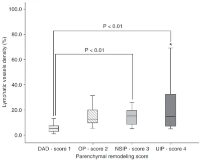
-chymal remodeling score in DAD, OP, NSIP, and UIP. In other words, the organizing fibrosis present as obliterative (score 1) in DAD, the intraluminal plugs (score 2) observed in OP, the septal inflammatory thickening and minimal organizing fibrosis (score 3) of NSIP, and the mural organizing fibrosis and fibroblast foci (score 4) found in UIP were significantly related to the density of lymphatic vessels (Figure 2).
Fluctuation of lymphatic vessels according to pulmonary parenchymal remodeling stage score
Lymphatic D2-40+ in the four categorized lesions is indicated in Figure 2. Lymphatic D2-40+ differed significantly in the lesions of the four categories. D2-40 expression
increased with the progression from DAD (6.66 ± 1.11),
OP (15.35 ± 3.75), NSIP (15.49 ± 1.80), up to UIP (23.45
± 5.24).
Pulmonary function tests and survival
Thirty-five patients died during the 180-month follow-up period (20 AIP, 1 COP, 5 NSIP, and 9 IPF). All patients studied had a restrictive lung function pattern characterized by a decrease in TLC (mean values were 57% of predicted values for AIP, 55% for NSIP and 75% for IPF) and an increase in FVC/FEV1 ratio (mean values were 97% of predicted values for AIP, 88% for COP, 109% for NSIP, and 118% for IPF). The mean predicted values of DLCO were
decreased in patients with NSIP (51%) and IPF (53%). The pulmonary function of patients with AIP, COP, NSIP, and IPF and with different degrees of organizing fibrosis and lymphatic density did not differ significantly between groups. We analyzed the survival curves by comparing patient characteristics (baseline data and physiological test) and distribution and fluctuation of lymphatic vessels
according to pulmonary parenchymal remodeling stage.
Kaplan-Meier survival curves showed that patients with a 9.3% median lymphatic vessel density in lung parenchyma presented higher survival than patients with a <9.3% median lymphatic vessel density in lung parenchyma (125 vs 45
months, respectively). Multivariate analyses by the Cox regression model showed statistical significance (log likeli
Lymphatic in progressive pulmonary remodeling 469
FVC (OR = 0.9), whereas a high risk of death was found for male patients (OR = 6.73) and with lymphatic density <9.3% (OR = 11.03). Regression plots of survival probability versus
follow-up time in months in patients with IIPs showed that
the group with lymphatic density >9.3% had better survival than the group with <9.3% (Figure 3).
Discussion
The present study using morphometry revealed a
large increase of the lymphatics in the interlobular and subpleural space, in which massive fibrosis increased with the development of new lymphatics in the lungs of patients with UIP. In contrast, in the lungs of patients with DAD, OP and cellular NSIP, a few lymphatics had extended into the alveolar lesions from the superficial lymphatics, especially into granulation tissues within the alveolar space. Although massive fibrotic diseases developed in IPF, including fi
-broblastic foci, lymphatic vessel density was increased in the lungs of UIP, probably draining excessive proteins and
fluid. These results suggest that the increase of superficial lymphatics may improve alveolar clear
-ance, accelerate organ repair and avoid disease
progress in patients with IPF.
Lymphatic dilatation was not detected in the
early stages of the remodeling process, but a few dilated lymphatics first appeared in the peripheral region adjacent to viable epithelial cells in the early process of fibroblastic foci. Lymphatic dila
-tation subsequently increased during the mural fibrosis period and was maintained thereafter
during scar formation. In addition, LDE increased in the early stage of intraluminal granulation of
OP and septal inflammatory thickening of NSIP, abruptly decreased in stages with DAD, but with a subsequent increase with the process of mural fibrosis in UIP. From these results, it can be seen
that, during the entire process of parenchymal remodeling, the lymphatics mainly participate in
fibrosis maturation and scar formation through the drainage of excessive proteins and fluid.
In the lung tissue of IIPs investigated in the present study, D2-40+ lymphatics were abundant in the subpleural regions, as well as in the interlobu
-lar interstitium. These findings are consistent with the distribution patterns of pulmonary lymphatics confirmed by other methods (5). In the light of lymphatic distribution, the abundant lymphatics in the subpleural region detected by D2-40 im -munohistochemistry seem to correspond to the
subpleural lymphatic plexus (6). Previous stud
-ies describing lymphatic distribution in sections
of normal renal tissue (16,20) and other reports
have also demonstrated the usefulness of D2-40
immunohistochemistry for detecting lymphatic capillaries in the renal interstitium (21).
Parenchymal remodeling after pulmonary
in-jury is characterized by an increase in pulmonary
endothelialand epithelial permeability.This early
phase is followed by a subacute fibroproliferative
phase that may allow repair of the injured lung
or result in progressiveobliteration of the inter
-stitial and alveolar compartments ofthe lung by a fibroproliferative process that may be in fact established by 24 h after the injury (22). In this process of epithelial necrosis, extracellular matri
-ces are degraded by inflammatory cell proteases (23-25). Based on such early events after the onset
of parenchymal remodeling, it is supposed that
pre-existing vascular tissues in the affected area also disintegrate through the influence of ischemia and inflammatory cells. The present results have clearly demonstrated that D2-40+ lymphatic ves -sels were poorly detected in the affected areas
of early remodeling such as DAD, OP and NSIP
Figure 2. Lymphatic density according to score for the four lesions. Lym-phatic D2-40+ in 10 fields of view observed by light microscopy at 400X magnification. The values of lymphatic D2-40+ are 6.66 ± 1.11 in diffuse al -veolar damage (DAD), 15.35 ± 3.75 in organizing pneumonia (OP), 15.49 ± 1.80 in nonspecific interstitial pneumonia (NSIP), and 23.45 ± 5.24 in usual interstitial pneumonia (UIP). Lymphatic density increases from DAD score 1, OP score 2, NSIP score 3, up to UIP score 4 with a statistically significant difference. *P < 0.01 (ANOVA).
Lymphatic in progressive pulmonary remodeling 471
compared to the UIP late stage. Lymphatics in the periph-eral pulmonary interstitium consist of single-layered, thin
endothelial cells, which possess an undeveloped basement membrane with no pericytes around them (6). These char
-acteristics of lymphatic structures may cause vulnerability to ischemic damage and/or degradation by proteases, as is the case for blood capillaries.
In the present study, LDE surprisingly increased in the
late mural fibrosis stage of UIP, when newly formed blood capillaries were usually decreased, as previously described by Parra et al. (10). In general, lymphangiogenesis devel
-ops through budding, sprouting and/or remodeling from pre-existing vessels, which frequently occur in pathological
conditions, such as malignant tumors (26,27). In the process of cutaneous wound healing in the mouse model, lymphatics
appear in subcutaneous tissue along the wound edge at 3-5
days after injury (28). It is therefore hypothesized that newly
formed lymphatics first appear in the peripheral area of the lesion through sprouting from the pre-existing lymphatics
around the lesion, and the proliferation of lymphatics follows
after blood vessel angiogenesis.
In the current study, LDE significantly increased with
healing maturation from the NSIP stage up to the UIP stage.
These results indicate that lymphatics mainly participate in the maturation of fibrosis and/or scar formation through the
drainage of excessive proteins and fluid. Based on these
descriptions, including our results, it is considered that the
role of lymphatics during parenchymal remodeling may be maintained for a longer time than that of blood vessels and
that immunohistochemistry using a lymphatic endothelial
marker is useful in investigating the role of lymphatics in
IIPs.
Our study has clinical implications. In order to establish the relevance of these findings to patient evolution, lymphatic distribution in AIP/DAD, COP/OP, NSIP/NSIP, and IPF/UIP was evaluated in terms of physiological testing and survival. Clearly, multivariate analyses showed a low risk of death for female patients with OP and high FVC, but a high risk
for males with median age >55 years and low lymphatic
D2-40+ expression.
Finally, lymphatic distribution may serve as a surrogate marker for the identification of patients at greatest risk for
death in IIPS.
Acknowledgments
Research supported by CNPq, FAPESP (#07/52785-0, #08/53022-3 and #08/57130-5), Laboratório Diagnóstika, Hospital das Clínicas and Faculdade de Medicina, Univer
-sidade de São Paulo.
References
1. Aharinejad S, Bock P, Firbas W, Schraufnagel DE. Pulmo -nary lymphatics and their spatial relationship to venous sphincters. Anat Rec 1995; 242: 531-544.
2. Bernaudin JF, Fleury-Feith J. [Structure and physiology of the pleura and the pleural space]. Rev Pneumol Clin 2006; 62: 73-77.
3. Shinohara H. Distribution of lymphatic stomata on the pleural surface of the thoracic cavity and the surface topography of the pleural mesothelium in the golden hamster. Anat Rec 1997; 249: 16-23.
4. Azzali G. The lymphatic vessels and the so-called “lymphatic stomata” of the diaphragm: a morphologic ultrastructural and three-dimensional study. Microvasc Res 1999; 57: 30-43. 5. Marchetti C, Poggi P, Clement MG, Aguggini G, Piacentini C,
Icaro-Cornaglia A. Lymphatic capillaries of the pig lung: TEM and SEM observations. Anat Rec 1994; 238: 368-373. 6. Ohtani O, Ohtani Y. Organization and developmental
aspects of lymphatic vessels. Arch Histol Cytol 2008; 71: 1-22.
7. White ES, Lazar MH, Thannickal VJ. Pathogenetic mecha -nisms in usual interstitial pneumonia/idiopathic pulmonary fibrosis. J Pathol 2003; 201: 343-354.
8. Selman M, King TE, Pardo A. Idiopathic pulmonary fibrosis: prevailing and evolving hypotheses about its pathogenesis and implications for therapy. Ann Intern Med 2001; 134: 136-151.
9. Parra ER, Kairalla RA, de Carvalho CR, Capelozzi VL. Abnormal deposition of collagen/elastic vascular fibres and prognostic significance in idiopathic interstitial pneumonias.
Thorax 2007; 62: 428-437.
10. Parra ER, David YR, da Costa LR, Ab’Saber A, Sousa R, Kairalla RA, et al. Heterogeneous remodeling of lung ves -sels in idiopathic pulmonary fibrosis. Lung 2005; 183: 291-300.
11. Parra ER, Silverio da Costa LR, Ab’Saber A, Ribeiro de Carvalho CR, Kairalla RA, Fernezlian SM, et al. Nonhomo -geneous density of CD34 and VCAM-1 alveolar capillaries in major types of idiopathic interstitial pneumonia. Lung 2005; 183: 363-373.
12. Ebina M, Shimizukawa M, Shibata N, Kimura Y, Suzuki T, Endo M, et al. Heterogeneous increase in CD34-positive alveolar capillaries in idiopathic pulmonary fibrosis. Am J Respir Crit Care Med 2004; 169: 1203-1208.
13. Yamashita M, Iwama N, Date F, Chiba R, Ebina M, Miki H, et al. Characterization of lymphangiogenesis in various stages of idiopathic diffuse alveolar damage. Hum Pathol 2009; 40: 542-551.
14. Mandal RV, Mark EJ, Kradin RL. Organizing pneumonia and pulmonary lymphatic architecture in diffuse alveolar dam -age. Hum Pathol 2008; 39: 1234-1238.
15. Akishima Y, Ito K, Zhang L, Ishikawa Y, Orikasa H, Kiguchi H, et al. Immunohistochemical detection of human small lymphatic vessels under normal and pathological conditions using the LYVE-1 antibody. Virchows Arch 2004; 444: 153-157.
15: 434-440.
17. American Thoracic Society/European Respiratory Society. American Thoracic Society/European Respiratory Society International Multidisciplinary Consensus Classification of the Idiopathic Interstitial Pneumonias. Am J Respir Crit Care Med 2002; 165: 277-304.
18. Quanjer PH, Tammeling GJ, Cotes JE, Pedersen OF, Peslin R, Yernault JC. Lung volumes and forced ventilatory flows. Report Working Party Standardization of Lung Function Tests, European Community for Steel and Coal. Official Statement of the European Respiratory Society. Eur Respir J Suppl 1993; 16: 5-40.
19. Gundersen HJ, Bendtsen TF, Korbo L, Marcussen N, Moller A, Nielsen K, et al. Some new, simple and efficient stereo-logical methods and their use in pathostereo-logical research and diagnosis. APMIS 1988; 96: 379-394.
20. Fukunaga M. Expression of D2-40 in lymphatic endothelium of normal tissues and in vascular tumours. Histopathology 2005; 46: 396-402.
21. Ishikawa Y, Akasaka Y, Kiguchi H, Akishima-Fukasawa Y, Hasegawa T, Ito K, et al. The human renal lymphatics under normal and pathological conditions. Histopathology 2006; 49: 265-273.
22. Rocco PR, Negri EM, Kurtz PM, Vasconcellos FP, Silva GH,
Capelozzi VL, et al. Lung tissue mechanics and extracellular matrix remodeling in acute lung injury. Am J Respir Crit Care Med 2001; 164: 1067-1071.
23. Santos FB, Nagato LK, Boechem NM, Negri EM, Guimarães A, Capelozzi VL, et al. Time course of lung parenchyma remodeling in pulmonary and extrapulmonary acute lung injury. J Appl Physiol 2006; 100: 98-106.
24. Rocco PR, Facchinetti LD, Ferreira HC, Negri EM, Capelozzi VL, Faffe DS, et al. Time course of respiratory mechanics and pulmonary structural remodelling in acute lung injury. Respir Physiol Neurobiol 2004; 143: 49-61.
25. Negri EM, Hoelz C, Barbas CS, Montes GS, Saldiva PH, Capelozzi VL. Acute remodeling of parenchyma in pul-monary and extrapulpul-monary ARDS. An autopsy study of collagen-elastic system fibers. Pathol Res Pract 2002; 198: 355-361.
26. Ji RC. Lymphatic endothelial cells, lymphangiogenesis, and extracellular matrix. Lymphat Res Biol 2006; 4: 83-100. 27. Scavelli C, Weber E, Agliano M, Cirulli T, Nico B, Vacca A,
et al. Lymphatics at the crossroads of angiogenesis and lymphangiogenesis. J Anat 2004; 204: 433-449.