Characterization of bovine respiratory
syncytial virus isolated in Brazil
1Laboratório de Virologia Animal, Departamento de Microbiologia e Imunologia, Instituto de Biologia, Universidade Estadual de Campinas, Campinas, SP, Brasil 2Laboratório de Diagnóstico de Doenças Infecciosas por Técnicas de Biologia Molecular, Departamento de Clínica Médica, Faculdade de Ciências Médicas, Universidade Estadual de Campinas, Campinas, SP, Brasil
C.W. Arns1, J. Campalans1,2, S.C.B. Costa2, H.G. Domingues1, R.C.F. D’Arce1 and R.S. Almeida1
Abstract
This paper presents the first isolation of bovine respiratory syncytial virus in Brazil and its physicochemical, morphological and molecular characterization. The virus was isolated from 33 samples of nasotracheal secretions, successively inoculated into a Madin-Darby bovine kidney cell culture, which was characterized by physicochemical tests and morphological observation by electron microscopy. The Brazilian sample is an RNA pleomorphic, enveloped, thermolabile and non-hemagglutinating spicular virus. Reverse transcription, followed by nested polymerase chain reaction (nRT-PCR) assay was carried out using oligonucleotides B1, B2A, B3 and B4 for the fusion proteins (F) and B5A, B6A, B7A and B8 for the attachment protein (G). The nRT-PCR-F amplified a fragment of 481 bp corresponding to part of the gene that codes for protein F, whereas nRT-PCR-G amplified a fragment of 371 bp, in agreement with part of the G gene. The virus isolated from Brazilian samples in this study corresponded to the bovine respiratory syncytial virus, and RT-PCR proved to be useful for the diagnosis of bovine clinical samples.
Correspondence
C.W. Arns
Laboratório de Virologia Animal Departamento de Microbiologia e Imunologia, IB, UNICAMP Caixa Postal 6109 13081-970 Campinas, SP Brasil
E-mail: [email protected] Research supported by FAPESP (No. 96/6273-1).
Received August 13, 2001 Accepted November 5, 2002
Key words
·Bovine respiratory syncytial
virus
·BRSV ·Virus isolation ·Characterization ·Nested RT-PCR
Bovine respiratory syncytial virus (BRSV) is an important pathogen that causes acute respiratory distress syndrome, and has been detected in cattle since 1970 (1). BRSV is a single-stranded negative-sense RNA virus which belongs to the Pneumovirus genus, a
member of the Paramyxoviridae family. Its genome has approximately 15,140 nucleo-tides and encodes 10 different mRNA mol-ecules (2,3). BRSV infection commonly oc-curs in cattle and serological studies indicate that the virus has already infected more than 95% of Brazilian cattle up to 3 years old,
with 70% of the calves being infected within the first year of life (4). The mortality rate in some herds with confirmed acute infection ranges from 5 to 20% (5). This disease poses a serious problem due to financial losses caused by animal death, costs of treatment, and subsequent reduced profits.
infec-tions in the south and southeast regions of the country (6-9). A serological investiga-tion was performed with 864 serum samples from 65 different farms located in the south-ern Brazilian states, using serum neutraliza-tion test (SNT) and enzyme-linked immuno-sorbent assay (ELISA), and the clinical his-tory was analyzed. Both tests showed high frequencies of positive BRSV (68 and 75%, respectively). Due to the high frequency of seropositivity detected in previous studies, there is a need to obtain Brazilian BRSV isolates in order to carry out detailed studies. The isolation of BRSV is not commonly used for diagnosis since tissue samples con-taining high concentrations of BRSV anti-gen do not often yield the virus in cell cul-tures. In most cases, BRSV is obtained dur-ing isolation procedures for viral pathogens rather than procedures specifically carried out for BRSV (10). The transport of clinical specimens from the field greatly reduces the sensitivity of virus isolation, but has a mini-mal effect on the results of ELISA and re-verse transcription-polymerase chain reac-tion (RT-PCR). These methods therefore are more suitable for diagnostic applications than virus isolation (11).
The objectives of the present study were to isolate BRSV from nasal secretion samples of young calves from Brazilian herds with respiratory distress signs, to characterize the viral agent by electron microscopy and to determine its physicochemical and molecu-lar features, and to develop nested RT-PCR (nRT-PCR) as a diagnostic method for BRSV in cell culture.
Viral isolates were obtained from samples of nasotracheal secretion from live animals in the southern and southeastern regions of Brazil. A total of 33 samples were analyzed (20 from Rio Grande do Sul State and 13 from São Paulo State). The study involved 23 Brazilian dairy cattle herds (25 to 120 animals) and 10 beef cattle herds (80 to 700 animals) kept under extensive management. The samples were collected from herds
pre-senting positive serology and from animals with respiratory distress signs.
Two positive control BRSV strains, BRSV-88 from Germany and the American Type Culture Collection reference strain Lehmkuhl 375 from the United States were used for the nRT-PCR-G assay, and an avian pneumovirus (SHS-121-BR) of the same subfamily was used as negative control.
The material for viral isolation was taken with swabs and transferred to tubes contain-ing sterile Eagle’s minimum essential medi-um with 20% glycerin, 20% horse sermedi-um and 5% antibiotic. The nasal secretions were frozen-thawed, homogenized at 1:10 dilu-tion and centrifuged, and the supernatant was filtered through a 0.2-µm filter. This filtrate (0.1 ml) was inoculated simulta-neously into a continuous Madin-Darby bo-vine kidney (MDBK) cell layer on 24-well microplates (Corning, New York, NY, USA). Each plate contained a cell control and a positive control (viral samples of BRSV). After 5 days of incubation with no detection of cytopathogenic effects, blind passages were carried out up to the 20th cell passage. The following tests were used to deter-mine the physicochemical features of the isolated virus: sensitivity to chloroform, tem-perature and 5-iodine-2-deoxyuridine, pH stability, hemagglutination, and hemadsorp-tion activity according to the methodology described by von Mayr et al. (12). For this purpose, a cell monolayer with 24 h of growth at 37ºC was used in 96-well microplates (Corning).
Cam-pinas, SP, Brazil.
The oligonucleotides used for nRT-PCR amplification were those reported by Vilcek et al. (5) based on the sequences of the F and G genes of the BRSV.
Total RNA was isolated from MDBK cell cultures infected with BRSV. Two days after infection, when the cytopathogenic ef-fect reached approximately 20%, and mono-layers were scraped, centrifuged, and lysed with Trizol™ (Invitrogen, Carlsbad, CA,
USA).
Reverse transcription was performed with Superscript™ RNAse H Reverse
Transcript-ase (Invitrogen) in a final volume of 20 µl following the manufacturer’s instructions.
Nested PCR was performed in a volume of 50 µl including 4 µl cDNA, containing 1 µl 10 pmol of each primer, 5 µl 10X RT-PCR buffer (200 mM Tris-HCl, pH 8.4, 500 mM KCl), 2 µl 50 mM MgCl2, 2 µl 5 mM deoxy-nucleotide triphosphate mix, 0.3 µl Taq DNA polymerase (Invitrogen), and 34.7 µl of di-ethyl pyrocarbonate-treated distilled water. RT-PCR was performed in a thermal cycler (PTC-100, MJ Research, Waltham, MA, USA).
Amplified DNA was visualized by 1% ethidium bromide staining in UV light, after 1.8% agarose gel electrophoresis at 90 V for 30 min and documented with an instant cam-era (Polaroid DS 34).
One of 33 cases examined was BRSV positive after viral isolation. The isolated BRSV sample was taken from a 2-month-old Holstein calf from the State of Rio Grande do Sul, located close to Argentina and Uru-guay. Cough, nasal secretion and severe dys-pnea were observed. This sample was packed in dry ice and arrived at the laboratory 18 h later. Successful isolation was obtained in MDBK cell culture after nine blind pas-sages, with a perceptible cytopathic effect-like syncytial arrangement observed on the fourth day after inoculation. This viral sample, propagated and submitted to viral characterization tests, was named
BRSV-25-BR.
The isolation of BRSV is a laborious procedure with unpredictable results because animals that develop the disease are not the best choice for virus isolation. Tissue samples containing high concentrations of BRSV antigen frequently do not reproduce the vi-rus in cell cultures (10). Several factors are involved, mainly high virus lability, which requires special care from the harvesting of samples to the inoculation in cell cultures. The time elapsed between the two phases must be minimal, and the virus should be kept at low temperatures. Another difficulty is the large number of cellular passages needed to obtain a successful isolation. Al-though BRSV may be present in many samples, isolation is not easily achieved, probably because the cell lines have few receptors for virus absorption.
Several clinical samples from the same source were tested for viral isolation and submitted to the same laboratory procedures, but it was not possible to isolate the virus from all of them. The distance from the laboratory and the transportation conditions, as well as sample packaging must have had an influence on the low yield of viral isola-tion.
It may be argued that successful isolation was favored by the use of live animal samples, which contained enough viral particles with infecting capacity in their cells. In former studies with samples from autopsied animal tissues, the difficulty in obtaining these free and infective viruses seemed to be greater due to the time elapsed between death, au-topsy and inoculation in the cell cultures (6,9). Furthermore, it is easier to obtain tis-sue samples from animals autopsied during the acute phase of the disease (10).
affected, which is an indication that it is an RNA virus. Regarding temperature treatment, it was observed that exposure to 56º and 60ºC for a 30-min period inactivated the viral sample. The same effect was observed regarding the chloroform effect. From the data obtained, it was possible to conclude that BRSV-25-BR is an enveloped virus, and the above procedures eliminated its infectiv-ity.
The treatments at different pH values showed that the virus was stable over an extended pH range (6.0 to 9.0) at 4ºC, but was affected by exposure time at this tem-perature, especially after 180 min. Cyto-pathogenic effects could not be detected at pH 3.0 but there was a toxic effect on the cells.
The results of hemagglutination activity tests of the BRSV-25-BR sample using chicken, turkey, guinea pig, rabbit, mouse, bovine and sheep erythrocytes in 1% NaCl buffer and incubation of 30 and 60 min at 4ºC were negative. The same erythrocytes were used for the hemadsorption activity test. The viral incubation periods studied were 2, 4 and 6 h. The hemadsorption activ-ity was negative. The results confirmed that this viral sample did not agglutinate or ad-sorb any kind of erythrocyte under investiga-tion after the incubainvestiga-tion periods tested.
The methods used for virus characteriza-tion, such as thermostability, chloroform sen-sitivity, pH stability, hemagglutination and hemadsorption tests, indicated that the sample can be identified as the bovine RNA virus, an enveloped non-hemagglutinant virus, with
cytopathic effects characterized as syncytial formation. These are the characteristics of BRSV, which were described by Stott and Taylor (15) and Mallipeddi et al. (16).
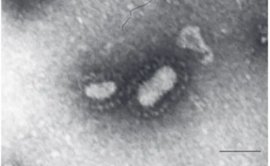
BRSV-25-BR was studied morphologi-cally by analysis of images generated by electron microscopy. The sample provided images of a pleomorphic virus, approximately 100-300 nm in size, showing the presence of spicules. A large number of defective or destroyed particles were observed, in agree-ment with data reported by Mallipeddi et al. (16). The images (Figure 1) showed the mor-phologic characteristics of BRSV and the physicochemical features allowed us to con-firm that the virus isolated from cattle in Brazil is a strain of this virus.
RT-PCR has been extensively applied in modern microbiology for molecular analy-sis, although only a few laboratories use this technique as a routine clinical test. Nested RT-PCR assays are useful tools, showing high sensitivity and specificity for BRSV detection in clinical material, better than virus growth in cell culture (11,17,18). Fur-thermore, comparative analysis revealed that nRT-PCR was ten times more sensitive than RT-PCR (5).
In the present study two nRT-PCR tests were developed based on the fusion protein gene (nRT-PCR-F) and on the attachment protein gene (nRT-PCR-G) of the BRSV, to be used for viral detection in infected cells. The primers were selected from two essen-tial genes (F and G) of the BRSV genome because both are enveloped by highly immu-nogenic proteins. The G protein is the most antigenically dissimilar protein among the BRSV strains (19), and for this reason is extensively used in comparative BRSV stud-ies.
The viral RNA was used as a template for cDNA synthesis with extension from a syn-thetic oligonucleotide primer. All bovine strains tested, BRSV-25-BR, BRSV-88 and Lehmkuhl 375, were detected by the PCR-F and PCR-G assays. An
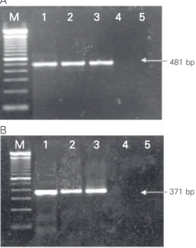
PCR product of the expected size, 481 bp corresponding to part of the F gene and 371 bp corresponding to part of the G gene, was observed (Figure 2). Amplification products were not observed for the negative control. The same regions were amplified in the Bra-zilian sample to confirm that the isolated virus was BRSV.
The optimal RT-PCR conditions were different for nRT-PCR-G and nRT-PCR-F and also differed between the first and the second RT-PCR-F. These conditions were adjusted in order to minimize nonspecifi-cally amplified bands.
The results obtained in this study indi-cate that it is possible to use this technique for a fast detection of BRSV; this advance is indeed of great clinical importance, produc-ing rapid results, in contrast to virus culture. The use of nRT-PCR with inner primers allowed us to enhance the sensitivity and specificity of the test. Because of its fast execution, sensitivity and specificity, nRT-PCR could be a useful tool for the diagnosis of clinical samples.
Nested RT-PCR-G seems to be a good option for routine disease diagnosis and for
481 bp
371 bp M
MM
MM 11111 22222 33333 44444 55555
M MM M
M 11111 22222 33333 44444 55555 A
B
Figure 2. Amplified products from viral samples proliferated in cell culture. Nested RT-PCR-F (A) and nested RT-PCR-G (B). Lanes 1, 2 and 3 represent BRSV-25-BR, BRSV-88 and Lehmkuhl 375 reaction products, respectively. Lane 4 represents SHS-121-BR, lane 5 is the blank, and M denotes the 100-bp DNA ladder lane.
analysis of the diversity of the BRSV ge-nome. The extensive application of the nRT-PCR-G technique in the future will provide information on BRSV genotypes circulating in Brazil and may contribute to the establish-ment of official control programs against this virus.
References
1. Paccaud MF & Jacquier C (1970). A respiratory syncytial virus of bovine origin. Archiv für die Gesamte Virusforschung, 30: 327-342. 2. Buchholz UJ, Finke S & Conzelmann KK (1999). Generation of
bo-vine respiratory syncytial virus (BRSV) from cDNA: BRSV NS2 is not essential for virus replication in tissue culture, and the human RSV leader region acts as a functional BRSV genome promoter. Journal of Virology, 73: 251-259.
3. Pastey MK & Samal SK (1995). Nucleotide sequence analysis of the non-structural NS1 (1C) and NS2 (1B) protein genes of bovine respi-ratory syncytial virus. Journal of General Virology, 76: 193-197. 4. Furze JM, Roberts SR, Wertz GW & Taylor G (1997). Antigenically
distinct G glycoproteins of BRSV strains share a high degree of genetic homogeneity. Virology, 231: 48-58.
5. Vilcek S, Elvander M, Ballagi-Pordany A & Belak S (1994). Develop-ment of nested PCR assays for detection of bovine respiratory syncytial virus in clinical samples. Journal of Clinical Microbiology, 32: 2225-2231.
6. Gonçalves IPD, Jost HC, Soglio AD, Simanke AT, Hotzel I & Moojen V (1993). Detection of bovine respiratory syncytial virus in calves of Rio Grande do Sul, Brazil. Ciência Rural, Santa Maria, 23: 389-390. 7. Campalans J & Arns CW (1997). Serological evidence of bovine
respiratory syncytial virus in Brazil. Virus Reviews and Research, 1-2:
50-56.
8. Driemeier D, Gomes MJP, Moojen V, Arns CW, Vogg G, Kessler L & Da Costa UM (1997). Manifestação clínico-patológica de infecção natural pelo vírus respiratório sincicial bovino (BRSV) em bovinos de criação extensiva no Rio Grande do Sul, Brasil. Pesquisa Veterinária Brasileira, 17: 77-81.
9. Flores EF, Weiblen R, Medeiros M, Botton SA, Irigoyen LF, Driemeier D, Schuch LF & Moraes M (2000). A retrospective search for bovine respiratory syncytial virus (BRSV) antigens in histological specimens by immunofluorescence and immunohistochemistry.
Pesquisa Veterinária Brasileira, 20: 139-143.
10. Dubovi EJ (1993). Diagnosing BRSV infection: A laboratory perspec-tive. Veterinary Medicine,88: 888-893.
11. West K, Bodgan J, Hamel A, Nayar G, Morley PS, Haines DM & Ellis JA (1998). A comparison of diagnostic methods for the detection of bovine respiratory syncytial virus in experimental clinical specimens.
Canadian Journal of Veterinary Research, 62: 245-250.
12. von Mayr A, Bachmann PA, Bibrack B & Wittmann G (1974). Züchtung von Viren in Zellkulturen. In: Virologische Arbeitsmethoden. Band I. Gustav Fischer Verlag, Stuttgart, Germany, 231-262.
14. Barth OM (1984). Estudos sobre a contrastação negativa de suspen-sões virais. Revista Brasileira de Biologia, 44: 71-80.
15. Stott EJ & Taylor G (1985). Respiratory syncytial virus. Brief Review.
Archives of Virology, 84: 1-52.
16. Mallipeddi SK, Samal SK & Mohanty SB (1990). Analysis of polypep-tides synthesized in bovine respiratory syncytial virus infected cells.
Archives of Virology, 115: 23-36.
17. Paton AW, Paton JC, Lawrence AJ, Goldwater PN & Harris RJ (1992). Rapid detection of Respiratory Syncytial Virus in nasopharyn-geal aspirates by reverse transcription and polymerase chain
reac-tion amplificareac-tion. Journal of Clinical Microbiology, 30: 901-904. 18. Valarcher JF, Bourhy H, Gelfi J & Schelcher F (1999). Evaluation of a
nested reverse transcription-PCR assay based on the nucleoprotein gene for diagnosis of spontaneous and experimental bovine respira-tory syncytial virus infections. Journal of Clinical Microbiology, 37: 1858-1862.