Effect of JJYMD-C, a novel synthetic
derivative of gallic acid, on proliferation
and phenotype maintenance in
rabbit articular chondrocytes
in vitro
G.J. Xu
1,2, Z.H. Lu
3*, X. Lin
4*, C.W. Lin
4, L. Zheng
2,3and J.M. Zhao
1,2 1Osteopathy Ward, The First Affiliated Hospital, Guangxi Medical University, Nanning, Guangxi, China 2Research Center for Regenerative Medicine, Guangxi Medical University, Nanning, Guangxi, China 3The Medical and Scientific Research Center, Guangxi Medical University, Nanning, Guangxi, China 4School of Chemistry and Chemical Engineering, Guangxi University, Nanning, Guangxi, ChinaAbstract
Tissue engineering encapsulated cells such as chondrocytes in the carrier matrix have been widely used to repair cartilage defects. However, chondrocyte phenotype is easily lost when chondrocytes are expandedin vitroby a process defined as ‘‘dedifferentiation’’. To ensure successful therapy, an effective pro-chondrogenic agent is necessary to overcome the obstacle of limited cell numbers in the restoration process, and dedifferentiation is a prerequisite. Gallic acid (GA) has been used in the treatment of arthritis, but its biocompatibility is inferior to that of other compounds. In this study, we modified GA by incorporating sulfamonomethoxine sodium and synthesized a sulfonamido-based gallate, JJYMD-C, and evaluated its effect on chondrocyte metabolism. Our results showed that JJYMD-C could effectively increase the levels of the collagen II, Sox9, and aggrecan genes, promote chondrocyte growth, and enhance secretion and synthesis of cartilage extracellular matrix. On the other hand, expression of the collagen I gene was effectively down-regulated, demonstrating inhibition of chondrocyte dedifferentiation by JJYMD-C. Hypertrophy, as a characteristic of chondrocyte ossification, was undetectable in the JJYMD-C groups. We used JJYMD-C at doses of 0.125, 0.25, and 0.5mg/mL, and the strongest response was observed with 0.25mg/mL.
This study provides a basis for further studies on a novel agent in the treatment of articular cartilage defects.
Key words: Sulfamonomethoxine sodium; Gallic acid; Pro-chondrogenic agent; Chondrocyte; Rabbit articular cartilage; Dedifferentiation
Introduction
Articular cartilage has poor regenerative capacity after injury (1-3). Catabolic factors such as pro-inflammatory cytokines, which can induce a gradual self-destruction of cartilage, are activated after injury, finally resulting in secondary osteoarthritis. The poor healing capability of cartilage and dense extracellular matrix (ECM), which prevent chondroprogenitors from migrating to the injury site, altogether contribute to irreversible cartilage loss (2,4). Tissue engineering encapsulated cells in carrier matrix have been widely used to repair cartilage defects (5-8) and are considered to be a promising strategy for regeneration of cartilage defects. Shaped cartilage has been regeneratedin
vitroand in immunocompromised animals by using auto-logous, allogeneic, or xenogeneic transplants (9). However, translation to immunocompetent animals or clinical use has proven to be difficult because post-injury inflammation and sustained inflammatory reactions may inhibit transplanted chondrocytes to synthesize sufficient ECM (10). Another difficulty is dedifferentiation of chondrocytes during expan-sion in vitro, which is necessary for cell-based therapy. Since dedifferentiated chondrocytes produce a non-carti-lage-specific ECM characterized by inferior mechanical properties, they are not suitable for cell-based therapy. Finding anti-inflammatory mediators that can also restore
Correspondence: L. Zheng and/or J.M. Zhao, Research Center for Regenerative Medicine, Guangxi Medical University, Nanning, Guangxi, 530021, China. Fax: ++86-077-1535-0975 and/or ++86-077-1535-0189. E-mail: [email protected] and/or [email protected]
*Z.H. Lu and X. Lin contributed equally to this work.
cartilage function is a prerequisite to support neo-cartilage formation and inhibit post-traumatic cartilage inflammation.
Gallic acid (GA) and its derivatives are a group of polyphenolic compounds known to affect several phar-macological and biochemical pathways, in addition to their strong anti-oxidation (11-13) and anti-inflammatory prop-erties (14,15). It was reported that GA could induce apoptosis in rheumatoid arthritis fibroblast-like synovio-cytes through regulation of apoptosis-related protein expression and reduction in the expression of pro-inflammatory mediators, such as pro-pro-inflammatory cytokines, chemokines, cyclooxygenase-2, and matrix metalloproteinase-9 (16). Another study reported that GA could attenuate the pro-inflammatory and pro-oxidant effects caused by tumor necrosis factor-a, interleukin-6, NADPH oxidase, and thioredoxin-interacting protein. It can also attenuate DNA damage and suppress hypergly-cemia-induced activation of inflammatory and pro-oxidant gene expression (17). The study by Kim et al. (18) showed that GA was a histone acetyltransferase inhibitor and could suppressb-amyloid neurotoxicity by inhibiting microglial-mediated neuroinflammation. However, GA was reported to suppress cell proliferation. The investiga-tion by Ou et al. (19) indicated that GA was an effective anti-atherogenic agent in vascular smooth muscle cells by attenuating cell cycle progression via AMP-activated protein kinase-mediated activation of endothelial nitric-oxide synthase (19). Being much more hydrophilic than its esters, GA displayed much weaker pharmacological effects in cell systems (11). Thus, modification of GA to improve its cytocompatibility is necessary.
Sulfamonomethoxine sodium has been used as a synthetic antibiotic for a long time because it is inexpensive and readily available. Most importantly, it easily penetrates through the cell membrane and into tissues and body fluids. Recently, new N-isopropoxy-arylsulfonamide-based hydro-xamate inhibitors were shown to be effective in a collagenin vitroassay and cartilage degradation model (20). These compounds contain several sulfonamide groups that could benefit cell growth. This study indicates that modification of the GA with a sulfa drug may promote chondrogenesis.
In this study, we synthesized a novel sulfonamide-based gallate to further examine its effect on chondrocyte metabolism. This study provides evidence for its applica-tion in cartilage tissue engineering.
Material and Methods
Synthesis of JJYMD-C
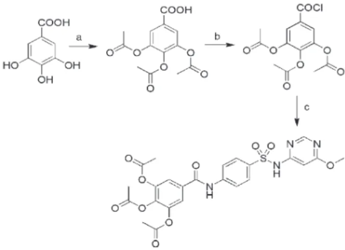
3,4,5-Triacetoxy-N-{4-[(6-pyrimidin-4-yl)sulfamoyl]-phenyl}benzamide (JJYMD-C) was prepared from GA and sulfamonomethoxine sodium. A scheme of the synthetic route is shown in Figure 1. After reaction c, distilled water was added to the mixture. The raw product (powder) was then precipitated and separated by vacuum filtration. The powder was recrystallized from methanol/tetrahydrofuran.
Articular cartilage cell culture
Articular chondrocytes were dissociated from knee joint cartilage slices of 1-week-old New Zealand rabbits by enzymatic digestion. In brief, two New Zealand rabbits were used, and cartilage slices were dissociated with trypsin (0.25% aqueous solution; Solarbio, China; 30 min; 376C) and then with type-II collagenase (2 mg/mL; Gibco, USA) in alpha-modified Eagle’s medium (a-MEM; Gibco; 3 h; 376C). After simple centrifugation (300g, 5 min, 376C), the chondrocytes were resuspended witha-MEM containing fetal bovine serum (20%, v/v; Gibco) and penicillin/streptomycin (1%, v/v; Solarbio) and cultured in a humidified incubator (5% CO2; 376C; 6 days). The culture medium was replaced every 3 days after plating. Rabbit chondrocytes (passage 1) were used for further studies.
JJYMD-C treatment
JJYMD-C dissolved in dimethylsulfoxide (DMSO; Sigma, USA) was prepared as stock solution and stored (-46C). This stock solution was then added to the cell cultures to provide different concentrations of JJYMD-C. Culture media containing different concentrations of JJYMD-C were replaced every 3 days.
Cytotoxicity assay
The assay for JJYMD-C cytotoxicity to chondrocytes was performed by the 3-(4,5-dimethylthiazol-2-yl)-2,5-diphenyltetrazolium bromide (MTT; Gibco) method. Chondrocytes were digested using trypsin/EDTA (ethy-lenediaminetetraacetic acid) solution (0.25%; Solarbio) when cells of passage 1 reached 60-70% confluence. After centrifugation (300g, 5 min, 376C), the cells were resuspended in fresh medium, counted using a hemocy-tometer, and then seeded on 96-well plates. The final cell number in each well was 56103. Concentrations of
JJYMD-C (0.00-2.5mg/mL) were then added to the cell cultures. After 3 days of culture, the cytotoxicity assay was carried out by MTT analysis. Briefly, a solution of MTT in phosphate-buffered saline, pH=7.0, was added to each well (final concentration: 5 mg/mL) and incubated (376C; 4 h). After the medium was removed, DMSO (200mL) was added to dissolve the MTT formazan formed by the metabolically viable cells. Absorbance (570 nm) of MTT was measured by an enzyme-labeled meter (Thermo Fisher Scientific, UK). All experiments were performed in triplicate.
Cell proliferation analysis and biochemical assay After culture (2, 4, and 6 days), the cells removed from the old media were digested with a proteinase K solution (Sigma) for the following biochemical assays. Intracellular secretion of glycosaminoglycans (GAGs) was assayed by the 1,9-dimethylmethylene blue dye (Sigma) and DNA content was quantified using the Hoechst 33258 dye (Sigma) assay. In each sample, absorbance (460 nm) of total intracellular DNA content, as indicated by the Hoechst 33258 dye, was measured using a spectro-fluorometer. Absorbance (525 nm) of total intracellular GAG secretion was measured spectrophotometrically and converted into concentration using a chondroitin sulfate (Sigma) standard curve. Production of GAGs by each chondrocyte was normalized to the total DNA content of all chondrocytes, which displayed their biosynthetic activity in the various culture media.
Safranin-O staining
Histology of chondrocytes was performed to assess GAG synthesis using safranin-O/Fast Green staining. After the cells were fixed in 95% ethanol (30 min), they were successively stained with 0.02% aqueous Fast Green for 5 min (Sigma) and 0.1% safranin O for 10 min (Sigma), immediately washed with tap water, and dried naturally at room temperature. The cells were then sealed with a neutral gum, observed, and photo-graphed under an inverted phase-contrast microscope (Zeiss Corporation, Germany).
Morphological examination
After 2, 4, and 6 days of incubation, the chondrocytes
were fixed in 95% ethanol before hematoxylin-eosin staining. Cells were initially stained with the nuclear (3 min) and then with the cytoplasm (5 s) dye. Subsequently, the cells were rinsed in tap water, dried naturally at room temperature and sealed with neutral gum. They were then examined and photographed using an inverted phase-contrast microscope (Zeiss Corporation).
Cell viability assay
Cell viability of chondrocytes was determined by fluorescein diacetate (FDA; Genway Biotech Inc., USA)/ propidium iodide (PI; Sigma) staining at days 2, 4, and 6. Briefly, FDA and PI stock solutions were added to the cells (final concentration: 2mM and 2mg/L, respectively), and they were incubated in the dark (376C; 5 min). Images were taken using a laser-scanning confocal microscope (Nikon A1, Japan).
Immunohistochemical staining
Secretion of type I and II collagens was characterized immunohistochemically using an immunohistochemical staining kit (Boster, China). To visualize collagen, the cells were first fixed in paraformaldehyde (4%, w/v) and treated with Triton X-100. To exclude endogenous peroxidase activity, the cells were then incubated at room temperature with H2O2 (3%; 10 min). In addition, the plates were blocked at room temperature with normal goat serum (10 min). Human anti-rabbit antibody (type I and II collagens) was added after dilution (1:200) and the cells were then incubated with the second antibody and biotin-labeled horseradish peroxidase. Subsequently, antibody binding was visualized with 3,39-diaminobenzidine tetra-hydrochloride (DAB kit, Boster) before brief counterstaining with hematoxylin. Cells were gradually dehydrated, sealed with neutral gum, observed, and photographed using an inverted phase-contrast microscope (Zeiss Corporation).
Quantitative real-time polymerase chain reaction (qRT-PCR) analysis
The genetic information for type I, II, and X collagens, aggrecan, and Sox9 genes was detected by qRT-PCR (Table 1). Total intracellular RNA was extracted employ-ing the RNA isolation kit (Tiangen Biotechnology, China) according to the manufacturer’s instructions. About
Table 1. Genes and primer design for quantitative real-time polymerase chain reaction analysis.
Genes Forward primer (59to 39) Reverse primer (59to 39)
GAPDH GTCATCATCTCAGCCCCCTC GGATGCGTTGCTGACAATCT
Aggrecan TTGCCTTTGTGGACACCAGT GAGCCAAGGACGTAAACCCA
Sox9 GACGCACATCTCGCCCAAC TCTCGCTTCAGGTCAGCCTT
Type I collagen CCCAGCCACCTCAAGAGAAG CGGGGCTCTTGATGTTCTCA
Type II collagen TCCGGAAACCAGGACCAAAG CTTTGTCACCACGGTCACCT
300 ng total RNA was used as a template and reverse-transcribed into cDNA using a reverse transcription kit (Fermentas Company, USA). qRT-PCR was performed using the Quantitative PCR Detection System (Realplex 4, Eppendorf Corporation, USA) with FastStart Universal SYBR Green Master Mix (Roche Company, Germany) at 956C (10 min) and 606C (1 min). The melting curve data were collected to verify PCR specificity. Each gene was analyzed in triplicate to diminish operation error. Relative gene expression levels were calculated using the 2-DDCt method relative to glyceraldehyde-3-phosphate dehydro-genase (GAPDH) gene expression.
Statistical analysis
Results are reported as means±SD. Statistical
differences were determined using one-way ANOVA followed by the Dunnett post hoc test. The level of significance was set at P,0.05.
Results
Preparation of JJYMD-C
The procedure for synthesis of JJYMD-C from GA and sulfamonomethoxine sodium is shown in Figure 1. JJYMD-C has the following properties: white powder, yield 59%, m.p. .2706C, MS-ESI: m/z: 557.0 [M-H]-, 1H-NMR (400 MHz, DMSO)d10.69 (s, 1H, -CO-NH), 8.41 (s, 1H, Py-H), 7.92 (m, 4H, 46Ar-H), 7.82 (s, 2H, Ar-H), 6.73
(s, 1H, Py-H), 3.83 (s, 3H, -OCH3), 2.33 (s, 3H, -CH3), 2.31 (s, 6H, 26-CH3).13C-NMR (125 MHz, DMSO)d169.99,
168.05, 166.99, 163.73, 158.56, 143.22, 142.85, 137.68, 132.45, 128.10, 120.79, 120.14, 90.94, 54.23, 20.34, 19.90.
Cytotoxicity assay
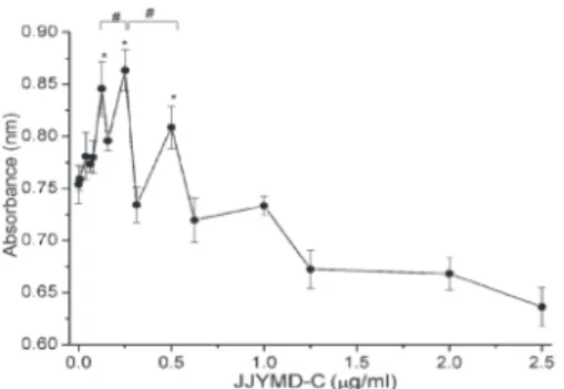
Cytotoxicity of JJYMD-C on articular chondrocytes was examined by the MTT assay. Articular chondrocytes were treated with three different increasing concentrations of JJYMD-C (range: 0.0-2.5mg/mL). As shown in Figure 2, absorbance values for JJYMD-C concentrations were comparable to those of the control in the range of 0.125-0.5mg/mL.
Cell proliferation
In this study, cell proliferation in the experimental and control groups was analyzed by measurements of DNA content. Comparatively, cells cultured with JJYMD-C (J1, J2, and J3: JJMYD-C of 0.125, 0.25, and 0.5mg/mL, respectively) grew more than those in the control group
Figure 2.Cytotoxicity analysis of chondrocytes after 3 days of treatment with different concentrations of JJYMD-C. Data are reported as means±SD for n=20. *P,0.05 compared to control; #P
,0.05 comparisons as indicated (one-way ANOVA followed by the Dunnettpost hoctest).
Figure 3.Quantification of cell proliferation by detection of DNA content and matrix production by glycosaminoglycan (GAG) analysis. A, Proliferation of chondrocytes culturedin vitrowith 0 (control), 0.125 (J1), 0.25 (J2), and 0.5mg/mL (J3) JJYMD-C for 2, 4, and 6 days.
(P,0.05), as indicated by higher DNA values (Figure 3A), at the same period. Furthermore, among different concentrations of JJYMD-C, the highest cell proliferation was achieved at 0.25mg/mL.
Secretion of GAGs
Extracellular production of GAGs by rabbit articular chondrocytes was measured by biochemical assay after 2, 4, and 6 days of culture. Regarding intracellular production of GAGs, given as a ratio of GAGs to DNA at different concentrations of JJYMD-C (Figure 3B), production of GAGs was time-dependent in all groups. Quantitatively, GAG production in culture media treated with JJYMD-C was increased significantly more than that in the control at the same times. JJYMD-C at 0.25mg/mL promoted the highest GAG synthesis among the three concentrations.
The safranin-O-positive stain in the JJYMD-C group indicated that GAGs were abundant and homogeneously
distributed around the chondrocytes (Figure 4).
Cell morphology
We assessed the morphology of articular chondro-cytes by inverted microscopy after treatment with JJYMD-C at concentrations of 0.125, 0.25, and 0.5mg/mL (Figure 5). No significant differences were observed in cartilagi-nous morphology between the experimental groups after 2, 4, and 6 days of culture in the presence of increasing concentrations of JJYMD-C. Compared with the control, the chondrocytes grew better in the presence of JJYMD-C and showed a distinctive proliferation tendency with gradually increasing culture time. In addition, JJYMD-C could enhance proliferation of chondrocytes at the concentration of 0.25mg/mL better than in other groups.
Cell viability assay
Viable cells and dead cells were visualized using the calcein-acetoxymethyl ester (AM)/PI double stain (Figure
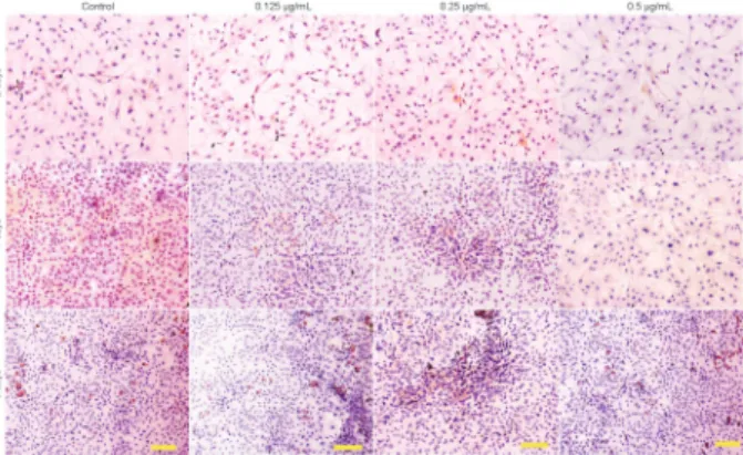
Figure 4. Safranin-O-staining showing the synthesis of extra-cellular matrix by chondrocytes culturedin vitrowith 0 (control), 0.125, 0.25, and 0.5mg/mL JJYMD-C for 2, 4, and 6 days. Scale bar: 100mm.
Figure 5.Hematoxylin-eosin staining showing the morphology of chondrocytes culturedin vitrowith 0 (control), 0.125, 0.25, and 0.5mg/mL JJYMD-C for 2, 4, and 6 days. Scale bar: 100mm.
Figure 6.Laser-scanning confocal microscopy images showing the viability of chondrocytes cultured in vitro with 0 (control), 0.125, 0.25, and 0.5mg/mL JJYMD-C for 2, 4, and 6 days. Scale
bar: 100mm.
Figure 7.Immunohistochemical staining revealed the presence of type I collagen. Chondrocytes culturedin vitrowith 0 (control), 0.125, 0.25, and 0.5mg/mL JJYMD-C for 2, 4, and 6 days. Scale
6). Calcein-AM/PI staining showed that survival in the JJYMD-C groups was more frequent than in the control group. Consistent with the results of cell proliferation (Figure 3), viable cells were far more frequent than dead cells in different concentrations of JJYMD-C. As evi-denced by the higher number of live cells, 0.25mg/mL JJYMD-C was superior to others.
Secretion of type I and type II collagens
Figures 7 and 8 show expression of type I and II collagens in the cytoplasm of chondrocytes at different treatment times with and without drug-treated culture media. Strongly positive staining is evident in large areas for cartilage-specific type II collagen and very sparse with light staining for type I collagen in the presence of JJYMD-C. In contrast, less positive staining of collagen II and less negative staining of collagen I were present in the control. At 0.25mg/mL, JJYMD-C could maintain the phenotype of chondrocytes better than at other concentrations.
Gene expression
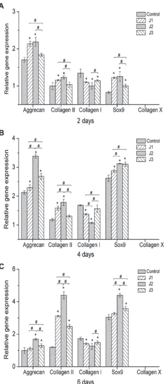
The effect of JJYMD-C on synthesis of ECM by chondrocytes was further investigated through gene expression of collagens I, II, and X, Sox9, and aggrecan (a proteoglycan composed of GAGs) after 2, 4, and 6 days of culture. As shown in Figure 9, expression of cartilage-specific genes, such as aggrecan, collagen II, and Sox9, was significantly boosted by JJYMD-C at the three concentrations compared with the control. Moreover, the highest expression of collagen II, aggrecan, and Sox9 in the presence of different concentrations of JJYMD-C was observed at 0.25mg/mL. JJYMD-C could up-regulate collagen II, aggrecan, and Sox9 expression. At the same time, collagen X expression was scarcely detectable in the presence of JJYMD-C, indicating that cell dedifferentiation and hypertrophy were not prominent. JJYMD-C at different concentrations induced lower
Figure 8.Immunohistochemical staining revealed the presence of type II collagen. Chondrocytes culturedin vitrowith 0 (control), 0.125, 0.25, and 0.5mg/mL JJYMD-C for 2, 4, and 6 days. Scale
bar: 100mm.
Figure 9.Quantitative comparison of extracellular matrix-related gene expression by quantitative real-time polymerase chain reaction. The chondrocytes were cultured with 0 (control), 0.125 (J1), 0.25 (J2), and 0.5mg/mL (J3) JJYMD-C for 2 days (A), 4 days (B), and 6 days (C) (n=3 for each experiment). The levels of gene expression in media containing JJYMD-C were analyzed by the 2-DDCT method, and glyceraldehyde-3-phosphate dehydrogenase was used as internal control. Data are reported as means±SD of three independent culture experiments. *P,0.05 compared to control; #P
expression of collagen I after culture for 2, 4, and 6 days compared with the control group. Moreover, the levels of collagen I with 0.25mg/mL JJYMD-C were lower than with the other two concentrations.
Among three different concentrations, JJYMD-C at a concentration of 0.25mg/mL demonstrated the highest expression of aggrecan and collagen II.
Discussion
GA, an endogenous plant polyphenol, which exists abundantly in tea, grapes, berries, and other plants, has important biological and pharmacological properties, such as cancer (21-24), oxidant (17,25), and anti-inflammatory (26) activities. It has been suggested that a more hydrophilic GA derivative would be more efficient in diffusing through the membrane polar phase (11,27). In this study, we synthesized a novel GA derivative, JJYMD-C, and examined its effect on chondrocyte metabolism. Our results indicated that JJYMD-C exhibited an effect of promoting chondrocyte growth compared with the control group, especially at the concentration of 0.25mg/mL (Figures 3, 5, 6, and 7). As demonstrated by biochemical assay and safranin-O staining (Figures 3 and 4), JJYMD-C could obviously promote GAG deposition in cultured chondrocytes, which play a pivotal role in maintaining cartilage load-bearing capacity (28). In addition, our results (Figures 8 and 9) indicated that JJYMD-C in the range of 0.125-0.5mg/mL enhanced the expression of collagen II, Sox9, and aggrecan genes, which are cartilage-specific markers (29,30). In JJMYD-C groups, 0.25mg/mL showed the highest expression of aggrecan and collagen II.
Expression of type I collagen, which indicates de-differentiation of chondrocytes (31-33), was significantly inhibited by JJYMD-C (Figures 8 and 9). In most cases, dedifferentiation occurred whereas the differentiated phenotype of chondrocytes consists primarily of type II collagen. Cartilage-specific proteoglycan is lost and replaced by a complex collagen phenotype consisting predominately of type I collagen with a low level of proteoglycan synthesis (34,35). Results from PCR, biochemical, and immunohistochemical analyses have shown that type I collagen expression was significantly down-regulated by JJYMD-C. Moreover, secretion of type X collagen, considered as characteristic of hypertrophic chondrocytes and endochondral ossification, was nearly undetectable in the presence of JJYMD-C (Figure 9) (36), implying that hypertrophy of chondrocytes would be
hardly induced by JJYMD-C. Therefore, dedifferentiation and hypertrophy may be prevented by JJYMD-C as evidenced by the reduced collagen I and the barely expressed collagen X.
As for the recommended dose of JJYMD-C, our results showed that chondrocyte proliferation is enhanced in the concentration range of 0.125-0.5mg/mL (Figure 2). This figure shows that among the three different con-centrations, that of 0.25mg/mL contributed to the highest cell proliferation and matrix secretion stimulation.
Due to the poor pharmacological effects and biological properties of GA (17,21,22,26), the changes achieved with JJYMD-C are significant. Epigallocatechin-3-gallate (EGCG), a GA derivative, was found to inhibit degradation of human cartilage proteoglycan and type II collagen, and selectively inhibits ADAMTS-1, ADAMTS-4, and ADAMTS-5 (disintegrins and metalloproteinases with thrombospondin motifs) (37,38). It was also reported that EGCG improves IL-1b-mediated suppression of TGF-b synthesis and enhances type II collagen and aggrecan core-protein synthesis in human articular chondrocytes (39,40). In this study, JJYMD-C, a novel GA derivative, could also support chondrocyte growth maintaining the chondrocytic phenotype. This implies that suitable modifications in GA may lead to improvements in its pharmacological effects.
In conclusion, we showed that JJYMD-C could effectively promote proliferation of chondrocytes, and enhance secretion and synthesis of cartilage ECM. Meanwhile, it could prevent chondrocyte dedifferentiation by up-regulating the expression levels of the aggrecan, collagen II, and Sox9 genes while down-regulating the expression of collagen I gene. Hypertrophy leading to chondrocyte ossification was not detected in the media containing JJYMD-C. The best performance was obtained with the concentration of 0.25mg/mL. Thus, JJYMD-C may be useful as a pro-chondrogenic agent for chon-drocyte-based therapy.
Acknowledgments
Research supported by the National Natural Science Foundation of China (Grant #81260277), the National Natural Science Foundation of China (Grant#81160221), the Guangxi Natural Science Foundation Program of China (Grant #2012GXNSFBA053114), and the Open Project of Guangxi Key Laboratory of Traditional Chinese Medicine Quality Standards (Grant Guizhongzhongkai #201304).
References
1. Menetrey J, Unno-Veith F, Madry H, Van Breuseghem I. Epidemiology and imaging of the subchondral bone in articular cartilage repair.Knee Surg Sports Traumatol Arthrosc2010; 18: 463-471, doi: 10.1007/s00167-010-1053-0.
Osteoarthritis Cartilage 2000; 8: 351-358, doi: 10.1053/ joca.1999.0309.
3. Wakitani S, Imoto K, Yamamoto T, Saito M, Murata N, Yoneda M. Human autologous culture expanded bone marrow mesenchymal cell transplantation for repair of cartilage defects in osteoarthritic knees. Osteoarthritis Cartilage2002; 10: 199-206, doi: 10.1053/joca.2001.0504. 4. Buckwalter JA, Mankin HJ. Articular cartilage: tissue design
and chondrocyte-matrix interactions. Instr Course Lect 1998; 47: 477-486.
5. Zhang S, Chen L, Jiang Y, Cai Y, Xu G, Tong T, et al. Bi-layer collagen/microporous electrospun nanofiber scaffold improves the osteochondral regeneration. Acta Biomater 2013; 9: 7236-7247, doi: 10.1016/j.actbio.2013.04.003. 6. Chang F, Ishii T, Yanai T, Mishima H, Akaogi H, Ogawa T,
et al. Repair of large full-thickness articular cartilage defects by transplantation of autologous uncultured bone-marrow-derived mononuclear cells.J Orthop Res2008; 26: 18-26, doi: 10.1002/jor.20470.
7. Giannini S, Buda R, Vannini F, Cavallo M, Grigolo B. One-step bone marrow-derived cell transplantation in talar osteochondral lesions.Clin Orthop Relat Res 2009; 467: 3307-3320, doi: 10.1007/s11999-009-0885-8.
8. Chung C, Burdick JA. Engineering cartilage tissue. Adv Drug Deliv Rev 2008; 60: 243-262, doi: 10.1016/j.addr. 2007.08.027.
9. Puelacher WC, Kim SW, Vacanti JP, Schloo B, Mooney D, Vacanti CA. Tissue-engineered growth of cartilage: the effect of varying the concentration of chondrocytes seeded onto synthetic polymer matrices.Int J Oral Maxillofac Surg 1994; 23: 49-53, doi: 10.1016/S0901-5027(05)80328-5. 10. Kojima K, Bonassar LJ, Roy AK, Vacanti CA, Cortiella J.
Autologous tissue-engineered trachea with sheep nasal chondrocytes.J Thorac Cardiovasc Surg2002; 123: 1177-1184, doi: 10.1067/mtc.2002.121161.
11. Lu Z, Nie G, Belton PS, Tang H, Zhao B. Structure-activity relationship analysis of antioxidant ability and neuroprotec-tive effect of gallic acid derivaneuroprotec-tives.Neurochem Int2006; 48: 263-274, doi: 10.1016/j.neuint.2005.10.010.
12. Shang L, Qin J, Chen LB, Liu BX, Jacques M, Wang H. Effects of sodium ferulate on human osteoarthritic chon-drocytes and osteoarthritis in rats. Clin Exp Pharmacol Physiol2009; 36: 912-918, doi: 10.1111/j.1440-1681.2009. 05171.x.
13. Nobre ME, Correia AO, Borges MB, Sampaio TM, Chakraborty SA, Goncalves DO, et al. Eicosapentaenoic acid and docosahexaenoic acid exert anti-inflammatory and antinociceptive effects in rodents at low doses.Nutr Res 2013; 33: 422-433, doi: 10.1016/j.nutres.2013.02.011. 14. Kroes BH, van den Berg AJ, Quarles van Ufford HC, van
Dijk H, Labadie RP. Anti-inflammatory activity of gallic acid. Planta Med 1992; 58: 499-504, doi: 10.1055/s-2006-961535.
15. Hsiang CY, Hseu YC, Chang YC, Kumar KJ, Ho TY, Yang HL. Toona sinensis and its major bioactive compound gallic acid inhibit LPS-induced inflammation in nuclear factor-kappaB transgenic mice as evaluated by in vivo bioluminescence imaging. Food Chem 2013; 136: 426-434, doi: 10.1016/j. foodchem.2012.08.009.
16. Yoon CH, Chung SJ, Lee SW, Park YB, Lee SK, Park MC. Gallic acid, a natural polyphenolic acid, induces apoptosis
and inhibits proinflammatory gene expressions in rheuma-toid arthritis fibroblast-like synoviocytes.Joint Bone Spine 2013; 80: 274-279, doi: 10.1016/j.jbspin.2012.08.010. 17. Kuppan G, Balasubramanyam J, Monickaraj F, Srinivasan
G, Mohan V, Balasubramanyam M. Transcriptional regula-tion of cytokines and oxidative stress by gallic acid in human THP-1 monocytes. Cytokine 2010; 49: 229-234, doi: 10.1016/j.cyto.2009.11.003.
18. Kim MJ, Seong AR, Yoo JY, Jin CH, Lee YH, Kim YJ, et al. Gallic acid, a histone acetyltransferase inhibitor, suppresses beta-amyloid neurotoxicity by inhibiting microglial-mediated neuroinflammation. Mol Nutr Food Res 2011; 55: 1798-1808, doi: 10.1002/mnfr.201100262.
19. Ou TT, Lin MC, Wu CH, Lin WL, Wang CJ. Gallic acid attenuates oleic acid-induced proliferation of vascular smooth muscle cell through regulation of AMPK-eNOS-FAS signaling.Curr Med Chem2013; 20: 3944-3953, doi: 10.2174/09298673113209990175.
20. Nuti E, Casalini F, Avramova SI, Santamaria S, Cercignani G, Marinelli L, et al. N-O-isopropyl sulfonamido-based hydroxamates: design, synthesis and biological evaluation of selective matrix metalloproteinase-13 inhibitors as poten-tial therapeutic agents for osteoarthritis.J Med Chem2009; 52: 4757-4773, doi: 10.1021/jm900261f.
21. Lo C, Lai TY, Yang JS, Yang JH, Ma YS, Weng SW, et al. Gallic acid inhibits the migration and invasion of A375.S2 human melanoma cells through the inhibition of matrix metalloproteinase-2 and Ras. Melanoma Res 2011; 21: 267-273, doi: 10.1097/CMR.0b013e3283414444.
22. Ho HH, Chang CS, Ho WC, Liao SY, Wu CH, Wang CJ. Anti-metastasis effects of gallic acid on gastric cancer cells involves inhibition of NF-kappaB activity and downregulation of PI3K/AKT/small GTPase signals. Food Chem Toxicol 2010; 48: 2508-2516, doi: 10.1016/j.fct.2010.06.024. 23. Subramanian V, Venkatesan B, Tumala A, Vellaichamy E.
Topical application of Gallic acid suppresses the 7,12-DMBA/Croton oil induced two-step skin carcinogenesis by modulating anti-oxidants and MMP-2/MMP-9 in Swiss albino mice. Food Chem Toxicol 2014; 66: 44-55, doi: 10.1016/j.fct.2014.01.017.
24. Cedo L, Castell-Auvi A, Pallares V, Macia A, Blay M, Ardevol A, et al. Gallic acid is an active component for the anticarcinogenic action of grape seed procyanidins in pancreatic cancer cells.Nutr Cancer2014; 66: 88-96, doi: 10.1080/01635581.2014.851714.
25. Singh JP, Singh AP, Bhatti R. Explicit role of peroxisome proliferator-activated receptor gamma in gallic acid-mediated protection against ischemia-reperfusion-induced acute kidney injury in rats.J Surg Res2014; 187: 631-639, doi: 10.1016/j.jss.2013.11.1088.
26. Kang MS, Jang HS, Oh JS, Yang KH, Choi NK, Lim HS, et al. Effects of methyl gallate and gallic acid on the production of inflammatory mediators interleukin-6 and interleukin-8 by oral epithelial cells stimulated with Fusobacterium nucleatum.J Microbiol2009; 47: 760-767, doi: 10.1007/s12275-009-0097-7.
28. Robinson D, Ash H, Yayon A, Nevo Z, Aviezer D. Characteristics of cartilage biopsies used for autologous chondrocytes transplantation. Cell Transplant 2001; 10: 203-208.
29. Tew SR, Li Y, Pothacharoen P, Tweats LM, Hawkins RE, Hardingham TE. Retroviral transduction with SOX9 enhances re-expression of the chondrocyte phenotype in passaged osteoarthritic human articular chondrocytes. Osteoarthritis Cartilage2005; 13: 80-89, doi: 10.1016/j.joca. 2004.10.011.
30. Uebersax L, Merkle HP, Meinel L. Insulin-like growth factor I releasing silk fibroin scaffolds induce chondrogenic differen-tiation of human mesenchymal stem cells.J Control Release 2008; 127: 12-21, doi: 10.1016/j.jconrel.2007.11.006. 31. Schnabel M, Marlovits S, Eckhoff G, Fichtel I, Gotzen L,
Vecsei V, et al. Dedifferentiation-associated changes in morphology and gene expression in primary human articular chondrocytes in cell culture.Osteoarthritis Cartilage2002; 10: 62-70, doi: 10.1053/joca.2001.0482.
32. Chen Y, Li S, Zhang X. [Taurine inhibits deposition of extracellular matrix in experimental liver fibrosis in rats]. Zhonghua Gan Zang Bing Za Zhi1999; 7: 165-167. 33. Ren K, Wang YC, Yang SJ. [Effects of taurine on
proliferation of rat cardiac fibroblast]. Yao Xue Xue Bao 2008; 43: 591-595.
34. Benya PD, Shaffer JD. Dedifferentiated chondrocytes reexpress the differentiated collagen phenotype when cultured in agarose gels. Cell 1982; 30: 215-224, doi: 10.1016/0092-8674(82)90027-7.
35. Karlsen TA, Shahdadfar A, Brinchmann JE. Human primary articular chondrocytes, chondroblasts-like cells, and dedif-ferentiated chondrocytes: differences in gene, microRNA, and protein expression and phenotype.Tissue Eng Part C Methods 2011; 17: 219-227, doi: 10.1089/ten.tec.2010. 0200.
36. Kwan KM, Pang MK, Zhou S, Cowan SK, Kong RY, Pfordte T, et al. Abnormal compartmentalization of cartilage matrix components in mice lacking collagen X: implications for function.J Cell Biol1997; 136: 459-471, doi: 10.1083/jcb. 136.2.459.
37. Adcocks C, Collin P, Buttle DJ. Catechins from green tea (Camellia sinensis) inhibit bovine and human cartilage proteoglycan and type II collagen degradation in vitro. J Nutr2002; 132: 341-346.
38. Vankemmelbeke MN, Jones GC, Fowles C, Ilic MZ, Handley CJ, Day AJ, et al. Selective inhibition of ADAMTS-1, -4 and -5 by catechin gallate esters. Eur J Biochem 2003; 270: 2394-2403, doi: 10.1046/j.1432-1033.2003.03607.x.
39. Andriamanalijaona R, Kypriotou M, Bauge C, Renard E, Legendre F, Raoudi M, et al. Comparative effects of 2 antioxidants, selenomethionine and epigallocatechin-gallate, on catabolic and anabolic gene expression of articular chondrocytes.J Rheumatol2005; 32: 1958-1967.