ISSN 0100-879X
BIOMEDICAL SCIENCES
AND
CLINICAL INVESTIGATION
www.bjournal.com.br
www.bjournal.com.br
Volume 43 (3) 182-267 March 2011
Braz J Med Biol Res, March 2011, Volume 44(3) 253-257
doi: 10.1590/S0100-879X2011007500006
Vancomycin-dependent Enterococcus faecium vanA: characterization
of the first case isolated in a university hospital in Brazil
G. Kerbauy, M.R.E. Perugini, L.M. Yamauchi and S.F. Yamada-Ogatta
Faculdade de Medicina de Ribeirão Preto Campus
Ribeirão Preto
Institutional Sponsors
The Brazilian Journal of Medical and Biological Research is partially financed by
analiticaweb.com.br S C I E N T I F I C
Vancomycin-dependent
Enterococcus
faecium
van
A: characterization of the first
case isolated in a university hospital in Brazil
G. Kerbauy
1, M.R.E. Perugini
2, L.M. Yamauchi
1and S.F. Yamada-Ogatta
11Laboratório de Biologia Molecular de Microrganismos, Departamento de Microbiologia,
Centro de Ciências Biológicas, Universidade Estadual de Londrina, Londrina, PR, Brasil 2Laboratório de Microbiologia Clínica, Departamento de Patologia,
Análises Clínicas e Toxicológicas, Centro de Ciências da Saúde, Universidade Estadual de Londrina, Londrina, PR, Brasil
Abstract
In this study, we report the characterization of a strain of Enterococcus faeciumvanA, which grows only in the presence of van-comycin (VDEfm-UEL). The bacterium was isolated from the feces of a female patient who had undergone surgical treatment of Reinke’s edema and was receiving intravenous vancomycin therapy for infection with methicillin/oxacillin-resistant Staphylococcus aureus, a postoperative complication. Antimicrobial dependence was further confirmed by the vancomycin E-test. VDEfm-UEL
was also shown to be resistant to ampicillin, ciprofloxacin, chloramphenicol, erythromycin, levofloxacin, penicillin, rifampicin,
and teicoplanin. The putative virulence genes efaA, gelE and esp were detected by PCR. The ddl gene from VDEfm-UEL was cloned and sequenced. Vancomycin dependence seems to be associated with the insertion of a nucleotide in that sequence,
which results in a frame-shift mutation, introducing a premature stop codon. This is the first report of vancomycin-dependent E. faecium isolation in a university hospital in Brazil.
Key words: Enterococcus faecium; Vancomycin; Dependence; vanA genotype; Virulence factors
Introduction
Correspondence: S.F. Yamada-Ogatta, Departamento de Microbiologia, Centro de Ciências Biológicas, Universidade Estadual de Londrina, Rodovia Celso Garcia Cid, PR 445, km 380, 86051-980 Londrina, PR, Brasil. Fax: +55-43-3371-4788. E-mail: [email protected]
Received August 5, 2010. Accepted January 10, 2011. Available online January 21, 2011. Published March 7, 2011.
Over the last decades, infections due to glycopeptide-resistant enterococci in healthcare-associated settings have been reported worldwide. Among enterococci, glycopeptide resistance is detected most commonly in Enterococcus fae-cium, which is often resistant to other classes of antibiotics (1,2), and this feature has resulted in limited therapeutic options. Besides showing antimicrobial resistance, en-terococci that require glycopeptides for growth have been isolated, particularly from patients on previous vancomycin therapy (3,4).
We report here the isolation and characterization of vancomycin-dependent Enterococcus faecium (VDEfm) isolated from a rectal swab of a patient who had received prolonged intravenous vancomycin therapy for the treatment of methicillin/oxacillin-resistant Staphylococcus aureus
(MRSA) infections. This is the first case of the isolation of
VDEfm in Brazil, which occurred at the University Hospital of Londrina, Paraná, Brazil.
Material and Methods
Case report
254 G. Kerbauy et al.
growing on VRE agar (Oxoid, UK) supplemented with 6
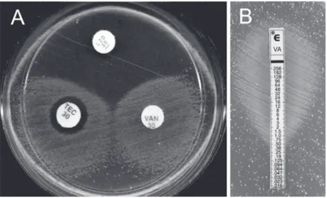
µg/mL vancomycin were identified as E. faecium, which was found to be growing only around the glycopeptide-impregnated discs in the agar diffusion assay (Figure 1A). The E-test further showed bacterial growth contiguous with the end of the strips containing the highest concentration of vancomycin (Figure 1B). The administration of antibiot-ics was discontinued because therapy was complete and
a negative blood culture was confirmed. The patient was
cured of infectious complications and left the hospital after 40 days of hospitalization. After 6 months, a rectal swab culture for VRE was negative, indicating that cessation of vancomycin had led to the clearance of VRE.
Hospital, surveillance and microorganism isolation The University Hospital of Londrina is a 353-bed tertiary care center that serves the city of Londrina, in addition to several localities in the States of Paraná, São Paulo, and Mato Grosso do Sul. The intensive care center houses 35 patients distributed among 17 beds for general cases, 6 beds for burn cases, 7 beds for neonatal cases, and 5 beds for pediatric cases. Surveillance cultures of stools were examined weekly for all patients housed in intensive care units and for all patients found to be colonized by, or infected with VRE. A rectal swab was obtained from the patient and the sample was transferred to VRE broth (Oxoid)
supple-mented with 6 µg/mL vancomycin, 6 µg/mL ciprofloxacin and
8 µg/mL colistin. After a 18-h incubation at 37°C, a 100-µL sample was spread on VRE agar supplemented with 6 µg/ mL vancomycin. The culture was further incubated at 37°C
for 24 h under aerobic conditions. Bacteria were stored at -80°C in 20% glycerol-brain heart infusion broth (Himedia, India) supplemented with 10 µg/mL vancomycin. The study protocol was approved by the Ethics Committee of the Universidade Estadual de Londrina (Protocol #186-09/ CEP-UEL). The patient gave written informed consent to participate in the study.
Phenotypic characterization
Species identification was based on colony morphology, Gram stain, catalase assay, and the profile determined by
the automated MicroScan WalkAway 96 Instrument (Dade MicroScan, USA). The disk diffusion method on Muller Hin-ton agar medium (Himedia) was carried out according to Clinical Laboratory Standard Institute (CLSI) (5) guidelines
in order to determine the profile of antimicrobial suscepti -bility to linezolide (30 µg), teicoplanin (30 µg), tigecycline (15 µg), and vancomycin (30 µg). As bacterial growth was detected only around the glycopeptide-impregnated discs, the isolate was tested for susceptibility to 12 other
antimicrobials (ampicillin, chloramphenicol, ciprofloxacin, erythromycin, gentamicin, levofloxacin, linezolide, penicil -lin, rifampicin, streptomycin, tetracycline, and tigecycline), using the automated broth microdilution panel of MicroScan WalkAway 96 according to manufacturer recommendations. The results reported here were those obtained after 24 h of incubation. The susceptibility breakpoints used were those recommended by the CLSI (5). E. faecalis ATCC 29212 and ATCC 51299 were used for quality control. The isolated bacterium was further tested by the vancomycin
E-test (AB BIODISK, Sweden). Genotypic characterization
The vancomycin-resistance genotype and the putative virulence genes cylA (activator of cytolysin, a secreted protein with hemolysin/bacteriocin activities), efaA (cell wall E. faecalis antigen A, an endocarditis-associated virulence factor), esp (enterococcal surface protein), and
gelE (gelatinase) were determined by PCR. The vancomy-cin resistance gene was determined using multiplex PCR as described by Petrich et al. (6). Genomic DNA of the enterococcal strain was extracted by the boiling method, and the virulence genes were determined as described by Ruzon et al. (7).
Cloning and sequencing of the ddl gene
The DNA fragments to be sequenced were amplified with
the following primers based on the ddl gene of E. faecium
BM 4339 (8): forward 5’ GAGTAAATCACTGAACGATT 3’ and reverse 5’ GGTTACGCAATCACTCCAGCCT 3’.
PCR was performed in a final volume of 20 µL containing
20 mM Tris-HCl, pH 8.4, 50 mM KCl, 1.5 mM MgCl2, 200
µM of each dNTP, 10 pmol of each primer, 2.5 U Pfx DNA polymerase (Invitrogen, Brazil), and 10 µL genomic DNA.
The PCR product was purified from agarose gel with the
QIAquick Gel Extraction kit (Qiagen, USA) and was inserted into the pCR®2.1 vector using the Original TA Cloning kit
(Invitrogen) according to manufacturer recommendations. The insert was sequenced with a 3730xl DNA analyzer (Applied Biosystems, USA) using the Big Dye® Terminator
v.3.1 Cycle Sequencing kit. A search for homologies in the GenBank/EMBL databases was carried out with the Blast algorithm (http://www.ncbi.nlm.nih.gov). The deduced amino acid sequence was analyzed with the ExPASy-Prosite program of the Swiss Institute of Bioinformatics, and the alignment of the sequences was carried out with ClustalW2 (http://www.cmbi.kun.nl/bioinf/tools/clustalw.shtml) of the EMBL-EBI software package.
Results and Discussion
The first VDE was isolated in 1994 from the urine of
a female patient receiving long-term vancomycin therapy (3). Since then, other cases of VDE isolation from differ-ent clinical specimens of patidiffer-ents receiving previous van-comycin therapy have been reported (4). A vanA-type E. faecalis vancomycin-dependent strain from a non-human source was also reported by Tanimoto et al. (9). In that case, VDE was isolated from chicken meat imported from China. For the cases of VDE-infected/colonized patients, 7 were on vancomycin to treat non-enterococcal infec-tions: 1 for diarrhea caused by Clostridium difficile, 5 for invasive staphylococcal infections, and 1 for bacteremia by Corynebacterium spp. resistant to ß-lactam antibiotics (4). In the remaining cases, VDE was considered to be
the primary cause of infection.
In the present study, we report the first case of VDE
isolation at a university hospital in Brazil (VDEfm-UEL). As reported in the other cases, the patient was receiving intravenous vancomycin therapy for non-enterococcal infec-tion. This isolation was possible because of the protocol for detecting VRE in a selective culture medium supplemented with vancomycin, which is utilized in the infection prevention and control laboratory of the hospital. Besides requiring glycopeptides, the isolated bacterium was also resistant
to ampicillin, ciprofloxacin, chloramphenicol, erythromycin, levofloxacin, penicillin, and rifampicin.
Although the isolated bacterium was considered to be colonizing Enterococcus according to CDC definitions
of healthcare-associated infections (10), it harbored the putative virulence genes efaA, gelE and esp, indicating a potential risk of infection. Low prevalence of the virulence markers has been shown for E. faecium isolates from differ-ent sources (11). In contrast to those results, we previously demonstrated a high prevalence of vancomycin-resistant E. faecium (VREfm) harboring virulence genes isolated from different sources at the University Hospital of Londrina, including the efaA, gelE and esp genes (7). Camargo et al. (12) showed that the esp gene was restricted to VREfm isolates from the southern region of Brazil, which also harbored the hyaluronidase gene (hyl). Studies with the
E. faecalisefaA- mutant showed attenuation of virulence
in a mouse peritonitis model when compared to the wild-type strain, suggesting that EfaA is a virulence factor (13). The chromosomal gelE gene encodes an extracellular zinc metalloprotease that can participate in the translocation of bacteria across intestinal cell layers (14) and can con-tribute to bacterial virulence in a mouse peritonitis model (15). The presence of the esp gene, which encodes an enterococcal surface protein, has been associated with colonization and persistence of enterococci in ascending
urinary tract infection in mice (16) and with biofilm forma -tion on an abiotic surface (17).
256 G. Kerbauy et al.
the ddl gene of E. faecium BM4147 (GenBank accession No. U39790). The alignment of the two nucleotide sequences showed several mutations in the ddl gene of VDEfm-UEL compared to that of the VRE BM4147 strain, most of them being silent point mutations. The insertion of an adenine at nucleotide position 710 of the ddl gene of VDEfm-UEL caused a frame-shift mutation, introducing a premature stop codon at position 733 of the sequence (Table 1) and thereby resulting in an inactive enzyme.
The isolation of an antibiotic-dependent strain alerts us to the importance of the control of healthcare-associated infections. This should be structured as an effective policy controlling the use of antibiotics in association with a laboratory capable of identifying microorganisms that show resistance to and also dependence on antibiotics.
This can be reflected in the appropriate management of
therapy and early adoption of precautionary measures to prevent the spread of infections by antibiotic resistant/ dependent microorganisms.
Acknowledgments
The present study was part of the M.Sc. dissertation of G. Kerbauy, who received a fellowship from CAPES. We thank Dr. A. Leyva for English editing of the manuscript, and Ediel Clementino da Costa and Jussevania Pereira Santos for technical support. Research supported by grants from Pro-Reitoria de Pesquisa e Pós-Graduação (PROPPG) of Universidade Estadual de Londrina (UEL).
References
1. Gales AC, Sader HS, Ribeiro J, Zoccoli C, Barth A, Pignatari AC. Antimicrobial susceptibility of Gram-positive bacteria isolated in Brazilian hospitals participating in the SENTRY Program (2005-2008). Braz J Infect Dis 2009; 13: 90-98. 2. Panesso D, Reyes J, Rincon S, Diaz L, Galloway-Pena
J, Zurita J, et al. Molecular epidemiology of vancomycin-resistant Enterococcus faecium: a prospective, multicenter study in South American hospitals. J Clin Microbiol 2010; 48: 1562-1569.
3. Fraimow HS, Jungkind DL, Lander DW, Delso DR, Dean JL. Urinary tract infection with an Enterococcus faecalis isolate that requires vancomycin for growth. Ann Intern Med 1994; 121: 22-26.
4. Bert F, Leflon-Guibout V, Le Grand J, Bourdon N,
Nicolas-Chanoine MH. [Emergence of vancomycin-dependent enterococci following glycopeptide therapy: case report and review]. Pathol Biol 2009; 57: 56-60.
5. CLSI - Clinical and Laboratory Standards Institute. Perfor-mance Standards for Antimicrobial Susceptibility Testing. Approved standard M100-S20-U. Wayne: CLSI; 2009. 6. Petrich AK, Luinstra KE, Groves D, Chernesky MA, Mahony
JB. Direct detection of vanA and vanB genes in clinical
specimens for rapid identification of vancomycin resistant
enterococci (VRE) using multiplex PCR. Mol Cell Probes
1999; 13: 275-281.
7. Ruzon FI, Paula SB, Kanoshiki RL, Pereira-Santos J, Ker-bauy G, Kobayashi RKT, et al. Virulence determinants in vancomycin-resistant Enterococcus faecium isolated from different sources at University Hospital of Londrina, Paraná, Brazil. J Microbiol 2010; 48: 814-821.
8. Gholizadeh Y, Prevost M, Van Bambeke F, Casadewall B, Tulkens PM, Courvalin P. Sequencing of the ddl gene and modeling of the mutated D-alanine:D-alanine ligase in glycopeptide-dependent strains of Enterococcus faecium.
Protein Sci 2001; 10: 836-844.
9. Tanimoto K, Nomura T, Hamatani H, Xiao YH, Ike Y. A vancomycin-dependent VanA-type Enterococcus faecalis
strain isolated in Japan from chicken imported from China.
Lett Appl Microbiol 2005; 41: 157-162.
10. Horan TC, Andrus M, Dudeck MA. CDC/NHSN surveillance
definition of health care-associated infection and criteria for specific types of infections in the acute care setting. Am J Infect Control 2008; 36: 309-332.
11. Vankerckhoven V, Huys G, Vancanneyt M, Snauwaert C, Swings J, Klare I, et al. Genotypic diversity, antimicrobial resistance, and virulence factors of human isolates and
Table 1. Point mutations in the ddl gene of vancomycin-dependent Enterococcus faecium.
Mutation (BM4147 → VDEfm-UEL) Position Mechanism Amino acid change
GGC → AGC 615 Transition Gly → Ser
- → A 710 Insertion Frame-shift
TGT → CGT 739 Transition Cys → Arg
C → - 799 Deletion Frame-shift
GGA → GTA 910 Transversion Gly → Val
ACG → CCG 988 Transversion Thr → Pro
probiotic cultures constituting two intraspecific groups of
Enterococcus faecium isolates. Appl Environ Microbiol 2008; 74: 4247-4255.
12. Camargo IL, Gilmore MS, Darini AL. Multilocus sequence typing and analysis of putative virulence factors in vanco-mycin-resistant and vancomycin-sensitive Enterococcus faecium isolates from Brazil. Clin Microbiol Infect 2006; 12: 1123-1130.
13. Singh KV, Coque TM, Weinstock GM, Murray BE. In vivo
testing of an Enterococcus faecalis efaA mutant and use
of efaA homologs for species identification. FEMS Immunol Med Microbiol 1998; 21: 323-331.
14. Zeng J, Teng F, Murray BE. Gelatinase is important for translocation of Enterococcus faecalis across polarized human enterocyte-like T84 cells. Infect Immun 2005; 73: 1606-1612.
15. Singh KV, Qin X, Weinstock GM, Murray BE. Generation and testing of mutants of Enterococcus faecalis in a mouse peritonitis model. J Infect Dis 1998; 178: 1416-1420. 16. Shankar N, Lockatell CV, Baghdayan AS, Drachenberg C,
Gilmore MS, Johnson DE. Role of Enterococcus faecalis
surface protein Esp in the pathogenesis of ascending urinary tract infection. Infect Immun 2001; 69: 4366-4372.
17. Heikens E, Bonten MJ, Willems RJ. Enterococcal surface
protein Esp is important for biofilm formation of Enterococ-cus faecium E1162. J Bacteriol 2007; 189: 8233-8240. 18. Courvalin P. Vancomycin resistance in Gram-positive cocci.
Clin Infect Dis 2006; 42 (Suppl 1): S25-S34.
19. Van Bambeke F, Chauvel M, Reynolds PE, Fraimow HS, Courvalin P. Vancomycin-dependent Enterococcus faecalis