ISSN 0100-879X
BIOMEDICAL SCIENCES
AND
CLINICAL INVESTIGATION
www.bjournal.com.br
www.bjournal.com.br
Volume 43 (5) 381-496 May 2011
Braz J Med Biol Res, May 2011, Volume 44(5) 477-482
doi:
10.1590/S0100-879X2011007500051
Reduction of the amount of intestinal secretory IgA in fulminant
hepatic failure
Dong-yan Liu, Weiguo Jiang and Pei Liu
Faculdade de Medicina de Ribeirão Preto Campus
Ribeirão Preto
Institutional Sponsors
The Brazilian Journal of Medical and Biological Research is partially financed by
analiticaweb.com.br S C I E N T I F I C
Reduction of the amount of intestinal secretory
IgA in fulminant hepatic failure
Dong-yan Liu
1, Weiguo Jiang
2and Pei Liu
31Research Center, China Medical University Affiliated Shengjing Hospital and Key Laboratory of Congenital Malformation Research, Ministry of Health, Shenyang, China 2Pathobiology Department, China Medical University Affiliated Shengjing Hospital, Shenyang, China 3China Medical University Affiliated Shengjing Hospital,Shenyang, China
Abstract
Intestinal barrier dysfunction plays an important role in spontaneous bacterial peritonitis. In the present study, changes in the intestinal barrier with regard to levels of secretory immunoglobulin A (SIgA) and its components were studied in fulminant hepatic
failure (FHF). Immunohistochemistry and double immunofluorescent staining were used to detect intestinal IgA, the secretory component (SC) and SIgA in patients with FHF (20 patients) and in an animal model with FHF (120 mice). Real-time PCR was used to detect intestinal SC mRNA in the animal model with FHF. Intestinal SIgA, IgA, and SC staining in patients with FHF was significantly weaker than in the normal control group (30 patients). Intestinal IgA and SC staining was significantly weaker in the animal model with FHF than in the control groups (normal saline: 30 mice; lipopolysaccharide: 50 mice; D-galactosamine: 50 mice; FHF: 120 mice). SC mRNA of the animal model with FHF at 2, 6, and 9 h after injection was 0.4 ± 0.02, 0.3 ± 0.01, 0.09 ± 0.01, respectively. SC mRNA of the animal model with FHF was significantly decreased compared to the normal saline group (1.0 ± 0.02) and lipopolysaccharide group (0.89 ± 0.01). The decrease in intestinal SIgA and SC induced failure of the intestinal immunologic barrier and the attenuation of gut immunity in the presence of FHF.
Key words: Fulminant hepatic failure; Secretory IgA; IgA; Secretory component; Intestine
Introduction
Correspondence: Pei Liu, China Medical University Affiliated Shengjing Hospital, Shenyang 110004, China.
E-mail: [email protected]
Received August 31, 2010. Accepted March 31, 2011. Available online April 25, 2011. Published May 16, 2011. Fulminant hepatic failure (FHF) is a rare but potentially
fatal complication of acute hepatitis. Spontaneous bacte-rial peritonitis (SBP) has been defined as culture positivity with a neutrophilic response (≥250 cells/mm3) in ascitic
fluid (1,2). SBP, which occurs in 15-25% of patients with cirrhosis (3,4), has been reported to occur in FHF (5-8). It has been shown that mortality is 2.5-fold higher and morbidity 2-fold higher in patients with SBP than in those without SBP (8).
Secretory IgA (SIgA), which is composed of IgA, se-cretory component (SC) and the J chain, is an important component of the intestinal immunologic barrier. IgA is a predominant Ig in mucosal secretions and serves as the first line of humoral defense on intestinal mucosal sur -faces. The majority of these IgA proteins are monomers, while the rest are predominantly polymeric to facilitate efficient transport to the mucosal secretions mediated via the polymeric Ig receptor (PIgR). SC is the extracellular component of the PIgR responsible for the transcytosis
of newly synthesized IgA (polymeric IgA). SIgA and its components, SC and IgA, are important components of the intestinal immunologic barrier. Up to now, investiga-tions of the intestinal barrier in patients with FHF have focused mainly on the mechanical barrier and barrier to the microorganism. In the present study, SIgA, SC and IgA were measured in patients with FHF and in an animal model of FHF in order to elucidate the state of the intestinal immunologic barrier.
Material and Methods
Tissue samples
478 Dong-yan Liu et al.
experimental procedures were approved by the Ethics Committee of China Medical University before the begin-ning of the study.
Animal model of FHF
Male BALB/c mice weighing 18-22 g were randomly divided into four groups. group 1 (30 mice): normal saline (NS); group 2 (50 mice): lipopolysaccharide (LPS); group 3 (50 mice): D-galactosamine (D-GalN); group 4 (120 mice): FHF (LPS and GalN; Sigma, USA). Mice were injected intraperitoneally with LPS (10 µg/kg) and/or GalN (800 mg/ kg). Mice of group 4 were killed by decapitation 2, 6, 9, 12, and 24 h after injection. The NS, LPS and GalN mice were killed by decapitation 9 h after the injection of NS, LPS or GalN (9). The study was approved by the animal Ethics Committee of China Medical University.
Immunohistochemistry of tissue samples
Intestinal IgA and SC of patients and mice with FHF were determined by immunostaining. Paraffin-embedded intestinal sections of patients and mice with FHF were de -paraffinized, rehydrated, and stained with goat anti-human IgA or anti-mouse IgA (Sigma) and mouse anti-human SC (SigA; Sigma) or anti-mouse SC mAb (Bethyl, USA) by sequential incubation. The slides were rinsed three times with 0.02 M PBS, pH 7.4, after each incubation, and sec -tions were counterstained with hematoxylin. The sec-tions from the same patient or the mouse were processed without the primary antibody and then examined by the procedure detailed above as a control for nonspecific binding of the secondary antibody. The absorbance values of IgA and SC were determined with the Image analysis software (China) after scanning.
Double immunofluorescent staining of intestinal SIgA
Paraffin-embedded intestinal sections of patients with FHF were deparaffinized, rehydrated, and double-stained by immunofluorescence with rhodamine-conjugated goat anti-human IgA (Southern Biotech, USA) and fluorescein-conjugated mouse anti-human SC (SigA; ICN, USA) by sequential incubation. The slides were rinsed three times with 0.02 M PBS, pH 7.4, after each incubation, and sec -tions were counterstained with hematoxylin. Sec-tions from the same patient or the mouse were processed without the primary antibody and then examined using the procedure detailed above as a control for nonspecific binding of the secondary antibody.
Quantitative real-time polymerase chain reaction of intestinal SC mRNA of mice with FHF
Total RNA was measured as described earlier in Refs. 10,11. Briefly, total RNA was extracted from mouse gut tis -sue with FHF using an RNA Mini Kit from Takara (Takara Biotechnology Co., Ltd., China). The quality of extracted
RNA was determined by the number and size of the bands obtained with agarose gel electrophoresis. cDNA was syn-thesized using 100 ng RNA. The levels of individual RNA transcripts were quantified by the quantitative real-time polymerase chain reaction (PCR). The primers of SC were as follows: PIgR-F: 5’-CAGACATTAGCATGGCAGACTT CAA-3’; PIgR-R: 5’-TGCCGAGTAGGCCATGTCAG-3’; GAPDH-F: 5’-AATGGTGAAGGTCGGTGTG-3’; GAPDH-R: 5’-TGAAGGGGTCGTTGATGG-3’.
The primers and fluorescent probes for SC and stan -dardwere purchased from Takara. The PCR conditions were a preliminary cycle at 95°C for 10 s, followed by 45 cycles at 95°C for 5 s and at 60°C for 20 s, followed by 1 min at 60°C and 5 s at 95°C. We also confirmed that the efficiency of amplification for each target gene (GAPDH) was 100% in the exponential phase of PCR. The levels of SC mRNA and GAPDH mRNA were determined ac -cording to the standard (Takara). The mRNA levels were normalized to GAPDH mRNA by dividing SC gene copies of the samples by the SC gene copies of GAPDH. The intestinal RNA level of the NS group was assumed to be 1, with which the intestinal RNA levels of other groups were compared.
Statistical analysis
The statistical differences between treatment groups were determined by the t-test using the SPSS 10.0 software.
Results
Intestinal IgA and SC staining in patients
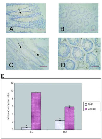
In the normal control group, SC staining of the cytoplasm and membranes of intestinal epithelial cells was brown (Figure 1A). SC staining of the cytoplasm and of the mem-branes of intestinal epithelial cells in patients with FHF was pale and considerably weaker than in the normal control group (Figure 1B). IgA staining of the cytoplasm of plasma cells within the lamina propria and of intestinal epithelial cells was brown in the normal control group (Figure 1C). However, in patients with FHF, IgA staining of the cytoplasm of plasma cells within the lamina propria was pale brown and IgA staining of intestinal epithelial cells was pale. IgA staining in patient samples was notably weaker than that of the normal control group (Figure 1D). The mean ab -sorbance values of SC and IgA in patients with FHF were significantly lower than those of the normal control group (P < 0.01; Figure 1E).
Double immunofluorescence staining
the intestinal epithelial cell cytoplasm and membranes as well as plasma cells within the lamina propria was green (Figure 2B). The staining results proved that intestinal SC, IgA and SIgA were decreased compared to the normal control group.
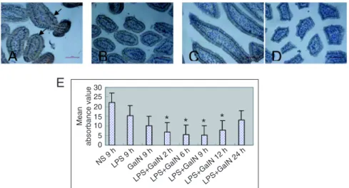
Intestinal IgA staining in the mouse model of FHF
IgA staining of the cytoplasm and/or membranes of intestinal epithelial cells, as well as of plasma cells within the lamina propria, was weaker in the mouse FHF group (Figure 3D) compared to the NS (Figure 3A), LPS (Figure
Figure 1. Secretory component (SC) and IgA staining of intestinal epithelial cells
in patients with fulminant hepatic failure (FHF). A, SC staining of intestinal epithe-lial cells in the normal control group (30 patients) was brown (see arrow). B, SC
staining of intestinal epithelial cells in patients with FHF (20 patients) was pale and remarkably weaker than in the normal control group. The staining results indicated that intestinal SC was significantly decreased in patients with FHF. C, IgA stain-ing of plasma cells within the lamina propria and of intestinal epithelial cells in the normal control group was brown (see arrows). D, IgA staining of the cytoplasm of plasma cells within the lamina propria was pale brown and IgA staining of intestinal
epithelial cells in patients with FHF was pale and remarkably weaker than in the normal control group. The staining results indicated that intestinal IgA in patients with FHF was significantly decreased. Magnification bars = 50 µm. E, The absor
-bance values of SC and IgA were notably less in patients with FHF compared to the normal control group. Data are reported as means ± SD. *P < 0.01 compared
480 Dong-yan Liu et al.
3B) and GalN groups (Figure 3C). The mean absorbance values of IgA in the mouse FHF group were significantly decreased from 2 to 12 h compared to the NS control group (P < 0. 01; Figure 3E).
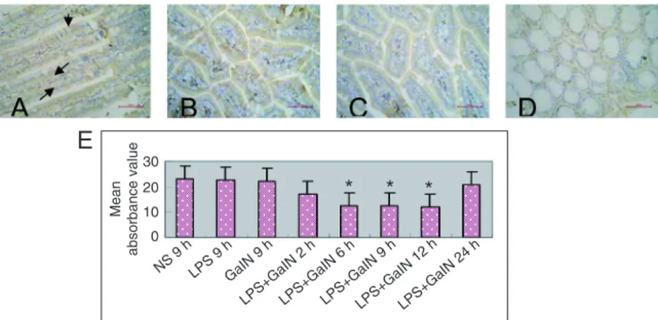
Intestinal SC staining in mice with FHF
SC staining of the cytoplasm and/or the membranes of intestinal epithelial cells was significantly weaker in the mouse FHF group (Figure
4D) compared to the NS group (Figure 4A), LPS group (Figure 4B), and GalN group (Figure 4C). The mean absorbance values of SC in the mouse FHF group were significantly decreased from 2 to 12 h compared to the NS group (P < 0.01; Figure 4E).
SC mRNA of intestinal tissues in the animal model of FHF
The relative content of intes -tinal SC mRNA in mice with FHF at 2, 6, and 9 h after injection was 0.4 ± 0.02, 0.3 ± 0.01, 0.09 ± 0.01, respectively. In the mice with FHF, intestinal SC mRNA was significantly lower at 2 to 9 h compared to the NS group (1.0 ± 0.02) and LPS group (0.89 ± 0.01; P < 0.01). In the GalN group, intestinal SC mRNA (0.2 ± 0.00) was also lower compared to the NS group and LPS group (P < 0.01; Figure 5).
Discussion
SBP is a common illness in patients with cirrhosis and ascites, which occurs without any apparent focus of infection. Bacterial translo-cation, gut motility alterations, and intestinal barrier dysfunction, along with bacterial overgrowth and changes in intestinal permeability play important roles in SBP, and it is evident from a number of studies that the gut is a major source of these bacteria (12). Intestinal SIgA levels are inversely correlated with bacterial overgrowth, bacterial translocation and changes in intes-tinal permeability (13). SIgA may weaken bacterial translation and restrict bacteria through intestinal
epithelia (14). Secretory IgA is the first line of defense against microorganisms through immune exclusion that prevents interaction of neutralized antigens with the intestinal epithe-lium. SIgA plays an important role in the immune balance of the intestinal epithelial barrier (15-17). SC plays a protective role in preventing the proteolytic degradation of polymeric IgA, thus enhancing the mucosal immunity provided by IgA at these sites. SC is a key defense against bacteria (18-21) and
Figure 2. IgA and secretory IgA (SIgA) staining of the intestinal epithelium by double
immu-nofluorescence. A, The secretory component (SC) and SIgA stained orange in the cytoplasm
and SC, IgA and SIgA on the membranes stained yellow in the normal intestinal epithelial cells. SIgA of the plasma cells within the lamina propria stained orange. IgA and SIgA of the plasma cells within the lamina propria stained yellow (see blue arrows). B, In patients with
fulminant hepatic failure (FHF), the color of the intestinal epithelial cell cytoplasm, membranes and plasma cells within the lamina propria was green. The staining results indicated that
intestinal SC, IgA and SIgA were notably decreased compared to the normal control group.
Magnification bars = 50 µm.
Figure 3. IgA staining of the intestinal epithelium in the animal model with fulminant hepatic
failure (FHF). A, In the normal saline (NS) group (30 mice), IgA staining of intestinal epithelial cell cytoplasm, membranes and plasma cells within the lamina propria was deep brown (ar-rows). B, In the lipopolysaccharide (LPS) group (50 mice), IgA staining of intestinal epithelial cell cytoplasm, membranes and plasma cells within the lamina propria was brown. C, In the galactosamine (GalN) group (50 mice), IgA staining of the intestinal epithelial cell cytoplasm, membranes and plasma cells within the lamina propria was pale brown. D, In the FHF group
(120 mice), IgA staining of some intestinal epithelial cell cytoplasm and membranes was pale.
Magnification bars = 50 µm. E, The absorbance values of IgA in the animal model with FHF (2, 6, 9, and 12 h) were significantly decreased compared to the normal saline (NS), lipopolysac
-charide (LPS) and galactosamine (GalN) groups. Data are reported as means ± SD. *P < 0.01
parasites (22) as well as playing a critical role in the immunologically mediated neutralization of cholera toxin (23). In vitro, free SC has been shown to fix the complement component C3b, suggesting a role for SC in enhancing local immune responses (24).
In liver diseases, notably alco-holic cirrhosis of the liver, intestinal SIgA is decreased (25). In the pres-ent study, intestinal IgA, SC and SIgA were markedly decreased in patients with FHF, this possibly being the reason why gut immunity is weakened in patients with FHF. Changes in intestinal SIgA, IgA and SC in the animal model of FHF were consistent with those of the patients with FHF. SC mRNA was simultane -ously detected by real-time PCR. A decrease in intestinal SC mRNA was observed in the animal model with FHF. These results confirm that at the mRNA and protein levels, SC is decreased in the animal model of FHF. It has been reported that in mildly inflamed ulcerative colitis le
-sions, SC levels showed no change but tended to decrease with increasing degree of inflammation. Decreased SC expression thus seemed to be related to intensified inflammatory activity (26). Therefore, we think that in patients with FHF, damaged intestinal mucus results in lower levels of intestinal SC and SIgA. Malnourished animals exhibit low levels of hepatic and intestinal pIgR. The concentration of biliary SC was also low in malnourished animals (27). In patients with FHF, the disease is usually complicated by malnutrition. Therefore, malnutrition may cause low concentrations of intestinal SC in FHF.
In conclusion, intestinal IgA and SC (SIgA) were lower in patients with FHF, with consequent failure of the intestinal im -munologic barrier and attenuation of gut immunity.
Acknowledgments
Research supported by the National Natural Science Foundation of China (#30670947, #30871158) and the Educa -tion Department Founda-tion of Liaoning Province (#05L466, #2008783).
Figure 4. Staining of the secretory component (SC) of the intestinal epithelium in the mouse
model of fulminant hepatic failure (FHF). A, In the normal saline (NS) group (30 mice), the SC staining of the intestinal epithelial cell cytoplasm and membranes was deep brown (see arrows). B, In the lipopolysaccharide (LPS) group (50 mice), SC staining of the intestinal epi-thelial cell cytoplasm and membranes was brown. C, In the galactosamine (GalN) group (50 mice), SC staining of the intestinal epithelial cell cytoplasm and membranes was pale brown.
D, In the FHF group (120 mice), SC staining of intestinal epithelial cells was pale. E, The absorbance values of SC in the animal model with FHF (2, 6, 9, and 12 h) were significantly
decreased compared to the normal saline (NS), lipopolysaccharide (LPS) and galactosamine
(GalN) groups. Data are reported as means ± SD. *P < 0.01 compared to NS 9 h, LPS 9 h,
and GalN 9 h (t-test).
Figure 5. Relative content of the intestinal secretory component (SC) mRNA. Relative content of intestinal SC mRNA in the
ani-mal model with fulminant hepatic failure (FHF) was significantly
lower compared to the normal saline (NS) and lipopolysaccharide
(LPS) groups. The relative content of intestinal SC mRNA in the
galactosamine (GalN) group was also lower compared to the NS
and LPS groups. Data are reported as means ± SD. *P < 0.01
482 Dong-yan Liu et al.
References
1. Chu CM, Chiu KW, Liaw YF. The prevalence and prognostic significance of spontaneous bacterial peritonitis in severe
acute hepatitis with ascites. Hepatology 1992; 15: 799-803.
2. Valla D, Flejou JF, Lebrec D, Bernuau J, Rueff B, Salzmann JL, et al. Portal hypertension and ascites in acute hepatitis: clinical, hemodynamic and histological correlations. Hepa-tology 1989; 10: 482-487.
3. Pinzello G, Simonetti RG, Craxi A, Di Piazza S, Spano C, Pagliaro L. Spontaneous bacterial peritonitis: a prospec-tive investigation in predominantly nonalcoholic cirrhotic patients. Hepatology 1983; 3: 545-549.
4. Runyon BA. Spontaneous bacterial peritonitis: an explosion of information. Hepatology 1988; 8: 171-175.
5. Thomas FB, Fromkes JJ. Spontaneous bacterial peritonitis
associated with acute viral hepatitis. J Clin Gastroenterol 1982; 4: 259-262.
6. Poddar U, Chawla Y, Dhiman RK, Vaiphei K, Vasishta RK,
Dilawari JB. Spontaneous bacterial peritonitis in fulminant hepatic failure. J Gastroenterol Hepatol 1998; 13: 109-111.
7. Dhiman RK, Makharia GK, Jain S, Chawla Y. Ascites and
spontaneous bacterial peritonitis in fulminant hepatic failure.
Am J Gastroenterol 2000; 95: 233-238.
8. Poddar U, Thapa BR, Prasad A, Sharma AK, Singh K. Natu
-ral history and risk factors in fulminant hepatic failure. Arch Dis Child 2002; 87: 54-56.
9. Dongyan Liu, Peng Ding, Pei Liu. The change of secretory IgA of gut induced by TNF-α in liver necrosis mice. Zhong hua Gan zang Bing Za Zhi 2010; 18: 306-308.
10. Schneeman TA, Bruno ME, Schjerven H, Johansen FE, Chady L, Kaetzel CS. Regulation of the polymeric Ig recep
-tor by signaling through TLRs 3 and 4: linking innate and
adaptive immune responses. J Immunol 2005; 175: 376-384.
11. Bruno ME, Kaetzel CS. Long-term exposure of the HT-29 human intestinal epithelial cell line to TNF causes sustained up-regulation of the polymeric Ig receptor and proinflamma -tory genes through transcriptional and posttranscriptional mechanisms. J Immunol 2005; 174: 7278-7284.
12. Ramachandran A, Balasubramanian KA. Intestinal dys -function in liver cirrhosis: Its role in spontaneous bacterial peritonitis. J Gastroenterol Hepatol 2001; 16: 607-612. 13. Deitch EA, Xu D, Qi L, Berg R. Elemental diet-induced
im-mune suppression is caused by both bacterial and dietary factors. J Parenter Enteral Nutr 1993; 17: 332-336.
14. Macpherson AJ, Slack E. The functional interactions of
commensal bacteria with intestinal secretory IgA. Curr Opin Gastroenterol 2007; 23: 673-678.
15. Corthësy B. Secretory immunoglobulin A: well beyond
im-mune exclusion at mucosal surfaces. Immunopharmacol Immunotoxicol 2009; 31: 174-179.
16. Nicholas JM, Stephen JF. Secretory IgA: Arresting microbial
pathogens at epithelial borders. Immunol Invest 2010; 39:
383-406.
17. Boullier S, Tanguy M, Kadaoui KA, Caubet C, Sansonetti
P, Corthésy B, et al. Secretory IgA-mediated neutralization of Shigella flexneri prevents intestinal tissue destruction by
down-regulating inflammatory circuits. J Immunol 2009; 183:
5879-5885.
18. Dallas SD, Rolfe RD. Binding of Clostridium difficile toxin A
to human milk secretory component. J Med Microbiol 1998;
47: 879-888.
19. de Araujo AN, Giugliano LG. Lactoferrin and free secretory
component of human milk inhibit the adhesion of entero -pathogenic Escherichia coli to HeLa cells. BMC Microbiol
2001; 1: 25.
20. Giugliano LG, Ribeiro ST, Vainstein MH, Ulhoa CJ. Free secretory component and lactoferrin of human milk inhibit
the adhesion of enterotoxigenic Escherichia coli. J Med Microbiol 1995; 42: 3-9.
21. Hammerschmidt S, Talay SR, Brandtzaeg P, Chhatwal GS. SpsA, a novel pneumococcal surface protein with specific
binding to secretory immunoglobulin A and secretory com-ponent. Mol Microbiol 1997; 25: 1113-1124.
22. Davids BJ, Palm JE, Housley MP, Smith JR, Andersen YS,
Martin MG, et al. Polymeric immunoglobulin receptor in intestinal immune defense against the lumen-dwelling pro-tozoan parasite Giardia. J Immunol 2006; 177: 6281-6290.
23. Uren TK, Wijburg OL, Simmons C, Johansen FE, Brandzaeg
P, Strugnell RA. Vaccine-induced protection against gastro-intestinal bacterial infections in the absence of secretory antibodies. Eur J Immunol 2005; 35: 180-188.
24. Marshall LJ, Perks B, Ferkol T, Shute JK. IL-8 released
constitutively by primary bronchial epithelial cells in culture forms an inactive complex with secretory component. J Im-munol 2001; 167: 2816-2823.
25. Pelletier G, Briantais MJ, Buffet C, Pillot J, Etienne JP. Se-rum and intestinal secretory IgA in alcoholic cirrhosis of the liver. Gut 1982; 23: 475-480.
26. Rognum TO, Brandtzaeg P, Elgjo K, Fausa O. Heteroge
-neous epithelial expression of class II (HLA-DR) deter -minants and secretory component related to dysplasia in ulcerative colitis. Br J Cancer 1987; 56: 419-424.
27. Ha CL, Woodward B. Depression in the quantity of intestinal
secretory IgA and in the expression of the polymeric
im-munoglobulin receptor in caloric deficiency of the weanling