DOI: 10.5935/2359-4802.20170064
Mailing Address: Maria Carolina Basso Sacilotto
Avenida Arlindo Joaquim de Lemos, 865, Apto: 32. Postal Code: 13100450, Vila Lemos, Campinas, SP – Brazil E-mail: [email protected]; [email protected]
A Simpler and Shorter Neuromuscular Electrical Stimulation Protocol Improves
Functional Status and Modulates Inflammatory Profile in Patients with End-Stage
Congestive Heart Failure
Maria Carolina Basso Sacilotto, Carlos Fernando Ramos Lavagnoli, Lindemberg Mota Silveira-Filho, Karlos Alexandre de Souza Vilarinho, Elaine Soraya Barbosa de Oliveira, Daniela Diógenes de Carvalho, Pedro Paulo Martins de Oliveira, Otávio Rizzi Coelho-Filho, Orlando Petrucci Junior
Universidade Estadual de Campinas (UNICAMP), Campinas, SP – Brazil
Manuscript received September 04, 2016; revised manuscript November 21, 2016; accepted April 05, 2017.
Abstract
Background: Neuromuscular electrical stimulation (NMES) using a stimulation wave for 5 days/week over 8 weeks has been used as a treatment option for congestive heart failure (CHF) patients who are unable to tolerate aerobic exercise.
Objective: We assessed the impact of a shorter NMES protocol using a Russian stimulation wave on the functional
status, quality of life (QoL) and inflammatory profile of end-stage CHF patients.
Methods: Twenty-eight patients with end-stage CHF (53 ± 11 years) were randomized to the NMES or control
group. Treatment was an NMES training program with Russian stimulation wave, applied for 50 minutes to both quadriceps femoral muscles twice weekly over seven weeks. The stimulation intensity was chosen to elicit muscle contractions in the NMES group and current input up to sensory threshold in the control group. Distance in the 6-minute walk test (6MWT) and QoL score by the Minnesota Living with Heart Failure Questionnaire were evaluated before, immediately after and one month after NMES protocol completion. Peripheral leukocytes were obtained to measure the gene expression levels of inflammatory cytokines.
Results: The NMES group showed increases in the 6MWT (324 ± 117 vs. 445 ± 100 m; p = 0.02) and QoL score
(64 ± 22 vs. 45 ± 17; p < 0.01) immediately but not 1 month after protocol completion, as well as increased gene expression levels of IL-1β, IL-6 and IL-8 after protocol completion.
Conclusion: Using a shorter and fewer sessions NMES protocol improved the QoL score and functional class
of severe CHF patients, and modulated the gene expression levels of some cytokines. This protocol might be a good alternative for patients with severe CHF and limitations in protocol adherence. (Int J Cardiovasc Sci. 2017;30(6)484-495)
Keywords: Heart Failure; Exercise Tolerance; Electric Stimulation Therapy / adverse effects; Exercise;
Rehabilitation; Heart Transplantation.
Introduction
Exercise intolerance is a challenging issue for patients with severe congestive heart failure (CHF)1,2, who
commonly exhibit fatigue and dyspnea. Patients with stable CHF should perform aerobic exercise training protocols to decrease muscle weakness caused by lack of physical activity, as well as to improve functional status, exercise capacity and quality of life (QoL).3-5
However, some patients are unable to perform aerobic exercise training protocols due to exercise intolerance or an unwillingness to participate in such programs.2,6
As an alternative to muscular training therapy in patients with CHF or chronic obstructive pulmonary disease (COPD), neuromuscular electrical stimulation (NMES) may be used to improve functional status, exercise capacity, endothelial function and QoL.2,3,6-8 NMES has
in patients with spinal cord lesions9 and athletes after a
bout of running.10 However, the effects of NMES on the
inflammatory profiles of CHF patients have not been well documented.
Common NMES training protocols involve treatments over 5 to 6 days/week for 6 to 8 weeks. These intensive therapy protocols may preclude some CHF patients from adhering to this treatment modality2,3 because,
similarly to other exercise training protocols, patients may be unwilling to attend the outpatient clinic daily for treatment.2,3,7,8,11 Most studies of
NMES-treated CHF patients have employed functional electrical stimulation,2,7,13,14 which causes muscle
contractions. Another NMES electric waveform modality is the Russian current, which involves the application of a 2500-Hz current at a burst frequency of 50 Hz. This modality effectively produces muscular strength in normal individuals15-17 and may be more comfortable
than other current modalities for patients during NMES sessions.15,18,19
The Russian current has a sine waveform, which has been reported as more comfortable for the patient during the electrical delivery to the muscle. Of note, the Russian current has an average frequency with a deeper stimulus effect and may be more tolerable by the patient.20-22
The unique characteristics of the Russian current might increase patient adherence to the NMES protocol. Therefore, the aim of this work was assess the effectiveness, compared to the literature, of a shorter NMES protocol consisting of twice-weekly training for 7 weeks using a Russian current, in terms of the functional status, QoL score, and inflammatory profile in patients with CHF.
Methods
Patient selection
Institutional Review Board approval was obtained for the study, and all patients provided written informed consent to participate. To be included in the study, patients must be on the awaiting heart transplant list, NYHA functional class III or IV, receiving optimal oral pharmacological treatment, and have stable CHF symptoms in the last 3 months. Exclusion criteria were neurologic or orthopedic disease that could limit the patient’s ability to accomplish the 6-minute walk test (6MWT). This study is registered at www.clinicaltrials. gov under the number NCT02313714.
Protocol for NMES
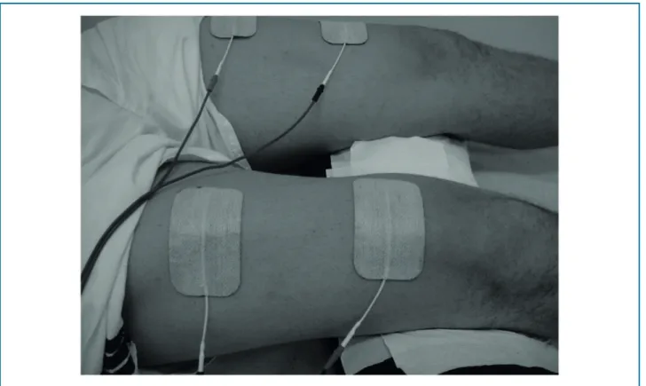
This study was carried out at an outpatient clinic of a university hospital with a room temperature maintained between 22 and 24 °C. NMES was applied by 2 adhesive electrodes on the skin over the upper lateral aspect of the quadriceps muscle, 5 cm below the inguinal fold and 3 cm above the upper patella border of both lower extremities (Figure 1).
In the NMES group, the stimulator (Neurodyn 2000, Ibramed, Amparo, SP, Brazil) was configured to deliver a direct electrical current with a 2500-Hz sinusoidal current at a burst frequency of 50 Hz (Russian current) for 3 seconds, followed by 9 seconds of rest. The intensity of stimulation was adjusted to achieve visible contraction. The stimulus intensity is adjusted according to patient tolerance. However, in order to be sure that muscle fiber contraction is occurring, the visual evaluation of at least one visible contraction is used. From this levels of contraction and patient´s comfort we adjusted higher levels of intensity and the highest level of intensity is adjusted according to patient´s tolerance.23
During electrical stimulation sessions, the legs were positioned in a light knee flexion position in both groups. In the control group, the same setup was used with 2 adhesive electrodes in the identical position as above, and the stimulator was configured to deliver a direct electrical current with the same specifications (Russian current). The intensity of stimulation was set low, to achieve a sensation of stimulus but with no visual muscular contraction.
Patients in both groups were evaluated weekly. The protocol was applied for 50 minutes twice a week, Wednesdays and Fridays, for 7 weeks.
Distance in the 6MWT
The 6MWT was performed in a 30-meter-long indoor hallway of the university hospital. Patients were trained and oriented to walk as fast as possible with encouragement. Only after the patient was considered trained was the walked distance recorded. All 6MWT sessions were carried out by the same observer (MCBS) in accordance with the American Thoracic Society (ATS) instructions.24 To ensure the patient’s safety and
Figure 1 – Placement of adhesive electrodes over the quadriceps femoral muscle. A single set of double adhesives was used as a simple way of applying neuromuscular electrical stimulation.
immediately before the first NMES session, 1 day after the last NMES session and 1 month after termination of the NMES protocol.
QoL by the Minnesota Living with Heart Failure Questionnaire (LHFQ)
A Portuguese language version of the LHFQ25 was
utilized to assess the QoL score 1 day before starting the NMES protocol, 1 day after completing the 7-week NMES protocol, and 1 month after protocol termination. The LHFQ is a questionnaire tool consisting of 21 questions, including physical and emotional variables. This questionnaire provides one score, with a scale from 0 to 105. Higher scores are associated with a worse QoL status. The LHFQ is a very frequently used score to assess QoL and is a reliable way to measure changes over time in the QoL of patients with CHF.13-26
Gene expression by peripheral leukocytes
Blood samples were drawn 1 day before NMES protocol initiation and 1 day after the last NMES session
(7 weeks). Peripheral leukocytes were isolated from blood by centrifugation at 1100 rpm for 10 minutes in a refrigerated centrifuge (4 oC). Total RNA was isolated from the cellular pellet by using TRIzol ® LS reagent (Ambion, Life Technologies, USA). Total RNA was quantified by using the 260/280 nm absorbance ratio data. The High-Capacity cDNA Reverse Transcription kit (Applied Biosystems, Carlsbad, CA, USA) with 1 µg of total RNA was used for reverse transcription reactions. To assess different levels of gene expression, quantitative real-time PCR was performed with the Taqman Fast Advanced Master mix (Applied Biosystems) and commercially available Taqman primers for IL-1α, IL-8, TNF, IL-10 and IL-6 (Applied Biosystems). The GADPH gene was used as an internal control.
Outcomes
Statistical analyses
All continuous data with Gaussian distribution are reported as means ± standard deviations (SDs). All continuous data with no Gaussian distribution are reported as median and range. All variables were assessed for normality distribution using Shapiro-Wilk test.
Discrete variables are described as the frequency of the corresponding population. Analyses of the 6MWT and QoL score were performed by two-way analysis of variance and the Tukey post hoc test for detecting which time points were different. Correlations between the 6MWT and QoL score were calculated by linear regression analysis. Analyses of demographics were performed using t test or Mann-Whitney where was appropriated. Analyses of baseline characteristics and medication were performed using chi-square test.
A simple random sampling was used to allocate patients to the 2 groups. An online tool was employed for the group draw (www.graphpad.com). The sample size was based on previous studies12,13,26 and on the availability
of inpatient/outpatient hospital facility. A P value smaller than 0.05 was considered statically significant.
All calculations were conducted and figures were constructed with GraphPad Prism software (version 6 for Mac OS X, GraphPad Software, La Jolla, CA, USA).
Results
Overall results
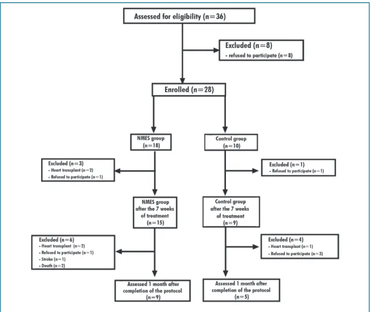
Of the 36 patients with severe CHF who were on the awaiting heart transplant list and initially assessed for the study, 8 patients refused to participate. The remaining 28 patients with end-stage CHF were randomly divided into 2 groups. Patients in the NMES group (n = 18) underwent treatment with the 7-week NMES protocol using the Russian current. Patients in the control group (n = 10) were tested with a similar protocol using an ineffective electric current. One month after termination of the treatment protocol, 9 patients in the NMES group and 5 patients in the control group were assessed. Figure 2 shows the flowchart of patient enrollment for the entire cohort. All patients were medicated for CHF, and the medication was not changed during the study period. The groups did not show differences regarding the distribution of
sex, age, demographics, medication, body mass index or functional class (Table 1).
Safety of NMES application
During the 7-week protocol, no NMES-related complications were observed in either group. One month after protocol termination, 1 patient in the NMES group had experienced cerebral vascular stroke, 2 patients in the NMES group had died due to CHF complications, and 2 patients in the NMES group and 1 patient in the control group had undergone heart transplant (Figure 2).
Improvement of the 6MWT with the 7-week NMES treatment
Compared to before treatment, 6MWT was increased immediately after the 7-week treatment in the NMES group (324 ± 117 vs. 445 ± 100 m; p = 0.02) but returned to baseline values by 1 month after protocol termination (324 ± 117 vs. 317 ± 194; p = 0.89). The control group showed no differences in 6MWT values between before and after treatment (Table 2).
Improvement of the QoL scores with the 7-week NMES treatment
Compared to pretreatment values, the NMES group showed improvement in QoL scores after completion of the 7-week NMES protocol (64 ± 22 vs. 45 ± 17; p < 0.01), whereas the control group showed no differences. However, the QoL scores in the NMES group had returned to the baseline values by 1 month after protocol termination (64 ± 22 vs. 51 ± 20; p = 0.07) (Table 2).
Association of QoL score with pretreatment 6MWT
Table 1 – Baseline characteristics, demographics and medication
Characteristic NMES group Control group p-Value
(n = 18) (n = 10)
Gender
Male 15 (83) 9 (90)
0.93**
Female 3 (17) 1 (10)
Age, years 54 ± 10 50 ± 12 0.39*
Height, cm 163 ± 28 172 ± 6 < 0.01*
Weight, kg 86 ± 22 80 ± 13 0.13*
BMI, kg/m2 27 ± 5 26 ± 5 0.85*
Ejection fraction, % 30 ± 10 34 ± 10 0.90*
CHF etiology
Idiopathic 6 (33) 4 (40)
0.51**
Ischemic 8 (44) 2 (20)
Valvar 2 (11) 2 (20)
Chagas’s disease 1 (6) 2 (20)
Viral 1 (6) 0 (0)
Functional class
III 13 (72) 9 (90)
0.54**
IV 5 (28) 1 (10)
Medications
Diuretics 18 (100) 10 (100) 0.71**
Espironolactone 15 (83) 9 (90) 0.86**
Digoxin 11 (61) 5 (50) 0.68**
Beta-blocker 16 (89) 10(100) 0.79**
Antiarrhythmic 0 (0) 1 (10) 0.71**
Antiplatelet 11 (61) 60 (60) 0.89**
Statin 9 (50) 2 (20) 0.33**
Ca2+ channel blockers 3 (17) 0 (0) 0.52**
Nitrates 4 (22) 2 (20) 0.86
ACE inhibitors 13 (72) 9 (90) 0.22
Data are reported as n (%) or mean ± standard deviation. BMI: body mass index; diuretics include furosemide and bumetamide; ACE: angiotensin-converting enzyme. (*) P value using t test. (**) P value using Chi-square test.
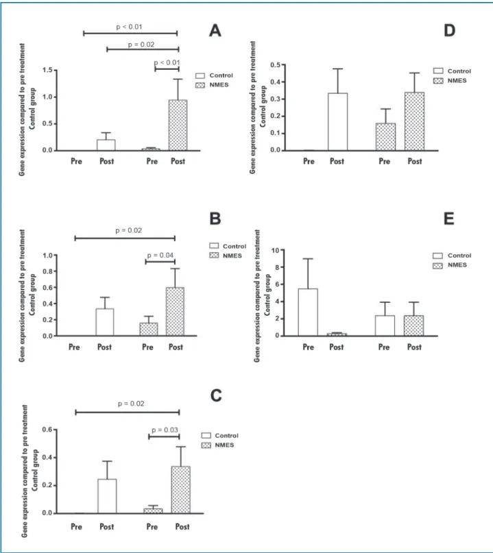
Modulation of gene expression with the 7-week NMES treatment
Compared to pretreatment values in the control group, the gene expression levels of IL-1β, IL-6, and IL-8
Figure 2 ––Flowchart of patient enrollment and reasons for patient drop-out from the study.
Table 2 – Hemodynamic values, functional capacity and quality of life
Parameter
Pretreatment Immediately posttreatment 1 month posttreatment
NMES Control NMES Control NMES Control
(n = 18) (n = 10) (n = 15) (n = 9) (n = 9) (n = 5)
Resting HR, bpm 74 ± 14 74 ± 9 68 ± 16 72 ± 7 73 ± 11 73 ± 8
Resting SBP, mmHg 101 ± 11 110 ±10 115 ± 66 103 ± 16 101 ± 8 108 ± 13
Resting DBP, mmHg 66 ± 8 70 ± 8 67 ± 7 67 ± 8 69 ± 6 68 ± 8
LHFQ 64 ± 22 51± 25 45 ± 17† 52 ± 25 51 ± 20 48 ± 24
6MWT, m 324 ± 117 393 ± 151 445 ± 100† 353 ± 159 317 ± 194 366 ± 92
Figure 4 –Fold differences in gene expression levels of cytokines before and after treatment in the control and NMES groups compared to the pretreatment levels in the control group. (A) IL-1β, (B) IL-6, (C) IL-8, (D) TNFα, and (E) IL-10.
Discussion
We found that the application of a shorter NMES protocol, exclusively to the quadriceps femoral muscles improved the functional capacity and QoL
an alternative solution for greater patient adherence to a rehabilitation program.
NMES using a Russian current is a well-known and safe modality for treating patients and improving their comfort during NMES sessions.15,18,19 NMES has
beneficial effects for functional capacity in patients with different diseases and conditions, including CHF, severe COPD, stroke, and osteoarthritis, as well as patients in intensive care units.3,28 The 6MWT was used
to determine functional capacity in this study and has been commonly utilized for this purpose in previous reports.26,29 The 6MWT has a good correlation with peak
oxygen consumption (VO2), and changes in the 6MWT can be used to predict mortality.6,27
The novelty of the present work lies in the less-frequent application of NMES through a simple pair of adhesive electrodes, which are placed on the skin over the quadriceps femoral muscle in each limb. All previous studies have used protocols with FES (Functional Electrical Stimulation) current ranging from 5 to 7 days per week.
All previous studies have enrolled patients with CHF of functional classes II and III, whereas the present study enrolled patients with functional classes III and IV on the awaiting heart transplant list.
Of note, we did not find in the literature studies that had evaluated a shorter protocol and using this specific type of current (Russian current) in a rehabilitation programs with patients in this specific clinical profile. Additionally, we did not find data reporting a longer training protocol that evaluated the functional capacity and quality of life following the discontinuation of electrical stimulus. We believe that a shorter protocol may improve patient adherence to the rehabilitation program, which is a day by day problem on those patients.
Previous studies of NMES protocols generally have utilized 8 adhesive electrodes in each limb.2,3
Using a more intense NMES protocol design of 8 adhesive electrodes applied for 30 minutes/day, 5 days/week for 6 weeks, Parissis et al.2 reported improvements in
QoL, functional status, emotional status and endothelial function in patients with stable systolic CHF (NYHA class II or III).2 We demonstrated similar QoL and functional
status results in sicker patients (NYHA functional class III or IV), using an less-demanding protocol with 1 pair of adhesive electrodes applied for 50 minutes/day, 2 days/week for 7 weeks. Banerjee et al.30 observed
increases in the 6MWT and VO2 after 8 weeks of
NMES training in patients with CHF and low left-ventricular ejection fraction. They utilized a more complex stimulation apparatus, composed of adhesive electrodes applied at multiple muscles (quadriceps, hamstrings, calves and gluteal muscles) in each limb.30
Other authors have demonstrated similar results to the present manuscript regarding the functional capacity after training with more intense NMES protocols, such as 5 days/week for 30 to 60 minutes/day over 5 to 8 weeks.13,31,32
Several papers have demonstrated an improvement of QoL after NMES, as assessed by different questionnaires, such as the Beck Depression Inventory, Zung Self-Rating Depression Scale12 and Kansas City Cardiomyopathy
Questionnaire.12 The LHFQ, which was used in the
present study,26 is increasingly being applied for the
assessment of QoL in patients with CHF.8,13,33 The QoL
score was improved immediately after completion of the NMES protocol, consistent with several reports in the literature using different NMES protocols.2,7,8
The QoL score has consistently demonstrated to improve with NMES and low-intensity aerobic exercise programs; however, the durability of these effects has not been frequently reported. We found that the QoL scores had deteriorated to near-baseline levels by 1 month after protocol completion. Patients who walked longer distances during the 6MWT before NMES treatment were more likely to have better QoL scores immediately after completing the NMES protocol in the NMES group. However, the pretreatment 6MWT and QoL values were not correlated with each other in either group. These findings support the use of the 6MWT as a useful tool for assessing QoL after NMES treatment in this subset of patients. Several studies have reported improvements of the 6MWT and QoL score after NMES, but, to our knowledge, a relationship between both has not been reported.7,33
Some reports have demonstrated favorable effects of either aerobic and isometric exercises on inflammatory modulation in, for example, healthy individuals34 and
patients with paraplegia or rheumatic disease.35 In CHF
patients, data regarding the inflammatory profile after any physical exercise protocol have been inconsistent.36
Overall, it seems that physical activity may decrease serum TNF levels, with little to no effect on serum IL-6 and IL-10 levels.39 Additionally, very few papers have
IL-6, C-reactive protein (CRP) or insulin-like growth factor-1 (IGF-1) after 6 weeks of NMES application to the quadriceps and calf muscles. A single report studying CHF patients in functional class II or III assessed the endothelial function and immune response after NMES therapy for 5 days/week over 6 weeks.26 This study
observed decreased levels of TNF, soluble intercellular adhesion molecule (sICAM), and soluble vascular cell adhesion molecule (sVCAM-1) between before and after NMES treatment. Interestingly, no differences in the serum levels of IL-6 were observed in the control or NMES group.
The present work is the first to describe gene expression from peripheral leukocytes, which may resemble the inflammatory profile, before and after NMES treatment. Using a shorter NMES protocol, we observed increased gene expression levels of IL-6, IL-1β and IL-8. Measuring gene expression by peripheral leukocytes is a useful and reliable method for assessing diseases, such as asthma and seasonal diseases.38,39
The observed increased gene expression of these cytokines may represent a response to the local stress caused by NMES, which is similar to the stress caused by intense physical exercise.40,41 The chronic exercise
may decrease the inflammatory response. However, in the present protocol is hard to tease out the chronic stimulation (the entirely protocol) from the acute phase. It is possible to say that the NMES modulates the inflammatory response, but we can not say whether or not increase or decrease it.
No previous study has followed patients for 1 month after completion of the NMES protocol. Surprisingly, we observed that the beneficial effects observed immediately after NMES termination were not permanent. The functional capacity and QoL scores returned to baseline values by 1 month after NMES protocol termination. These findings encourage the use of other strategies when implementing the shorter NMES protocol. Nevertheless, the most important aspect of our shorter protocol is that it could facilitate access to a different rehabilitation program. The protocol is composed of only a few sessions per week and does not cause fatigue or dyspnea comparable to conventional therapy (i.e. aerobic exercise training). These factors might stimulate treatment adherence and produce equivalent results to more intense NMES protocols or aerobic exercise training protocols.12,30,32,42-45
Although the present findings are motivating, they must be considered in the context of the study
limitations. As a prospective study, we kept adherence to the randomization draw before the study initiated. The randomization generated blocks of studied groups and not a single patient selection. This trial included a relatively small number of patients with a different group size, although most studies on the same subject have enrolled similar numbers.6,12,13,26,31,32
We observed a more frequent negative events in the treatment group compared to the control group after termination of the training period. It is not possible to exclude whether is related to the termination of the protocol or random. It is important to highlight that there are no data in the literature evaluating patients after termination of the training period and this is the first work that tries to show this information. However, it seems important to keep special attention on the patients after terminating the electrical stimulation protocol period. Moreover, some patients discontinued treatment before protocol completion due to cardiac transplant or death.
Conclusion
Using a shorter and simpler NMES protocol improved the QoL score and functional status in patients with end-stage CHF. The clinical improvement of these patients was accompanied by a increase in gene expression of some cytokines in peripheral leukocytes. This modified treatment might be an interesting alternative for physical rehabilitation in these very sick patients, and may provide similar results compared to longer protocols described in the literature.
Author contributions
1. Dickstein K, Cohen-Solal A, Filippatos G, McMurray JJ, Ponikowski P, Poole-Wilson PA, et al; ESC Committee for Practice Guidelines (CPG). Esc Guidelines for the Diagnosis and Treatment of Acute and Chronic Heart Failure 2008: The Task Force for the Diagnosis and Treatment of Acute and Chronic Heart Failure 2008 of the European Society of Cardiology. Developed in Collaboration with the Heart Failure Association of the Esc (HFA) and Endorsed by the European Society of Intensive Care Medicine (ESICM).Eur Heart J. 2008;29(19):2388-442.
2. Parissis J, Karavidas A, Farmakis D, Papoutsidakis N, Matzaraki V, Arapi S, et al. Efficacy and safety of functional electrical stimulation of lower limb muscles in elderly patients with chronic heart failure: a pilot study. Eur J Prev Cardiol. 2015;22(7):831-6.
3. Sillen MJ, Franssen FM, Delbressine JM, Vaes AW, Wouters EF, Spruit MA. Efficacy of lower-limb muscle training modalities in severely dyspnoeic individuals with copd and quadriceps muscle weakness: results from the Dices Trial. Thorax.2014;69(6):525-31.
4. McMurray JJ, Adamopoulos S, Anker SD, Auricchio A, Bohm M, Dickstein K, et al; ESC Committee for Practice Guidelines. Esc Guidelines for the Diagnosis and Treatment of Acute and Chronic Heart Failure 2012: The Task Force for the Diagnosis and Treatment of Acute and Chronic Heart Failure 2012 of the European Society of Cardiology. Developed in Collaboration with the Heart Failure Association (Hfa) of the Esc.Eur Heart J.2012;33(14):1787-847. Erratum in: Eur Heart J. 2013;34(2):158.
5. Downing J, Balady GJ. The role of exercise training in heart failure.J Am Coll Cardiol.2011;58(6):561-9.
6. Deboeck G, Muylem AV, Vachiery JL, Naeije R. Physiological response to the 6-minute walk test in chronic heart failure patients versus healthy control subjects. Eur J Prev Cardiol.2013;21(8):997-1003.
7. Smart NA, Dieberg G, Giallauria F. Functional electrical stimulation for chronic heart failure: a meta-analysis.Int J Cardiol.2013;167(1):80-6.
8. Giallauria F, Vigorito C, Piepoli MF, Stewart Coats AJ. Effects of cardiac contractility modulation by non-excitatory electrical stimulation on exercise capacity and quality of life: an individual patient's data meta-analysis of randomized controlled trials.Int J Cardiol.2014;175(2):352-7.
9. Paulson TA, Bishop NC, Smith BM, Goosey-Tolfrey VL. Inflammation-mediating cytokine response to acute handcycling exercise with/without functional electrical stimulation-evoked lower-limb cycling.J Rehabil Res Dev.2014;51(4):645-54.
10. ScottJP, Sale C, Greeves JP, Casey A, Dutton J, Fraser WD. Effect of exercise intensity on the cytokine response to an acute bout of running. Med Sci Sports Exerc.2011;43(12):2297-306.
11. Nuhr MJ. Beneficial effects of chronic low-frequency stimulation of thigh muscles in patients with advanced chronic heart failure. EurHeart J.2004;25(2):136-43.
12. Karavidas A, Parissis JT, Matzaraki V, Arapi S, Varounis C, Ikonomidis I, et al. Functional electrical stimulation is more effective in severe symptomatic heart failure patients and improves their adherence to rehabilitation programs. J Card Fail.2010;16(3):244-9.
13. Karavidas A, Driva M, Parissis JT, Farmakis D, Mantzaraki V, Varounis C, et al. Functional electrical stimulation of peripheral muscles improves endothelial function and clinical and emotional status in heart failure patients with preserved left ventricular ejection fraction. Am Heart J.2013;166(4):760-7.
14. Sbruzzi G, Ribeiro RA, Schaan BD, Signori LU, Silva AM, Irigoyen MC, et al. Functional electrical stimulation in the treatment of patients with chronic heart failure: a meta-analysis of randomized controlled trials. Eur J Cardiovasc Prev Rehabil.2010;17(3):254-60.
15. WardAR, Oliver WG, Buccella D. Wrist extensor torque production and discomfort associated with low-frequency and burst-modulated kilohertz-frequency currents. Phys Ther.2006;86(10):1360-7.
16. Dantas LO, Vieira A, Siqueira Jr AL, Salvini TF, Durigan JL.Comparison between the effects of 4 different electrical stimulation current waveforms on isometric knee extension torque and perceived discomfort in healthy women. Muscle Nerve.2015;51(1):76-82.
17. Bellew JW, Beiswanger Z, Freeman E, Gaerte C, Trafton J. Interferential and burst-modulated biphasic pulsed currents yield greater muscular force than russian current. Physiother Theory Pract. 2012;28(5):384-90.
18. Broderick BJ, Kennedy C, Breen PP, Kearns SR, O. Laighin G. Patient tolerance of neuromuscular electrical stimulation (eenm) in the presence of orthopaedic implants. Med Eng Phys.2011;33(1):56-61.
19. Ward AR, Robertson VJ, Sensory, motor, and pain thresholds for stimulation with medium frequency alternating current. Arch Phys Med Rehabil. 1998;79(3):273-8.
20. Bennie SD, Petrofsky JS, Nisperos J, Tsurudome M, Laymon M. Toward the optimal waveform for electrical stimulation of human muscle. Eur J Appl Physiol.2002;88(1-2):13-9.
21. Baker LL, Bowman BR, McNeal DR. Effects of waveform on comfort during neuromuscular electrical stimulation. Clin Orthop Relat Res. 1988(233):75-85.
22. Bowman BR, Baker LL. Effects of waveform parameters on comfort during transcutaneous neuromuscular electrical stimulation. AnnBiomedEng.1985;13(1):59-74.
23. Maffiuletti NA. Physiological and methodological considerations for the use of neuromuscular electrical stimulation. Eur J Appl Physiol.20101;110(2):223-34.
24. ATS Committee on Proficiency Standards for Clinical Pulmonary Function Laboratories.ATSstatement: guidelines for the six-minute walk test. Am J Respir Crit Care Med.2002;166(1):111-7.
25. Carvalho VO, Guimaraes GV, Carrara D, Bacal F, Bocchi EA. Validation of the Portuguese version of the Minnesota living with Heart Failure Questionnaire. Arq Bras Cardiol.2009;93(1):39-44.
26. Karavidas AI, Raisakis KG, Parissis JT, Tsekoura DK, Adamopoulos S, Korres DA, et al. Functional electrical stimulation improves endothelial function and reduces peripheral immune responses in patients with chronic heart failure. Eur J Cardiovasc Prev Rehabil.2006;13(4):592-7.
27. McDermott MM, Guralnik JM, Criqui MH, Liu K, Kibbe MR, Ferrucci L. Six-minute walk is a better outcome measure than treadmill walking tests in therapeutic trials of patients with peripheral artery disease. Circulation.2014;130(1):61-8.
28. Laufer Y, Shtraker H, Elboim Gabyzon M. The effects of exercise and neuromuscular electrical stimulation in subjects with knee osteoarthritis: a 3-month follow-up study. Clin Interv Aging.2014;9:1153-61.
29. Banerjee P. Electrical muscle stimulation for chronic heart failure: an alternative tool for exercise training?. Curr Heart Fail Rep.2010;7(2):52-8.
30. Banerjee P, Caulfield B, Crowe L, Clark AL. Prolonged electrical muscle stimulation exercise improves strength, peak VO2, and exercise capacity in patients with stable chronic heart failure. J Card Fail.2009;15(4):319-26.
References
Potential Conflict of Interest
No potential conflict of interest relevant to this article was reported.
Sources of Funding
This study was funded by FAPESP.
Study Association
31. Deley G, Eicher JC, Verges B, Wolf JE, Casillas JM. Do low-frequency electrical myostimulation and aerobic training similarly improve performance in chronic heart failure patients with different exercise capacities?. J Rehabil Med.2008;40(3):219-24.
32. Deley G, Kervio G, Verges B, Hannequin A, Petitdant MF, Salmi-Belmihoub S, et al. Comparison of low-frequency electrical myostimulation and conventional aerobic exercise training in patients with chronic heart failure. Eur J Cardiovasc Prev Rehabil.200512;(3):226-33.
33. Smart NA, Giallauria F, Dieberg G. Efficacy of inspiratory muscle training in chronic heart failure patients: a systematic review and meta-analysis. Int J Cardiol.2013;167(4):1502-7.
34. Wethal T, Roysland R, Torbjorn O, Kjekshus J. Exercise induced vasodilatation in healthy males; a marker of reduced endothelial function. Scand Cardiovasc J.2015;49(3):123-9.
35. Benatti FB, Pedersen BK. Exercise as an anti-inflammatory therapy for rheumatic diseases-myokine regulation. Nat Rev Rheumatol.2015;11(2):86-97.
36. Smart NA, Steele M. The effect of physical training on systemic proinflammatory cytokyne expression in heart failure patients: a systematic review. Congest Heart Fail. 2011;17(3):110-4.
37. Vivodtzev I, DebigaréR, Gagnon P, Maingury V, Saey D, DubéA, et al. Functional and muscular effects of neuromuscular electrical stimulation in patients with severe COPD: a randomized clinical trial. Chest. 2012;141(3):716-25.
38. Kozmus CE, Potocnik U. Reference genes for real-time qPCR in leukocytes from asthmatic patients before and after anti-asthma treatment. Gene. 2015;570(1):71-7.
39. Goldinger A, Shakhbazov K, Henders AK, McRae AF, Montgomery GW, Powell JE. Seasonal effects on gene expression. PLoS One. 2015;10(5):e0126995.
40. Wlec SS, Clanton TL. The regulation of interleukin-6 implicates skeletal muscle as an integrative stress sensor and endocrine organ. Exp Physiol. 2013;98(2):359-71.
41. Peake J, Della Gatta P, Suzuki K, Nieman DC. Cytokine expression and secretion by skeletal muscle cells: regulatory mechanisms and exercise effects. Exerc Immunol Rev.2015;21:8-25.
42. Youssef MK. The impact of neuromuscular electric stimulation versus aerobic exercise in rehabilitation of patients with chronic heart failure. J Arab Soc Med Res. 2014:9(1):40-7.
43. Deftereos S,Giannopoulos G,Raisakis K,Kossyvakis C,Kaoukis A, Driva M, et al. Comparison of muscle functional electrical stimulation to conventional bicycle exercise on endothelium and functional status indices in patients with heart failure.Am J Cardiol. 2010;106(11):1621-5.
44. Dobsak P, Novakova M, Fiser J, Siegelova J, Balcarkova O, Spinarova L, et al. Electrical stimulation of skeletal muscles an alternative to aerobic exercise training in patients with chronic heart failure? Int Heart J. 2006;47(3):441-53.