O
RIGINALA
RTICLE Revista Brasileira de FisioterapiaAnalysis of cardiovascular system responses
to forced vital capacity in COPD
Análise das respostas do sistema cardiovascular ao teste de capacidade vital
forçada na DPOC
Felipe A. R. Mendes1, Isadora L. Moreno2, Marina T. Durand3, Carlos M. Pastre2, Ercy M. C. Ramos2, Luiz C. M. Vanderlei2
Abstract
Background: The forced vital capacity (FVC) test is routinely performed to evaluate pulmonary function in patients with chronic obstructive pulmonary disease (COPD). However, the influence of the FVC maneuver on the cardiovascular system of patients with COPD is poorly understood. Objectives: To analyze the behavior of heart rate (HR), blood pressure (BP) and heart rate variability (HRV) during the FVC test in COPD patients. Methods: Nineteen men with COPD (72±7 years, GOLD stage I=3, II=5, III=7 and IV=4 patients) performed the FVC test while having their HR monitored. HRV was assessed in time (rMSSD) and frequency domains (LF, HF and LF/HF) at rest, before and after the best FVC maneuver. BP was measured at rest, immediately before and at the end of the test, as well as 10 minutes after the end of the test. Results: At the beginning of the FVC maneuver, HR decreased (p<0.001) and then increased gradually until the end of the test (p<0.001). After the end of maneuver, HR continued to increase until it reached a peak (p<0.001), and then it fell quickly to below at-rest values (p<0.001) prior to returning to baseline. The BP and HRV indices did not change during the assessment.
Conclusion: The FVC test influences the behavior of COPD patient HR without changing autonomic control or BP.
Keywords: chronicobstructive pulmonary disease; spirometry; vital capacity; heart rate; autonomic nervous system.
Resumo
Contextualização: O teste de capacidade vital forçada (CVF) é rotineiramente realizado na avaliação da função pulmonar de pacientes com doença pulmonar obstrutiva crônica (DPOC). Entretanto, permanece pouco compreendida a influência do teste de CVF sobre o sistema cardiovascular de pacientes com DPOC. Objetivos: Analisar o comportamento da frequência cardíaca (FC), pressão arterial (PA) e variabilidade da frequência cardíaca (VFC) no teste de CVF na DPOC. Métodos: Dezenove homens com DPOC (72 ± 7 anos, no estágio de gravidade GOLD I=3, II=5, III=7 e IV=4 pacientes) realizaram a manobra de CVF e tiveram sua FC monitorada durante todo o exame, e a VFC analisada nos domínios do tempo (rMSSD) e da frequência (BF, AF e BF/AF) durante o repouso, antes e após a melhor manobra de CVF. A PA foi analisada no repouso, imediatamente ao final da manobra de CVF e 10 minutos após o término de todos os testes. Resultados: Ao início da manobra de CVF, a FC reduziu (p<0,001) e, em seguida, aumentou progressivamente até o final do teste (p<0,001). Após término da manobra, a FC continuou a aumentar até atingir um pico (p<0,001) e depois caiu rapidamente a valores inferiores aos de repouso (p<0,001) e retornou ao seu valor basal. A PA e os índices da VFC não sofreram alterações nos períodos analisados. Conclusão: O teste de CVF influencia o comportamento da FC, sem alterar o seu controle autonômico, bem como a PA em pacientes com DPOC nos períodos analisados.
Palavras-chave: doença pulmonar obstrutiva crônica; espirometria; capacidade vital; frequência cardíaca; sistema nervoso autônomo.
Received: 04/02/2010 – Revised: 15/06/2010 – Accepted: 16/11/2010
1 Physical Therapy Department, Speech Therapy and Occupational Therapy, School of Medicine, Universidade de São Paulo (USP), São Paulo, SP, Brazil 2 Physical Therapy Department, Faculty of Science and Technology,Universidade Estadual Paulista (UNESP), Presidente Prudente, SP, Brazil 3 Department of Physiology, USP, Ribeirão Preto, SP, Brazil
Correspondence to: Faculdade de Ciências e Tecnologia – FCT/UNESP, Departamento de Fisioterapia, Rua Roberto Simonsen, 305, Cidade Universitária, Caixa Postal 957, CEP 19060-900, Presidente Prudente, SP, Brasil, e-mail: lcmvanderlei@fct.unesp.br
Introduction
Chronic obstructive pulmonary disease (COPD) is linked with high mortality and morbidity, is progressive and irrevers-ible, and is characterized by obstruction of airflow1. Besides
pulmonary involvement, cardiovascular events are commonly observed in this population2, although the mechanisms
re-sponsible for these manifestations have not yet been fully elucidated. It is postulated that chronic systemic inflamma-tion and/or cardiovascular autonomic neuropathy may be involved3-6, since there is evidence that COPD patients present
dysautonomia, sympathetic hyperactivity, reduced vagal tone and heart rate variability (HRV). These phenomena are highly associated with the appearance of arrhythmias and a risk of sudden death in this population7-13.
An important method for diagnosing, classifying and following the progression of COPD is spirometry1,14, which is considered the
best means of assessing pulmonary function15-17 and is routinely
performed via the forced vital capacity (FVC) maneuver18,19.
It is known that respiratory patterns exert great influ-ence on the cardiovascular system20,21 and that intrathoracic
pressure, right atrium variations, changes in autonomic modulation and displacement of thoracic organs occur during forced expiratory maneuvers, which can induce the appearance of cardiac arrhythmias5,22. Much of the
influ-ence of breathing on the cardiovascular system occurs due to complex interaction with autonomic modulation of the cardiorespiratory system, which is generated by the close relationship between central neural control of these sys-tems, the modulation of baroreceptors and chemoreceptors, changes in venous return and blood pressure (BP), and the activation of pulmonary and thoracic stretch receptors23.
Due to cardiovascular involvement, the greater suscepti-bility of COPD patients to arrhythmic events, the significant influence of respiratory pattern on the cardiovascular system and the importance of spirometry in the clinical routine of these patients, the hypothesis was raised that the performance of forced respiratory maneuvers such as the FVC test influ-ences cardiovascular system and autonomic cardiac control variables in this population. Thus, the aim of this study was to analyze heart rate (HR), blood pressure and heart rate variabil-ity in COPD patients undergoing FVC testing.
Methods
Study design
Cross-sectional observational study.
Subjects
A sample of 29 male patients with a clinical diagnosis of COPD who were undergoing treatment at the Center for Re-search and Care in Physical Therapy and Rehabilitation of the Universidade Estadual Paulista “Júlio Mesquita Filho” (UNESP), Presidente Prudente, SP, Brazil were selected.
The inclusion criteria were: COPD, a body mass index (BMI)<30 kg/m2, the use of no medicine that might influence
autonomic activity, no infections or inflammations of any kind, no metabolic disease or cardiopulmonary disorder that might interfere in cardiac autonomic control, and the abil-ity to perform spirometry according to American Thoracic Society (ATS) criteria for acceptability and reproducibility24.
Patients were required to have undergone outpatient medical treatment for at least the previous six months, to have been clinically stable for at least the previous 30 days and to have been using appropriate medication.
All procedures used in this study were approved by the Re-search Ethics Committee of the Faculdade de Ciências e Tec-nologia (FCT), UNESP, Presidente Prudente, SP, Brazil (protocol nº 014/2005) and were in accordance with resolution 196/96 of The Ministry of Health. All volunteers were properly informed about the procedures and objectives of this study and, after having agreed, signed an informed consent form.
Method
In order to reduce the subjects’ anxiety level during data collection sessions, the equipment was prepared and the en-vironmental conditions controlled prior to their arrival, with a minimum of personnel circulating in the laboratory. All tests were carried out in the morning in order to avoid circadian influence, with the temperature maintained between 20 and 25ºC and the humidity between 50 and 60%.
Patients were instructed to avoid alcoholic beverages and stimulants such as coffee and tea, and to have had a light breakfast 2 hours before performing the protocol. Those who used maintenance medication (bronchodilators, mucolytics, anti-inflammatories, etc.) were also instructed to interrupt their use 12 hours before beginning the protocol.
On the assessment day, the volunteers met the research-ers, were familiarized with the materials and were instructed again about the procedures to be performed. Anthropomet-ric measurements were obtained following the methods of Lohman, Roche and Martorell25. Weight was measured on a
digital balance (Plenna TIN 00139 MAXIM, São Paulo, Bra-zil) and height was measured with a stadiometer (ES 2020 - Sanny, São Paulo, Brazil). BMI was calculated according to the following formula: weight (kg)/height (m)2.
Next, the transmitter belt was positioned over the patients’ precordium region, and a Polar S810i heart rate receiver (Polar Electro, Kempele, Finland) was attached to the wrist. The patients then remained seated for 15 minutes in absolute rest, after which the FVC test was carried out. Between each test maneuver, the patients rested for at least 5 minutes after their HR had returned to baseline in order to stabilize it for the variability analysis.
Evaluation protocol
Spirometry
The FVC test was performed with a spirometer (MIR - Spi-robank version 3.6, Rome, Italy) according to criteria estab-lished by the ATS24, with predictive values based on Knudson
et al.26. During the test, the volunteers were seated at 90º of hip
flexion without posterior trunk support and with their arms relaxed and upright. Patients were instructed to perform two complete respiratory cycles at tidal volume followed by a deep inspiration until reaching total pulmonary capacity and then by an abrupt and continuous expiration until reaching residual volume. The test was carried out with a minimum of three and a maximum of eight repetitions.
Monitoring the cardiovascular system
Only the best curve of the pulmonary function test, which was chosen by the highest FVC value obtained among the reproducible curves, was used to analyze the behavior of the HR and HRV indices.
HR behavior was registered beat-by-beat during the entire experimental protocol with the Polar S810i frequency meter (Polar Electro, Kempele, Finland), which has been previously validated for capturing data for HRV analysis27,28. Data were
col-lected at a sampling frequency of 1000 Hz and transferred to a computer through the Polar® interface via infrared signal. The
QRS detection timing accuracy of the Polar® interface was set
at 1 ms29. Data was recorded with Polar Precision Performance
3.0 software and displayed in a tachogram. The program identi-fies the QRS complexes, and the resulting signal is processed through a standard filter in the above-described equipment30.
The RR intervals were manually edited, and areas with ectopic beats and artifacts were removed.
HRV analysis was performed on the following sections: a) the final 300s of the initial rest; b) 300s before the beginning of the best FVC test; and c) 300s after the return of HR to baseline value after the best FVC test. Only temporal series of more than 256 RR intervals were used for analysis31.
The RR intervals were exported for analysis by HRV Analy-sis Software (Biosignal AnalyAnaly-sis and Medical Imaging Group, University of Kuopio, Finland)32,33. With this program, HRV
calculation was performed in the temporal and frequency domains. In the temporal domain, the rMSSD index was evalu-ated, which corresponds to the square root of the mean sum of the squared differences between adjacent normal RR intervals, registered in an time interval, divided by the number of RR in-tervals minus one, expressed in ms34.
In the frequency domain, Fast Fourier transform (FFT) spec-tral analysis, a non-parametric method, was used. The specspec-tral power was determined according to the Task Force of the Euro-pean Society of Cardiology and the North American Society of Pacing Electrophysiology31 as follows: a) high frequency (HF) was
estimated between 0.15 and 0.40 Hz, b) low frequency (LF) was estimated between 0.04 and 0.15 Hz, and c) very low frequency (VLF) was estimated between 0.003 and 0.04 Hz. The LF/HF ra-tio was considered as a marker of sympatho-vagal balance35. The
HF and BF values were given in absolute units (ms2) and
nor-malized units (un). The nornor-malized unit was calculated as: HF or LF / (total spectral power - VLF) × 100.
BP was measured indirectly with an aneroid sphygmoma-nometer (Welch Allyn - Tycos, New York, USA) and a stetho-scope (Littmann, Saint Paul, USA) immediately after the initial rest and 10 minutes after the completion of all FVC tests.
Statistical analysis
Data are expressed as mean and standard deviation. Nor-mality was verified by the Kolmogorov-Smirnov test. Analysis of variance (ANOVA) for repeated measures with post-hoc Student-Newman-Keuls was used to compare data. The signifi-cance level was set at 5% for all tests. The program Sigma Stat 3.5 (SPSS Inc, USA) was used for statistical analysis.
The study power was calculated according to the number of analyzed volunteers and with a significance level of 5% (two tailed), thus guaranteeing a test power greater than 80% for detecting differences between variables.
Results
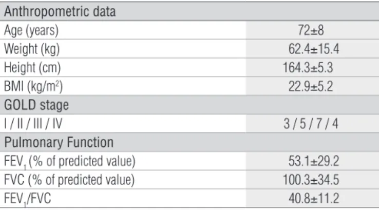
Of the 29 eligible patients, ten did not meet the inclusion cri-teria. With respect to the analyzed patients’ comorbidities, two had a history of gout and two of sinusitis or typhoid fever. The anthropometric characteristics and pulmonary function of the 19 patients who concluded the study are expressed in Table 1.
To analyze the behavior of the cardiovascular system, the best FVC test was chosen because the comparison of HR and HRV index values revealed no differences between them, re-gardless of the number of tests performed (ANOVA, p≥0.05).
The predominant HR behavior during the FVC tests was initially a decrease (79%), followed by a progressive increase with oscillations (95%), and the highest value was reached after
Even after the end of the FVC test, HR continued to increase an av-erage of 21.3±13.9 bpm (75.9±14.6 vs. 97.2±21.8; p<0.001) and then reduce an average of 4.9±7.6 bpm (75.9±14.6 vs. 71.0±11.2, p <0.001) compared to resting values. The HR peak reached after the end of the FVC test was an average 9.5±8.3 bpm higher than obtained at the end of the test (87.7±16.8 vs. 97.2±21.8; p<0.001 ).
Despite an average increase of 6.3±20.1 mmHg (136.8±21.4 vs. 143.16±30.0) in systolic BP from the beginning to the end of the test and upon returning to baseline value after 10 min-utes of rest (133.2±23.8 mmHg), there were no differences between the analyzed periods (p≥0.05). Diastolic BP remained unchanged during the periods analyzed (83.2±12.9 mmHg vs 85.3±13.9 mmHg vs 83.2±13.8 mmHg; p≥0.05).
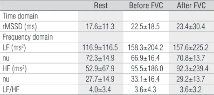
The values of HRV indices can be observed in Table 3. There were no statistically significant differences in HRV indices in the temporal (rMSSD) or the frequency (LF, HF, and LF / HF ratio) domains between the analyzed periods (p≥0.05).
Discussion
The results of this study showed that the FVC test produced significant changes in COPD patient HR but did not change the BP or HRV index values during the analyzed periods.
The HR response to the FVC test was characterized by a reduction at the beginning of the inspiratory phase and by an increase at its end, with the HR peak being reached after the end of the test. The HR then decreased below resting HR values and subsequently returned to baseline value.
The initial reduction and subsequent increase in HR, which were observed in this study during the execution of the inspiratory phase of the FVC test, were similar to the pattern observed by Martins et al.36 during the sustained inspiratory
breath-holding maneuver.
It is postulated that the initial decrease in HR during the inspiratory phase of the test may be related, at least in part, to the activation of bronchopulmonary C-fibers. These fibers, when stimulated, produce reflex bradycardia and hypotension and may be related to the observed reduction in HR37-39.
The subsequent progressive increase in HR may be at-tributed to the decrease in intrathoracic pressure that, con-sequently, produces a reduction in both the stroke volume of Figure 1. Example of heart rate (HR) behavior during FVC test in a
COPD patient.
76
62 97
120
65
45 60 75 90 105 120
HR (bpm)
Time Di
Df HR peak
1 2
HR (bpm)
97
1 2
HR peak
Di Df
76
65 62
120
Time
120
105
90
75
60
45
bpm=beats per minute; Di=Initial HR decrease in FVC test; Df=Decreased after the end of the FVC test; HRpeak=the highest value of heart rate reached; Dotted lines 1 and 2=beginning and end of the FVC test, respectively.
Table 1. Anthropometric characterization, pulmonary function and severity of 19 patients with COPD.
Data are expressed as mean ± standard deviation. BMI=body mass index; kg=kilogram;
cm=centimeter; GOLD=Global Initiative for Obstructive Lung Disease; I=mild;
II=moderate; III=severe; IV=very severe; FEV1=forced expiratory volume in one second;
FVC=forced vital capacity.
Anthropometric data
Age (years) 72±8
Weight (kg) 62.4±15.4
Height (cm) 164.3±5.3
BMI (kg/m2) 22.9±5.2
GOLD stage
I / II / III / IV 3 / 5 / 7 / 4
Pulmonary Function
FEV1 (% of predicted value) 53.1±29.2 FVC (% of predicted value) 100.3±34.5
FEV1/FVC 40.8±11.2
Table 2. Values of heart rate delta obtained during the FVC maneuver in relation to initial heart rate.
SD=standard deviation; CI95=confidence interval of 95%; HRI=initial heart rate (HR); HRQI=initial HR decrease in FVC test; HRF=HR at the end of the FVC test; HRP=the highest HR value
reached after finishing the FVC test; HRQF=HR decreased after the end of the FVC test.
∆HRQI - HRI ∆HRF - HRI ∆HRP - HRI ∆HRQF - HRI
Mean±SD -6.10±8.5 11.8±10.9 21.3±13.9 -4.9±7.6
Median -2 10 17 -2
[CI95] [-10.2 – -1.9] [6.5 – 17.0] [14.6 – 27.9] [-8.6 – -1.2]
[Minimum – Maximum] [-32 – 0] [-10 – 38] [3 – 60] [-27 – 0]
the end of the maneuver (84%). The HR then decreased below the initial resting HR (58%) and later returned to baseline. One example of this behavior can be observed in Figure 1.
Table 2 shows the delta HR values obtained during the FVC maneuver in relation to initial HR. It was observed that at the be-ginning of the inspiratory phase of the FVC test, HR was reduced an average of 6.10±8.5 bpm (75.9±14.6 vs. 69.8±10.0 p<0.001) and then increased an average of 11.8±10.9 bpm until the end of the test, compared to the resting values (75.9±14.6 vs. 87.7±16.8; p<0.001).
the left ventricle and in BP. This, in turn, would produce an increase in HR mediated by the activation of baroreceptors40.
Another aspect to be considered is the stimulation of pulmo-nary stretch receptors induced by an increase in tidal volume during inspiration41, which produces cardiac acceleration with
subsequent tachycardia38.
During the expiratory phase of the FVC test, the HR contin-ued to increase gradually, which may be related to an increase in intrathoracic pressure and a decrease in venous return which, in turn, promotes changes in myocardial contractility, vasomotor tone and baroreflex control42-44. The reduction of
vagal activity followed by the increase in sympathetic activity that occurs during expiration can also increase HR42.
After the end of the FVC test, the HR continued to increase until it reached its highest value, which can be explained by the release of the expiratory effort43,44. During a sudden decrease
in intrathoracic pressure, a large amount of blood is seques-tered to the pulmonary vascular bed, which reduces the left ventricular filling and causes a rise in HR44. Moreover, the lower
conduction velocity and the slow uptake and neutralization of noradrenaline by the sympathetic nervous system causes a gradual decay in the stimulus for increased HR, even after ces-sation of the expiratory effort45.
The decrease in HR after the accomplishment of the FVC maneuver is similar to that observed by Marães et al.43 after
the release of strain during the Valsalva maneuver (phase IV), which they attributed to increased reflex vagal activity and inhibited sympathetic activity due to great stimulation of baroreceptors during the expiratory phase and which was followed by vasodilation and a reduction in HR to below base-line values43.
Despite the influence of FVC maneuvers on HR fluctuations, the analysis of HRV indices showed no significant changes in the analyzed periods. This is similar to the results of Marães et al.43, who analyzed HRV before and after the Valsalva
maneu-ver in middle-aged individuals and observed no changes in the rMSSD index. The lack of change in HRV indices may be related to the short duration of the maneuver, the advanced age of the volunteers or the probable autonomic dysfunction present in these individuals. Paschoal, Petrelluzzi and Gonçalves13 and
Acharya et al.46 observed that advancing age may reduce heart
rate variability. Other studies have verified that COPD patients have depressed HRV when compared to healthy subjects matched by age and sex7,8,47-49.
Furthermore, the FVC maneuver did not modify BP be-havior during the analyzed periods. However, it should be pointed out that an indirect (auscultatory) method was used to measure BP, which, although reliable, did not allow measure-ment during the entire period of FVC performance, but only before and after the maneuver. Therefore, the possibility that
the test promoted changes in this variable during its execution should not be discarded.
The results of this study may benefit health professionals in the clinical setting, particularly physiotherapists, since according to COFFITO SECEX communication 123/9650 they are sufficiently qualified and specialized to perform spirometry.
Spirometry is considered the gold standard for diagnos-ing and evaluatdiagnos-ing COPD because it is the most reproducible, standardized and objective way of measuring airflow limita-tion1. Thus, understanding cardiovascular system responses
to the FVC test, performed by spirometry, provides physical therapists with a better means of monitoring COPD patients during its performance.
A limitation of this study was the lack of a healthy control group, which might have clarified if the results were due solely to the FVC test or were influenced by the disease. Additionally, the inclusion of patients whose COPD severity varied widely could be considered another limiting factor, although most of our patients (63%) were classified according to GOLD as stage II and III. Some studies have indicated an association between low FEV1 and a high risk of cardiovascular disease51,52, although
Camillo et al.53 observed no association between the degree of
bronchial obstruction and the values of HRV indices.
Another aspect to be considered is the way that HR pro-cesses are analyzed during FVC. During the maneuver, the time necessary for completing the inspiratory and expiratory movements varies between subjects, which complicates the analysis of HR response over time, and thus it was decided only to describe what happened to it during the maneuver.
This study represents a description of cardiovascular sys-tem responses while performing the FVC maneuver, and the methodology allowed inferences to be made about the mecha-nisms involved in these responses. However, more studies in-vestigating these mechanisms should be carried out.
Table 3. HRV indices of COPD patients at rest, before and after the FVC maneuver.
rMSSD=square root of the mean of the squared differences between adjacent normal RR intervals recorded in a time interval, divided by the number of RR intervals minus one; HF=high frequency; LF=low frequency; nu=normalized unit; ms=milliseconds. (Repeated measures ANOVA with post-hoc Student-Newman-Keuls; p>0.05).
Rest Before FVC After FVC
Time domain
rMSSD (ms) 17.6±11.3 22.5±18.5 23.4±30.4 Frequency domain
LF (ms2) 116.9±116.5 158.3±204.2 157.6±225.2
nu 72.3±14.9 66.9±16.4 70.8±13.7 HF (ms2) 52.9±67.9 95.5±186.0 92.3±239.4
nu 27.7±14.9 33.1±16.4 29.2±13.7
LF/HF 4.0±3.4 3.6±4.3 3.6±3.2
The present results suggest that the FVC test significantly influences the HR behavior of COPD patients, but did not change autonomic modulation, according to HRV indices, or BP during the studied periods.
Acknowledgment
This study was financed by the UNESP Development Foun-dation (FUNDUNESP - protocol nº. 00206/05).
1. Global Initiative for Chronic Obstructive Pulmonary Disease (GOLD). Global strategy for the diagnosis, management, and prevention of Chronic Obstructive Pulmonary Disease. Updated 2008. Disponível em: http://www.goldcopd.org. Acessado em 14 de dezembro de 2009.
2. Finkelstein J, Cha E, Scharf SM. Chronic obstructive pulmonary disease as an independent risk factor for cardiovascular morbidity. Int J Chron Obstruct Pulmon Dis. 2009;4:337-49.
3. Richens JL, Urbanowicz RA, Lunt EAM, Metcalf R, Corne J, Fairclough L, et al. Systems biology coupled with label-free high-throughput detection as a novel approach for diagnosis of chronic obstructive pulmonary disease. Respir Res. 2009;10:29.
4. Borghi-Silva A, Arena R, Castello V, Simões RP, Martins LE, Catai AM, et al. Aerobic exercise training improves autonomic nervous control in patients with COPD. Respir Med. 2009;103(10):1503-10.
5. Araújo CG, Vianna LC. How often does spirometry testing induce cardiac arrhythmias? Prim Care Respir J. 2009;18(3):185-8.
6. Celli BR. Update on the Management of COPD. Chest. 2008;133(6):1451-62.
7. Chhabra SK, De S. Cardiovascular autonomic neuropathy in chronic obstructive pulmonary disease. Respir Med. 2005;99(1):126-33.
8. Volterrani M, Scalvini S, Mazzuero G, Lanfranchi P, Colombo R, Clark AL, et al. Decreased heart rate variability in patients with chronic obstructive pulmonary disease. Chest. 1994;106(5):1432-7.
9. Gunduz H, Talay F, Arinc H, Ozyildirim S, Akdemir R, Yolcu M, et al. Heart rate variability and heart rate turbulence in patients with chronic obstructive pulmonary disease. Cardiol J. 2009;16(6):553-9.
10. Yildiz P, Tükek T, Akkaya V, Sözen AB, Yildiz A, Korkut F, et al. Ventricular arrhythmias in patients with COPD are associated with QT dispersion. Chest. 2002;122(6):2055-61.
11. Senior RM, Lefrak SS, Kleiger RE. The heart in chronic obstructive pulmonary disease: Arrhythmias. Chest. 1979;75(1):1-2.
12. Tükek T, Yildiz P, Atilgan D, Tuzcu V, Eren M, Erk O, et al. Effect of diurnal variability of heart rate on development of arrhythmia in patients with chronic obstructive pulmonary disease. Int J Cardiol. 2003;88(2-3):199-206.
13. Paschoal MA, Petrelluzzi KFS, Gonçalves NVO. Estudo da variabilidade da frequência cardíaca em pacientes com doença pulmonar obstrutiva crônica. Rev Ciên Med. 2002;11(1):27-37.
14. Schneider A, Gindner L, Tilemann L, Schermer T, Dinant GJ, Meyer FJ, et al. Diagnostic accuracy of spirometry in primary care. BMC Pulm Med. 2009;9:31. doi: 10.1186/1471-2466-9-31.
15. Papaioannou AI, Loukides S, Gourgoulianis KI, Kostikas K. Global assessment of the COPD patient: Time to look beyond FEV1? Respir Med. 2009;103(5):650-60.
16. Bednarek M, Gorecka D, Wielgomas J, Czajkowska-Malinowska M, Regula J, Mieszko-Filipczyk G, et al. Smokers with airway obstruction are more likely to quit smoking. Thorax. 2006;61(10):869-73.
17. Barreiro TJ, Perillo I. An approach to interpreting spirometry. Am Fam Physician. 2004;69(5):1107-14.
18. Modrykamien AM, Gudavalli R, McCarthy K, Liu X, Stoller JK. Detection of upper airway obstruction with spirometry results and the flow-volume loop: a comparison of quantitative and visual inspection criteria. Respir Care. 2009;54(4):474-9.
19. Ziora K, Ziora D, Oswiecimska J, Roczniak W, Machura E, Dworniczak S, et al. Spirometric parameters in malnourished girls with anorexia nervosa. J Physiol Pharmacol. 2008;59 Suppl 6:801-7.
20. Pinsky MR. Cardiovascular issues in respiratory care. Chest. 2005;128(5 Suppl 2):592S-7S.
21. Yasuma F, Hayano J. Respiratory sinus arrhythmia: Why does the heartbeat synchronize with respiratory rhythm? Chest. 2004;125(2):683-90.
22. Fields CL, Byrd RP Jr, Ossorio MA, Roy TM, Michaels MJ, Vogel RL. Cardiac arrhythmias during performance of the flow-volume loop. Chest. 1993;103(4):1006-9.
23. Oliveira RB, Vianna LC, Ricardo DR, de Almeida MB, Araújo CGS. Influence of different respiratory maneuvers on exercise-induced cardiac vagal inhibition. Eur J Appl Physiol. 2006;97(5):607-12.
24. Standardization of spirometry, 1994 update. American Thoracic Society. Am J Respir Crit Care Med. 1995;152(3):1107-36.
25. Lohman TG, Roche AF, Martorell R. Anthropometric standardization reference manual. Champaign: Human Kinetics Books; 1988.
26. Knudson RJ, Lebowitz MD, Holberg CJ, Burrows B. Changes in the normal maximal expiratory flow-volume curve with growth and aging. Am Rev Respir Dis. 1983;127(6):725-34.
27. Porto LG, Junqueira LF Jr. Comparison of time-domain short-term heart interval variability analysis using a wrist-worn heart rate monitor and the conventional electrocardiogram. Pacing Clin Electrophysiol. 2009;32(1):43-51.
28. Vanderlei LCM, Silva RA, Pastre CM, Azevedo FM, Godoy MF. Comparison of the Polar S810i monitor and the ECG for the analysis of heart rate variability in the time and frequency domains. Braz J Med Biol Res. 2008;41(10):854-9.
29. Jurca R, Church TS, Morss GM, Jordan AN, Earnest CP. Eight weeks of moderate intensity exercise training increases heart rate variability in sedentary postmenopausal women. Am Heart J. 2004;147(5):e21.
30. Kiviniemi AM, Hautala AJ, Kinnunen H, Tulppo MP. Endurance training guided individually by daily heart rate variability measurements. Eur J Appl Physiol. 2007;101(6):743-51.
31. Heart rate variability: standards of measurement, physiological interpretation and clinical use. Task Force of the European Society of Cardiology and the North American Society of Pacing and Electrophysiology. Circulation. 1996;93(5):1043-65.
32. Gall B, Parkhouse W, Goodman D. Heart rate variability of recently concussed athletes at rest and exercise. Med Sci Sports Exerc. 2004;36(8):1269-74.
33. Pichon A, de Bisschop C, Diaz V, Denjean A. Parasympathetic Airway Response and Heart Rate Variability Before and at the end of Methacholine Challenge. Chest. 2005;127(1):23-9.
34. Antila K. Quantitative characterization of heart rate during exercise. Scand J Clin Lab Invest Suppl. 1979;(153):3-68.
35. Pagani M, Lombardi F, Guzzetti S, Rimoldi O, Furlan R, Pizzinelli P, et al. Power spectral analysis of heart rate and arterial pressure variabilities as a marker of sympatho-vagal interaction in man and conscious dog. Circ Res. 1986;59(2):178-93.
36. Martins FP, Barbosa PRB, Oliveira ILC, Gomes Filho JBM. Modelo linear da resposta de adaptação dos intervalos rr à manobra de apnéia inspiratória sustentada. Rev SOCERJ. 2006;19(4):292-301.
37. Kubin L, Alheid GF, Zuperku EJ, McCrimmon DR. Central pathways of pulmonary and lower airway vagal afferents. J Appl Physiol. 2006;101(2):618-27.
38. Kaufman MP, Iwamoto GA, Ashton JH, Cassidy SS. Responses to inflation of vagal afferents with endings in the lung of dogs. Circ Res. 1982;51(4):525-31.
39. Lee LY, Pisarri TE. Afferent properties and reflex functions of bronchopulmonary C-fibers. Respir Physiol. 2001;125(1-2):47-65.
40. Larsen PD, Tzeng YC, Sin PY, Galletly DC. Respiratory sinus arrhythmia in conscious humans during spontaneous respiration. Respir Physiol Neurobiol. 2010;174(1-2):111-8.
41. Marshall JM. Peripheral chemoreceptors and cardiovascular regulation. Physiol Rev. 1994;74(3):543-94.
42. Lu K, Clark JW Jr, Ghorbel FH, Ware DL, Bidani A. A human cardiopulmonary system model applied to the analysis of the valsalva maneuver. Am J Physiol Heart Circ Physiol. 2001;281(6):H2661-79.
43. Marães VRFS, Santos MDB, Catai AM, Moraes FR, Oliveira L, Gallo Jr L, et al. Modulação do sistema nervoso autonômico na resposta da frequência cardíaca em repouso e à manobra de valsalva com o incremento da idade. Rev Bras Fisioter. 2004;8(2):97-103.
44. Freeman R. Assessment of cardiovascular autonomic function. Clin Neurophysiol. 2006;117(4):716-30.
45. Berne RM, Levy MN. Fisiologia. 3ª ed. Rio de Janeiro: Guanabara Koogan; 1996.
46. Acharya UR, Kannathal N, Sing OW, Ping LY, Chua T. Heart rate analysis in normal subjects of
References
various age groups. Biomed Eng OnLine. 2004;3(1):24.
47. Pantoni CBF, Reis MS, Martins LEB, Catai AM, Costa D, Borghi-Silva A. Estudo da modulação autonômica da frequência cardíaca em repouso de pacientes idosos com doença pulmonar obstrutiva crônica. Rev Bras Fisioter. 2007;11(1):35-41.
48. Bartels MN, Jelic S, Ngai P, Basner RC, DeMeersman RE. High-frequency modulation of heart rate variability during exercise in patients with COPD. Chest. 2003;124(3):863-9.
49. Chen WL, Chen GY, Kuo CD. Hypoxemia and autonomic nervous dysfunction in patients with chronic obstructive pulmonary disease. Respir Med. 2006;100(9):1547-53.
50. Conselho Regional de Fisioterapia e Terapia Ocupacional da 5ª região. O Fisioterapeuta pode realizar teste de esforço físico? O Fisioterapeuta pode realizar testes espirométricos? O Fisioterapeuta pode
realizar hidroginástica/hidroterapia em grupo (piscina) como terapia em processo ortopédico? 15/janeiro/1996. Disponível em: http://www.crefito5.com.br/web/pareceres_det.php?id=2. Acesso em 01/ 09/2004.
51. Sin DD, Wu L, Man SF. The relationship between reduced lung function and cardiovascular mortality: a population-based study and a systematic review of the literature. Chest. 2005;127(6):1952-9.
52. Engström G, Hedblad B, Janzon L, Valind S. Respiratory decline in smokers and ex-smokers–an independent risk factor for cardiovascular disease and death. J Cardiovasc Risk. 2000;7(4):267-72.
53. Camillo CA, Pitta F, Possani HV, Barbosa MV, Marques DSO, Cavalheri V, et al. Heart Rate Variability and Disease Characteristics in Patients with COPD. Lung. 2008;186(6):393-401.