Semiquantitative echocardiographic evaluation
of intrapulmonary vascular dilatations: correlation
with evaluation of shunt levels and pulmonary
function parameters*
Avaliação semiquantitativa ecocardiográfica de dilatações vasculares intrapulmonares em candidatos a transplante hepático: correlação com
avaliação de shunt e parâmetros funcionais pulmonares
Maria Angélica Pires Ferreira, Sérgio Saldanha Menna Barreto, Marli Maria Knorst, Mario Reis Álvares da Silva, Antonio Furlan Pinotti
Abstract
Objective: To correlate semiquantitative evaluation of intrapulmonary vascular dilatations (IPVD) with quantitative evaluation of shunt levels, as well as to describe clinical and pulmonary function findings in a sample of liver disease patients with IPVD. Methods: Patients presenting transthoracic echocardiography (TTE) positivity for IPVD underwent clinical evaluation, pulmonary function tests and pulmonary shunt quantification (scintigraphy with technetium-99m-labeled macroaggregated albumin and blood gas analysis after pure oxygen breathing).
Results: A total of 28 liver cirrhosis patients were studied (mean age, 47.5 years; 60.7% were Child-Pugh class B). A 4-point, ascending scale was used as a measure of IPVD intensity, which was scored as 1, 2, 3 and 4, respectively, in 13 (46.4%), 9 (32.1%), 2 (7.1%) and 4 (14.3%) of the patients. Patients were divided into a low-intensity group (scores 1 and 2) and a high-intensity group (scores 3 and 4). The mean shunt assessed using scintigraphy was 14.9% in the sample as a whole and was lower in the low-intensity group (11.7% vs. 26.3%; p = 0.01). The mean shunt by blood gas analysis was higher in the high-intensity group (8.3% vs. 16.3%; p < 0.001). Mean PaO2 was
lower in the high-intensity group. There was a negative correlation between DLCO and IPVD severity (r = −0.406,
p = 0.01). Conclusions: TTE is a safe, useful tool for assessing IPVD severity in liver disease patients. The IPVD intensity assessed using TTE correlated with the intrapulmonary shunt values obtained through the quantitative methods evaluated, as well as with pulmonary gas exchange abnormalities.
Keywords: Anoxia; Liver cirrhosis; Hepatopulmonary syndrome; Echocardiography.
Resumo
Objetivo: Verificar a relação entre a avaliação semiquantitativa de dilatações vasculares intrapulmonares (DVIP) e a avaliação quantitativa de shunt, e descrever achados clínicos e funcionais pulmonares em uma amostra de hepatopatas com DVIP. Métodos: Pacientes com ecocardiografia transtorácica (ETT) positiva para DVIP foram submetidos à avaliação clínica e de função pulmonar assim como à quantificação de shunt intrapulmonar (cinti-lografia com macroagregados de albumina marcados com tecnécio-99m e por gasometria com oxigênio a 100%).
Resultados: Foram estudados 28 pacientes cirróticos (média de idade, 47,5 anos; 60,7% dos casos classificados como Child-Pugh B). Uma escala de 4 pontos, em ordem ascendente, foi utilizada para medir a intensidade das DVIP, classificada de 1 a 4, respectivamente, em 13 (46,4%), 9 (32,1%), 2 (7,1%) e 4 (14,3%) dos pacientes. A amostra foi dividida em grupo baixa intensidade (escores 1 e 2) e grupo alta intensidade (escores 3 e 4). A média de shunt por cintilografia foi 14,9% na amostra total, sendo menor no grupo baixa intensidade (11,7% vs. 26,3%; p = 0,01). O grupo alta intensidade teve maiores valores de shunt através de gasometria (8,3% vs. 16,3%; p < 0.001). A PaO2 média foi inferior no grupo alta intensidade. A intensidade de DVIP e a DLCO
correlaciona-ram-se de forma inversa (r = −0,406, p = 0,01). Conclusões: A ETT é um método útil e seguro para avaliação da gravidade das DVIP em pacientes com hepatopatia. A classificação ecocardiográfica da intensidade das DVIP se correlacionou com valores de shunt intrapulmonar obtidos pelos métodos quantitativos avaliados, bem como com anormalidades nas trocas gasosas pulmonares.
Descritores: Anóxia; Cirrose hepática; Síndrome hepatopulmonar; Ecocardiografia.
* Study carried out at the Porto Alegre Hospital de Clínicas, Porto Alegre, Brazil.
Correspondence to: Maria Angélica Pires Ferreira. Rua Balduino Roehrig, 98, Três Figueiras. CEP 91330-140, Porto Alegre, RS, Brasil. Tel 55 51 2101-8491. E-mail: [email protected]
IPVD screening and diagnosis of hepatopulmo-nary syndrome, patients with negative screening results were not submitted to intrapulmonary shunt quantification using other methods. We excluded patients presenting any of the following characteristics: severe structural chronic pulmonary disease; moderate to severe COPD (stage II to IV according to the Brazilian Thoracic Association); severe cardiopathy; intracardiac shunt; voluminous pleural effusion seen on chest X-ray (more than one third of the hemithorax); diaphragmatic paralysis or other restrictive lung diseases of moderate to severe intensity; and tense refractory ascites.
The patients were evaluated as outpatients, all presenting clinical stability at the time of evaluation. Chest X-ray, arterial blood gas anal-ysis, spirometry (including a pharmacodynamic test) and DLCO (single-breath method) were performed, in accordance with the American Thoracic Society criteria. In addition, pulse oximetry and orthodeoxia investigation were performed. Orthodeoxia was performed using pulse oximetry, SpO2 being determined after 5 min in the horizontal supine position and after 5 min in a sitting position. Positivity was defined as a decrease in SpO2 ≥ 4%. Arterial blood gas analysis was performed through radial artery puncture, with the patient in a sitting position and on room air. Patients with a PaO2 < 80 mmHg on room air and an
alveo-Introduction
Hepatopulmonary syndrome, which consists of the triad of intrapulmonary vascular dilata-tions (IPVD), hypoxemia and liver disease, has been described in 5-29% of individuals with liver disease. Occurring in 5-47% of all cases of advanced liver disease, IPVD constitute the leading cause of severe hypoxemia in such cases. (1-6) However, not all patients with IPVD present hypoxemia; the clinical significance and prog-nosis of IPVD in the absence of gas exchange abnormalities remain undefined.(7)
The diagnosis of intrapulmonary shunt can be made through functional studies, such as pure oxygen inhalation tests and imaging studies. The latter include contrast-enhanced echocardiography, lung scintigraphy with radioisotope-labeled albumin and pulmonary angiography.(6,8-10) Contrast-enhanced transtho-racic echocardiography detects right-to-left shunts and is considered the method of choice for IPVD screening, since it presents greater sensitivity than does scintigraphy with macro-aggregated albumin.(3,9,11,12) Other advantages of echocardiography include its routine use for the diagnosis of pulmonary hypertension. In addition, it allows the differentiation between intracar-diac and intrapulmonary shunts. Furthermore, echocardiography makes it possible to evaluate IPVD intensity semiquantitatively.(3,11,12)
The present study aims at comparing levels of venous mixture (intrapulmonary shunt) obtained with two methods (radioisotope scintigraphy and blood gas analysis after pure oxygen breathing) and relating them to the IPVD level determined using contrast-enhanced echocardiography in a group of patients under evaluation for liver transplant. Our secondary objectives were to describe clinical and pulmonary function find-ings in liver transplant candidates with IPVD and to correlate these findings with IPVD intensity.
Methods
We included consecutive patients over the age of 16 with chronic liver disease or any type of portal hypertension who were under evalu-ation for liver transplant at the Porto Alegre Hospital de Clínicas, with positive screening for IPVD using transthoracic echocardiography. Since the literature indicates that transthoracic echocardiography is the method of choice for
Table 1 - Demographic data and data on the underlying disease in 28 liver transplant candidates with intrapulmonary vascular dilatations.
Demographic data and data on the underlying diseasea
Mean age in years (min-max) 47.5 (20-64) Male gender 19 (67.9) Female gender 9 (32.1) Mean time since diagnosis,
years (min-max)
2.6 (1-7)
Child-Pugh class A 5 (17.9) Child-Pugh class B 17 (60.7) Child-Pugh class C 6 (21.4) INR, mean ± SD 1.56 ± 0.52 Total bilirubin, mg/dL, mean ± SD 3.05 ± 2.67 Mean albumin, g/dL, mean ± SD 3.28 ± 0.74
INR: international normalized ratio. aValues expressed in
abnormal. This percentage is based on data in the literature.(3,9)
Blood gas analysis after pure oxygen breathing was performed using a Douglas bag for oxygen inhalation via a mask with a unidi-rectional expiratory valve. Patients remained in a sitting position, and the sample for arterial blood gas analysis was drawn from the radial artery using a plastic syringe at the end of a 20-min inhalation, using a technique previ-ously described for the collection, on room air, of samples for immediate analysis. The shunt fraction was calculated using the Berggren pulmonary shunt equation:
Qs/QT (%) = [(PAO2 − PaO2) × 0.003]/[C(a-v) O2] + [(PAO2 − PaO2) × 0.0031]
where Qs/QT (%) is the shunt fraction in percentage, PAO2 is the alveolar oxygen tension, and [C(a-v)O2] is the difference between arterial and venous oxygen content, which was consid-ered to be 4.5 mmHg in all cases, since all of the patients were clinically stable.(15) In most cases, arterial blood gas analysis, on room air, was performed simultaneously with the pure oxygen test.
The statistical analysis was performed using the program Statistical Package for the Social Sciences, version 13.0 (SPSS Inc., Chicago, IL, USA). Data are presented as number, percentage, mean and SD. Pearson’s correlation coefficient was applied in order to assess the correlations among variables with normal distribution, and Spearman’s rank correlation test was applied for variables with non-normal distribution. In order to compare the means of the two groups, we used the Student’s t-test for independent samples and for variables with normal distribu-tion. Nonparametric tests were used in order to compare groups of variables with non-normal lar-arterial oxygen gradient ≥ 15 mmHg were
considered hypoxemic, regardless of age.(13) The echocardiographic studies were performed using bidimensional transthoracic echocardi-ography. Isotonic saline solution submitted to manual agitation, on room temperature, was used as contrast. The cardiac chambers were examined immediately after injection of the contrast agent into a peripheral vein, and echo-genicity was evaluated for 60 s after injection. In order to determine the reproducibility of the test, which was videorecorded, a minimum of two injections were given. Echocardiographic positivity for IPVD was defined by the late passage (after the fourth beat) of the contrast agent into the left heart followed by its appear-ance in the right heart. As previously described by other authors,(12) a 4-point scale was used in order to score IPVD intensity as follows: 1 = passage of a small quantity of microbubbles into the left ventricle (LV); 2 = moderate passage of microbubbles into the LV; 3 = passage of a great number of microbubbles, without outlining the LV endocardium; and 4 = passage of a great number of bubbles with clear outline of the LV endocardium. Tests were independently performed and read by two echocardiographers, previously trained in identifying and grading IPVD using this scale.
Hepatopulmonary syndrome was diagnosed in patients with liver disease, IPVD being detected by echocardiography and alveolar- arterial oxygen gradient > 15 mmHg. This value was defined as proposed in the literature, due to its acceptable sensitivity for the identification of gas exchange abnormalities in such patients.(7,10,14)
Patients underwent perfusion scintigraphy with technetium-99m-labeled macroaggregated albumin (99mTc-MAA). The radioactive drug (2-4 mCi) was injected into a peripheral vein, with the patient in the sitting position. Immediately after the injection, images were obtained with the patient in the supine position. Shunt frac-tion was determined based on the ratio between the whole body and pulmonary activity of the radioactive drug, using software attached to a gamma camera. The commercial preparation DRN 4378 TechneScan Lyo-MAA (Mallinckrodt Medical B.V., Petten, Holland) was used. A scintillation gamma camera equipped with a low-energy, high resolution parallel-hole colli-mator was used. Values > 6% were considered
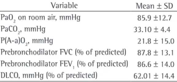
Table 2 - Pulmonary function in 28 liver transplant candidates with intrapulmonary vascular dilatations.
Variable Mean ± SD PaO2 on room air, mmHg 85.9 ±12.7 PaCO2, mmHg 33.10 ± 4.4 P(A-a)O2, mmHg 21.8 ± 15.0 Prebronchodilator FVC (% of predicted) 87.8 ± 13.1 Prebronchodilator FEV1 (% of predicted) 86.6 ± 14.0 DLCO, mmHg (% of predicted) 62.01 ± 14.4
Arterial blood gas analysis on room air was analyzed in 27 of the 28 patients, and in 1 case the test was excluded due to a collection error. Hypoxemia was identified in 8 patients (29.6%). In the sample as a whole, the mean prebron-chodilator FEV1 was 86.6% of predicted. Data related to arterial blood gases and pulmonary function are presented in Table 2.
All echocardiographic tests were considered technically adequate. There were no method-related complications. Among the 28 patients included in this study, echocardiography-deter-mined IPVD intensity was scored as 1, 2, 3 and 4 (ascending intensity) in 13 (46.4%), 9 (32.1%), 2 (7.1%) and 4 (14.3%) of the patients, respectively. None of the patients presented echocardiographic signs of pulmonary arterial hypertension.
Mean PaO2 was higher in the patients with IPVD scores of 1 or 2 (Student’s t-test, p = 0.01; Table 3). The correlation coefficient between PaO2 on room air and echocardiography-deter-mined IPVD score presented borderline statistical significance (r = −0.368; p = 0.05).
Hepatopulmonary syndrome was diag-nosed in 16 of the 28 patients studied (57.1% of the patients), taking into consideration the concomitance between IPVD and increased alve-olar-arterial oxygen gradient. However, when the 51 patients submitted to echocardiographic screening for IPVD were considered, the preva-lence of hepatopulmonary syndrome was 31.4% (16 of the 51 patients).
There was an inverse correlation between DLCO and echocardiography-determined IPVD score (r = −0.406; p = 0.01). Mean DLCOwas significantly higher among patients with IPVD distribution. Bland & Altman plots were used
in order to improve the visualization of the concordance between shunt values measured by scintigraphy with 99mTc-MAA and blood gas analysis after pure oxygen breathing. Bilateral tests were employed, and the level of statistical significance was set at 5% (p < 0.05). The study was approved by the ethics in research committee of the institution.
Results
Of the 51 patients submitted to contrast-enhanced echocardiography, 1 presented early contrast passage and was diagnosed with patent foramen ovale. Another 28 presented echocardiographic positivity for IPVD and were included in the study. Of those 28 patients, 9 were female. The mean age was 47.5 years. The most common etiology of liver disease, seen in 11 patients (39.3%), was viral hepatitis C. In the majority of the patients (60.7%), the severity of the liver disease was classified as Child-Pugh class B (Table 1).
Dyspnea was reported by 2 patients, 1 of which also presented platypnea. Both were diagnosed with hepatopulmonary syndrome and were hypoxemic at rest. Two cases of orthode-oxia were detected: one accompanied by dyspnea and platypnea; and one asymptomatic from a respiratory standpoint. Most of the patients (71.4%) reported current or previous smoking, and 9 (32.1%) were current smokers. The mean tobacco intake was 14.6 pack-years.
Non-voluminous unilateral pleural effu-sion was present in two cases. Minimal ascites was identified using echocardiography in seven cases.
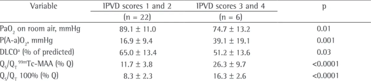
Table 3 - Blood gas analysis parameters, pulmonary diffusion and shunt fraction in 28 patients with low and high scores for echocardiography-determined intrapulmonary vascular dilatations.
Variable IPVD scores 1 and 2 IPVD scores 3 and 4 p (n = 22) (n = 6)
PaO2 on room air, mmHg 89.1 ± 11.0 74.7 ± 13.2 0.01 P(A-a)O2, mmHg 16.9 ± 9.4 39.1 ± 19.1 0.001 DLCOa (% of predicted) 65.0 ± 13.4 51.2 ± 13.6 0.03
QS/QT 99mTc-MAA (% Q) 11.7 ± 3.8 26.3 ± 9.7 <0.0001
QS/QT 100% (% Q) 8.3 ± 2.3 16.3 ± 2.6 <0.0001
P(A-a)O2: alveolar-arterial oxygen gradient; QS/QT 100%: shunt fraction using blood gas analysis after pure oxygen
brea-thing; QS/QT99mTc-MAA: shunt fraction using scintigraphy with technetium-99m-labeled macroaggregated albumin; and
Of the 28 patients with IPVD identified using contrast-enhanced echocardiography, the degree of shunting was quantified using scin-tigraphy and blood gas analysis in 21 (75%), only scintigraphy in 6 (21.4%) and only blood gas analysis in 1 (3.6%). Therefore, 27 scinti-graphic imaging tests and 22 blood gas analyses after pure oxygen breathing were performed. Shunt quantification could not be performed in 4 patients due to adherence problems (3 patients did not undergo the pure oxygen breathing test, and 1 patient did not undergo scintig-raphy). Another 3 patients were not submitted to the pure oxygen breathing test due to clinical complications, established after performance of the first two tests (slowly resolving pneumonia, aggravation of ascites and liver transplant).
For the 27 patients submitted to scintigraphy, the mean scintigraphy-determined shunting of cardiac output was 14.9 ± 9% (range, 6.9-39%; median, 12%). When scintigraphy was used, none of the patients presented a normal shunt frac-tion (< 6%). A positive correlafrac-tion was observed between echocardiography-determined IPVD score and scintigraphy-determined shunt frac-tion (r = 0.567; p < 0.001; Figure 1).
The mean shunt value obtained using the pure oxygen breathing test was 9.8 ± 3.9% (range, 4-19%). Final PaO2 in the test ranged from 317.8 mmHg to 615 mmHg (mean, 513.6 mmHg) and was significantly higher among the patients with IPVD scores of 1 or 2 than among those with IPVD scores of 3 or 4 (542.2 mmHg vs. 384 mmHg; p < 0.001). The median shunt was 9.5%. One patient (4.5%) presented a normal shunt fraction (< 5%) when the pure oxygen breathing test was used. That patient presented an echocardiography-determined IPVD score of 1 and normoxemia on room air. A statisti-cally significant correlation was found between echocardiography-determined IPVD score and the shunt value determined using blood gas analysis after pure oxygen breathing (rs = 0.609, p < 0.01).
Scintigraphy-determined shunt correlated significantly with that determined using blood gas analysis after pure oxygen breathing (r = 0.666; p < 0.001). The correlation between shunt values obtained using the two methods and the comparison of results using Bland & Altman plots are shown in Figure 2. The mean shunt difference determined using the two methods scores of 1 or 2 than among those with IPVD
scores of 3 or 4 (p = 0.03; Table 3).
There was no significant correlation between severity of the underlying disease, according to the Child-Pugh classification, and echocar-diography-determined IPVD score (r = −0.13; p = 0.48). Similarly, there was no correlation between time since diagnosis of liver disease and echocardiography-determined IPVD score.
40
30
20
10
0
0 1 2 3 4
Echocardiography-determined IPVD score (1-4)
QS
/Q
T
scintigraphy (%)
Figure 1 - Distribution of shunt values, as determined using scintigraphy with technetium-99m-labeled macroaggregated albumin, according to echocardiography-determined intrapulmonary vascular dilatations (IPVD) score (r = 0.567; p < 0.001).
0 5
5 10
10
15
15
20
20
25
25 40
35 30 25 20
20 15
15 10
10
-10 5
5
-5
0 0
r = 0.666, p < 0.001
30 a
b
Scintigraphy-determined
shunt (%)
Shunt determined using blood gas analysis after pure oxygen (O2) breathing
Shunt difference (scintigraphy
vs. pure o
xygen test)
Mean shunt determined using scintigraphy and pure oxygen test Figure 2 - Shunt values evaluated using scintigraphy with technetium-99m-labeled macroaggregated albumin and blood gas analysis after pure oxygen (O2) breathing. In A, correlation between shunt values evaluated by the two methods (r= 0.666; p < 0.001). In B, Bland & Altman plots were used in order to improve the visualization of shunt values evaluated by the two methods; mean of the differences, 4.3%;
who presented IPVD without hepatopulmonary syndrome, IPVD was scored as 1+.(14)
In the present study, echocardiography-determined IPVD score was found to correlate significantly with shunt fraction calculated using either of the two quantitative methods employed (blood gas analysis after pure oxygen breathing and scintigraphy).
All of the patients evaluated in the present study presented echocardiographic and scinti-graphic positivity. Nevertheless, in functional terms, there were no cases of total lack of response to pure oxygen, and final PaO2 after pure oxygen inhalation was > 300 mmHg in all cases. This is consistent with the theoretical model employed in order to explain the diffu-sion-perfusion mismatch related to impaired gas exchange among the hemoglobin molecules in the center of the dilated vessel. In addition to the distance between the alveoli and the hemo-globin molecules in the central stream of the vessel, the shorter transit time of red cells in the pulmonary capillary would contribute to the worsening of the disturbance. This abnormality in oxygenation can be significantly improved with pure oxygen, resulting in lower shunt indices obtained through blood gas analysis after pure oxygen breathing. Together with ventila-tion-perfusion mismatch and anatomical shunt, diffusion-perfusion mismatch is considered a central mechanism in the genesis of hypoxemia related to liver disease.(4,8,22)
Data in the literature show that the isotopic method frequently overestimates the shunt values obtained using blood gas analysis. One group of authors, evaluating 8 patients with hepatopulmonary syndrome, found individual differences between the methods ranging from 2% to 30%. Convergence of shunt values calcu-lated using the two methods was associated with anatomical shunt demonstrated using angiog-raphy, whereas the divergence was related to a combination of anatomical shunt and diffusion disturbance.(23)
According to the literature, scintigraphy is less sensitive than is transthoracic echocar-diography in the identification of IPVD. The scintigraphic positivity in all 27 of the patients submitted to the test might be due to differ-ences in the reading of the test, since there is an operator-dependent aspect in the demarcation of areas with abnormal uptake of the radioac-was 4.3 ± 3.8%. There was greater discrepancy
between the results of the two methods in three cases: scintigraphy overestimated shunt in two cases and underestimated shunt in one case.
Discussion
Of the pulmonary functional parameters analyzed, the principal abnormality observed was a decrease in DLCO, which correlated inversely with IPVD score. This finding is in accordance with the data in the literature.(16) We observed a high frequency of increased alveolar-arte-rial oxygen gradient and hypocapnia, both of which correlated significantly with IPVD score. The effects of smoking might have contrib-uted to the gas exchange alterations described here, although vascular abnormalities resulting from liver disease are possibly the predominant factors.(17-20)
Mean PaO2 was within the limits of normality. Taking into account the exclusion of patients with respiratory comorbidities, this reflects a low to moderate IPVD score found in most cases, since 78.5% of the cases presented echocardiography-determined scores of 1 or 2, and the majority presented scintigraphy-determined shunt < 20%. According to the literature, echocardiography is frequently posi-tive in patients with normal arterial blood gases, presumably reflecting an IPVD level insufficient to be reflected in gas exchange.(3,8,11,12)
Georgia: results of a nation-wide prevalence survey among sentenced inmates. Int J Tuberc Lung Dis. 2000;4(12):1104-10.
3. Centers for Disease Control (CDC). Prevention and control of tuberculosis in correctional institutions: recommendations of the Advisory Committee for the Elimination of Tuberculosis. MMWR Morb Mortal Wkly Rep. 1989;38(18):313-20, 325.
4. Oliveira HB, Cardoso JC. Tuberculose no sistema prisional de Campinas, Säo Paulo, Brasil. Rev Panam Salud Públ. 2004;15(3):194-9.
5. Abrahão RM, Nogueira PA, Malucelli MI. Tuberculosis in county jail prisoners in the western sector of the city of São Paulo, Brazil. Int J Tuberc Lung Dis. 2006;10(2):203-8.
6. Fukazawa K, Aritake S, Minemura S, Shinohara T, Nakazono T, Mori T. A tuberculosis outbreak in a mental hospital [Article in Japanese]. Nippon Koshu Eisei Zasshi. 2000;47(9):801-8.
7. MacIntyre CR, Kendig N, Kummer L, Birago S, Graham NM. Impact of tuberculosis control measures and crowding on the incidence of tuberculous infection in Maryland prisons. Clin Infect Dis. 1997;24(6):1060-7. 8. Stern V. Problems in prisons worldwide, with a particular
focus on Russia. Ann N Y Acad Sci. 2001;953:113-9. 9. Hutton MD, Cauthen GM, Bloch AB. Results of a 29-state
survey of tuberculosis in nursing homes and correctional facilities. Public Health Rep. 1993;108(3):305-14. 10. Chaves F, Dronda F, Cave MD, Alonso-Sanz M,
Gonzalez-Lopez A, Eisenach KD, et al. A longitudinal study of transmission of tuberculosis in a large prison population. Am J Respir Crit Care Med. 1997;155(2):719-25. 11. MacIntyre CR, Kendig N, Kummer L, Birago S, Graham
NM, Plant AJ. Unrecognised transmission of tuberculosis in prisons. Eur J Epidemiol. 1999;15(8):705-9. 12. From the Centers for Disease Control. Transmission
of multidrug-resistant tuberculosis among immunocompromised persons, correctional system--New York, 1991. JAMA. 1992;268(7):855-6.
13. Jones TF, Craig AS, Valway SE, Woodley CL, Schaffner W. Transmission of tuberculosis in a jail. Ann Intern Med. 1999;131(8):557-63.
14. Steenland K, Levine AJ, Sieber K, Schulte P, Aziz D. Incidence of tuberculosis infection among New York State prison employees. Am J Public Health. 1997;87(12):2012-4.
15. Bellin EY, Fletcher DD, Safyer SM. Association of tuberculosis infection with increased time in or admission to the New York City jail system. JAMA. 1993;269(17):2228-31.
16. Kendig N. Tuberculosis control in prisons. Int J Tuberc Lung Dis. 1998;2(9 Suppl 1):S57-63.
17. World Health Organization. Tuberculosis control in prisons: a manual for programme managers. Geneva: WHO; 2000.
18. Aerts A, Hauer B, Wanlin M, Veen J. Tuberculosis and tuberculosis control in European prisons. Int J Tuberc Lung Dis. 2006;10(11):1215-23.
19. Chiang CY, Hsu CJ, Hsu PK, Suo J, Lin TP. Pulmonary tuberculosis in the Taiwanese prison population. J Formos Med Assoc. 2002;101(8):537-41.
20. Nyangulu DS, Harries AD, Kang’ombe C, Yadidi AE, Chokani K, Cullinan T, et al. Tuberculosis in a prison population in Malawi. Lancet. 1997;350(9087):1284-7.
tive drug. However, the significant correlation between the scintigraphy-determined shunt values and those obtained using the other two methods, as well as that between shunt values and abnormalities in arterial oxygenation are aspects to be considered in weighing the results obtained. Regarding the role of radioisotope scintigraphy in the evaluation of patients with liver disease and hypoxemia, in agreement with others authors, we believe that this method can be useful in the evaluation of hypoxemic patients with low echocardiography-determined IPVD score, in the cases in which echocardiography presents technical difficulties, such as obesity or deformities in the rib cage, or in those cases of accompanying pulmonary disease which make the diagnosis of hepatopulmonary syndrome difficult.(3) The combined analysis of the severity of oxygenation disturbance and the assessment of shunt using the method with 99mTc-MAA can be useful in the risk stratification of patients with hepatopulmonary syndrome for mortality associ-ated with liver transplants, with worse prognosis associated with PaO2≤ 50 mmHg and scintigra-phy-determined shunt fraction ≥ 20%.(7) In the present study, patients with higher echocardiog-raphy-determined IPVD scores (3 or 4) presented advanced dilatation of the vascular bed, demon-strated by a great passage of particles of the radioactive drug in scintigraphy, with a mean shunt fraction of 26%.
Transthoracic echocardiography is considered less sensitive than transesophageal echocardiog-raphy in IPVD detection. Among the advantages of the transthoracic approach are the lower costs, the fact that sedation is unnecessary and, theoretically, the lower risk, especially for indi-viduals with esophageal varices.(2)
In conclusion, transthoracic echocardiog-raphy is a useful and safe method of assessing IPVD severity in individuals with advanced liver disease. Transthoracic echocardiography-determined IPVD intensity correlated with the intrapulmonary shunt values obtained through the quantitative methods evaluated, as well as with pulmonary gas exchange abnormalities.
References
1. World Health Organization. Guidelines for the control of tuberculosis in prisons. Geneva: WHO; 1998.
23. Fournet N, Sanchez A, Massari V, Penna L, Natal S, Biondi E, et al. Development and evaluation of tuberculosis screening scores in Brazilian prisons. Public Health. 2006;120(10):976-83.
24. Drobniewski F. Tuberculosis in prisons--forgotten plague. Lancet. 1995;346(8980):948-9.
21. Niero R. Tuberculose pulmonar em uma prisão: Casa de Detenção de São Paulo 1976-1980. Temas IMESC Soc Dir Saúde. 1986;3(1):25-38.
22. Sanchez A, Gerhardt G, Natal S, Capone D, Espinola A, Costa W, et al. Prevalence of pulmonary tuberculosis and comparative evaluation of screening strategies in a Brazilian prison. Int J Tuberc Lung Dis. 2005;9(6):633-9.
About the authors
Maria Angélica Pires Ferreira
Pulmonologist. Porto Alegre Hospital de Clínicas, Porto Alegre, Brazil.
Sérgio Saldanha Menna Barreto
Head of the Pulmonology Department of the Porto Alegre Hospital de Clínicas, Porto Alegre, Brazil.
Marli Maria Knorst
Associate Professor in the Department of Internal Medicine. Federal University of Rio Grande do Sul, Porto Alegre, Brazil.
Mario Reis Álvares da Silva
Hepatologist in the Gastroenterology Department. Porto Alegre Hospital de Clínicas, Porto Alegre, Brazil.
Antonio Furlan Pinotti