J Bras Pneumol. 2009;35(2):186-189
uncommon and include functioning paragan-gliomas (pheochromocytomas) and nonsecreting chemodectomas.(1)
Of all of the neurogenic mediastinal tumors, 40-65% originate from the peripheral nerve sheath. Schwannomas (neurilemmomas) and neurofibromas, both benign lesions, account for over 95% of the tumors in this group. Malignant peripheral nerve sheath tumors (malignant schwannomas) are extremely aggressive and account for the remaining 2-4%.(2)
Case report
A 61-year-old male patient sought treat-ment in the emergency room of our institution reporting subscapular pain for two months. The
Introduction
Neurogenic tumors are the most common mediastinal tumors, accounting for 20-35% of all of the mediastinal neoplasms.(1,2) Neurogenic tumors are also responsible for approximately 75% of all lesions in the posterior mediastinum, although they are typically benign and asympto-matic. When symptoms are present, they should raise the suspicion of a malignant lesion.(3)
Neurogenic mediastinal tumors can originate in any neural structure within the chest and are classified according to their origin: those origi-nating in the peripheral nerve sheaths, those originating in the sympathetic nervous system and those originating in the parasympathetic nervous system. Sympathetic tumors are classi-fied as neuroblastomas, ganglioneuroblastomas or ganglioneuromas. Parasympathetic tumors are
A rare case of synchronous malignant thoracic tumors*
Um caso raro de tumores torácicos malignos sincrônicos
Benoit Jacques Bibas, Marcos Madeira, Rodrigo Gavina, Leonardo Hoehl-Carneiro, Sergio Sardinha
Abstract
Malignant neurogenic mediastinal tumors in adults are uncommon and extremely aggressive. We report the case of a 61-year-old male patient with the simultaneous occurrence of malignant mediastinal schwannoma and bronchioloalveolar carcinoma. Although bronchioloalveolar carcinoma is present in 4-7% of the resected synchronous thoracic tumors, this association has never been reported in the literature. However, it is a common finding in patients presenting apparently inflammatory infiltrates and ground-glass opacities, as in the case presented here.
Keywords: Mediastinal neoplasms; Nerve sheath neoplasms; Neurilemmoma; Neoplasms, multiple primary; Adenocarcinoma, bronchiolo-alveolar.
Resumo
Tumores neurogênicos malignos do mediastino em adultos são raros e extremamente agressivos. Este artigo relata o caso de um paciente de 61 anos com a ocorrência simultânea de schwannoma maligno de mediastino e carcinoma bronquíolo-alveolar. Apesar do carcinoma bronquíolo-alveolar estar presente em 4-7% dos tumores torácicos sincrônicos ressecados, essa associação nunca foi apresentada na literatura. É, no entanto, um achado frequente em pacientes com infiltrados aparentemente inflamatórios e com opacidades em vidro fosco, como apresentado neste caso.
Descritores: Neoplasias do mediastino; Neoplasias da bainha neural; Neurilemoma; Neoplasias primárias múltiplas; Adenocarcinoma bronquíolo-alveolar.
* Study carried out at the Hospital Central da Polícia Militar do Rio de Janeiro – HCPM, Central Hospital of the Rio de Janeiro Military Police – Rio de Janeiro, Brazil.
Correspondence to: Benoit Jacques Bibas. Av. Epitácio Pessoa, 3350, apto. 201, Lagoa, CEP 22471-001, Rio de Janeiro, RJ, Brasil. Tel 55 21 2539-0994. E-mail: [email protected]
Financial support: None.
Submitted: 29 December 2007. Accepted, after review: 13 May 2008.
A rare case of synchronous malignant thoracic tumors
J Bras Pneumol. 2009;35(2):186-189
187
throughout both lung bases. The cardiac auscul-tation was normal. Examination of the abdomen and lower limbs revealed no alterations. The results of the blood workup and biochemical analyses were within the limits of normality. A CT scan of the chest (Figure 1a) revealed a well-defined mass, with regular borders, in the right costovertebral region, measuring 6 × 4.3 cm. Adjacent to the mass, we observed pulmonary infiltrates, apparently inflammatory and nons-pecific (Figure 1b).
A right posterolateral thoracotomy was performed. We observed a large unencapsu-lated mass, lobuunencapsu-lated and densely adhered to the costovertebral region, extending from T7 to patient reported that, in the beginning, the pain
was tolerable when controlled with common analgesics. However, it became increasingly intense. There were no symptoms other than pain, and the patient reported no cough, hoar-seness, fever, chills, night sweats, weight loss, hemoptysis, dyspnea or exposure to TB. He was a 30-pack-year smoker and a social drinker. The patient stated that he had no family history of neoplasms.
A physical examination revealed good general health and nutritional status, and vital signs were normal. The patient presented no lymph node enlargement. Upon pulmonary auscultation, there were fine inspiratory rales
a b
Figure 1 - In a), well-defined mass, with regular borders, located in the right costovertebral region, without
radiological signs of invasion of the spinal canal or adjacent structures. In b), area of ground-glass infiltrate (arrow) in the sixth segment of the right lower lobe, adjacent to the mediastinal mass.
a b
188 Bibas BJ, Madeira M, Gavina R, Hoehl-Carneiro L, Sardinha S
J Bras Pneumol. 2009;35(2):186-189
Malignant neurogenic mediastinal tumors are extremely aggressive and associated with a low five-year survival rate. Local invasion, hematogenous metastases and pulmonary metastases frequently occur.(2) Local recurrence and metastases are usual.(3) Findings suggestive of malignancy on CT scans are as follows: low density areas; compression of adjacent struc-tures; pleural abnormalities, such as pleural effusion or pleural nodules; and metastatic pulmonary nodules.(4) Erosion of bone struc-tures and pain are not uncommon findings and also suggest malignancy.(5) Radical surgical resection with wide margins is the treatment of choice. When complete resection is not feasible, a simple excision without wide margins or a subtotal excision followed by radiotherapy in high doses are possible alternatives.(3) Adjuvant radiotherapy and chemotherapy do not seem to increase survival but are useful in the treatment of the metastatic disease.(3) Surgical cure is rarely possible.(1)
The simultaneous occurrence of a malignant mediastinal schwannoma and bronchioloal-veolar carcinoma is unprecedented. Resection of multiple thoracic neoplasms accounts for approximately 4% of all lung cancer resections, and most cases occur between the sixth and seventh decade of life.(7) The genesis of synchro-nous tumors can be attributed to pathogenic factors intrinsic to the individual or can be related to a random phenomenon.(7) According to two authors,(8) synchronous thoracic tumors are defined as those that are diagnosed simul-taneously with the index tumor, are separated from the index tumor by areas of healthy lung parenchyma and do not share lymphatic drainage with the index tumor. Histopathological charac-teristics, morphology, location, vascular invasion and immunohistochemical analysis should also be taken into consideration in the differen-tiation of the tumors.(9) Different histology in clearly distinct neoplasms is pathognomonic of the primary nature of the lesions. However, this occurs in only 10-15% of the cases.(7,10)
Areas of ground-glass opacity on tomo-graphy scans of the chest are unspecific findings and may represent various conditions, such as pulmonary edema, alveolar proteinosis, alveo-litis, interstitial pneumonitis and neoplasms.(11) A diagnosis of neoplasia in ground-glass pulmonary infiltrates, as in this case, is common. One group T8 and infiltrating the vertebral bodies of these
ribs, as well as the heads of the seventh and eighth ribs. In addition, there was a macros-copic invasion of the lower pulmonary lobe. The tumor was excised en bloc, together with a wedge resection of the affected lung paren-chyma. Complete resection of the lesion was not possible and there was evident residual disease in the costochondral region as well as in the body of the seventh rib.
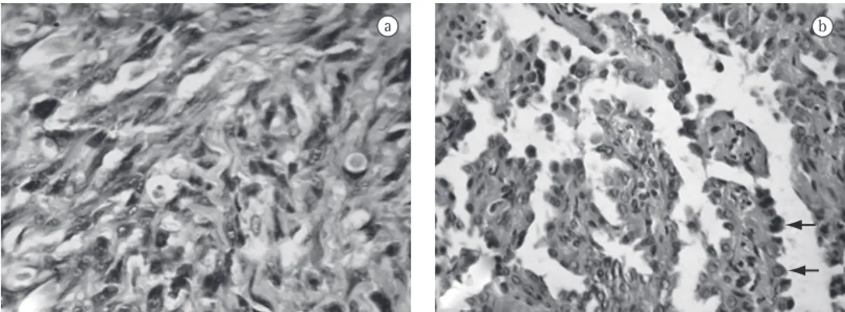
Histopathology showed spindle cell neoplasia, with extensive necrosis, moderate mitotic index and severe atypia, consistent with high-grade sarcoma, probably a malignant peripheral nerve sheath tumor (Figure 2a). The immu-nohistochemical assay was positive for S-100 protein, confirming the diagnosis of malignant schwannoma. We were surprised to find a bron-chioloalveolar carcinoma was in the parenchyma Of the resected lung (Figure 2b). The lesion was 2.5 cm along its longest axis and affected the resection margins. The two neoplasms were separated by healthy lung tissue, confirming the distinct origin of the lesions.
The patient received adjuvant radiothe-rapy. However, over the following months, he developed medullary compression and flaccid paralysis, dying four months after the surgery.
Discussion
A rare case of synchronous malignant thoracic tumors
J Bras Pneumol. 2009;35(2):186-189
189
References
1. Teixeira JP, Bibas RA. Surgical treatment of tumors of the mediastinum: the Brazilian experience. In: Martini N, Vogt-Moykopf I, editors. Thoracic surgery: Frontiers and uncommon neoplasms. International trends in general thoracic surgery. St. Louis: Mosby; 1989. p.211-225. 2. Wain JC. Neurogenic tumors of the mediastinum. Chest
Surg Clin North Am. 1992;2:121-36.
3. Macchiarini P, Ostertag H. Uncommon primary mediastinal tumours. Lancet Oncol. 2004;5(2):107-18. 4. Moon WK, Im JG, Han MC. Malignant schwannomas
of the thorax: CT findings. J Comput Assist Tomogr. 1993;17(2):274-6.
5. Shields TW. Benign and malignant neurogenic tumors of the mediastinum in adults. In: Shields TW, LoCicero III J, Ponn RB, editors. General Thoracic Surgery. Philadelphia: Lippincott Williams &Wilkins; 2000. p. 2313-2327.
6. Salama I, Malone PS, Mihaimeed F, Jones JL. A review of the S100 proteins in cancer. Eur J Surg Oncol. 2008;34(4):357-64.
7. Rostad H, Strand TE, Naalsund A, Norstein J. Resected synchronous primary malignant lung tumors: a population-based study. Ann Thorac Surg. 2008;85(1):204-9.
8. Martini N, Melamed MR. Multiple primary lung cancers. J Thorac Cardiovasc Surg. 1975;70(4):606-12. 9. Chang YL, Wu CT, Lee YC. Surgical treatment
of synchronous multiple primary lung cancers: experience of 92 patients. J Thorac Cardiovasc Surg. 2007;134(3):630-7. Erratum in: J Thorac Cardiovasc Surg. 2008;136(2):542.
10. Trousse D, Barlesi F, Loundou A, Tasei AM, Doddoli C, Giudicelli R, et al. Synchronous multiple primary lung cancer: an increasing clinical occurrence requiring multidisciplinary management. J Thorac Cardiovasc Surg. 2007;133(5):1193-200.
11. Nakajima R, Yokose T, Kakinuma R, Nagai K, Nishiwaki Y, Ochiai A. Localized pure ground-glass opacity on high-resolution CT: histologic characteristics. J Comput Assist Tomogr. 2002;26(3):323-9.
of authors(11) evaluated 20 cases of patients with ground-glass infiltrates submitted to surgical resection. Of those, 50% were diagnosed with bronchioloalveolar carcinoma and 10% were diagnosed with adenocarcinomas. In addition, 25% were diagnosed with atypical adenomatous hyperplasia, which is considered the precursor lesion of bronchioloalveolar carcinoma.
Although it is the most common malignant neoplasia of the peripheral nervous system, malignant schwannoma is still one of the least studied sarcomas. The five-year survival rate is low and is negatively affected by the size of the lesion, incomplete resection and concomitance with Von Recklinghausen disease.(3) Complete resection is usually impossible. Adjuvant radio-therapy and chemoradio-therapy can be useful in the treatment of the metastatic disease. The lesion is rare as are its symptoms. Therefore, a high level of clinical suspicion is recommended in order to correctly diagnose this rare neoplasia. It is likely that the combination of malignant schwannoma and bronchioloalveolar carci-noma was a random phenomenon and did not influence the outcome of the case, which was a consequence of the aggressive nature of the mediastinal lesion. However, the presence of neoplasia should always be suspected in patients presenting pulmonary infiltrates and localized ground-glass opacities that do not disappear or grow. In such cases, an aggressive approach should be adopted, with early biopsy and histo-pathological analysis.
About the authors
Benoit Jacques Bibas
Resident in General Surgery. Hospital Central da Polícia Militar do Rio de Janeiro – HCPM, Central Hospital of the Rio de Janeiro Military Police – Rio de Janeiro, Brazil.
Marcos Madeira
Captain Physician in the Thoracic Surgery Department. Hospital Central da Polícia Militar do Rio de Janeiro – HCPM, Central Hospital of the Rio de Janeiro Military Police – Rio de Janeiro, Brazil.
Rodrigo Gavina
Captain Physician in the Thoracic Surgery Department. Hospital Central da Polícia Militar do Rio de Janeiro – HCPM, Central Hospital of the Rio de Janeiro Military Police – Rio de Janeiro, Brazil.
Leonardo Hoehl-Carneiro
Lieutenant Physician in the Pathological Anatomy Department. Hospital Central da Polícia Militar do Rio de Janeiro – HCPM, Central Hospital of the Rio de Janeiro Military Police – Rio de Janeiro, Brazil.
Sergio Sardinha