ORIGINAL
RES
EAR
CH
Correspondence to: Roberta de Oliveira Cacho – Faculdade de Ciências da Saúde do Trairi, Rua Vila Trairi, s/n – Centro – CEP 59200-000 – Santa Cruz (RN), Brazil – E-mail: [email protected]
Presentation: Apr. 2014 – Accepted for publication: July 2014 – Financing source: Reuni (scientific initiation scholarship) – Conflict of interests: nothing to declare.
ABSTRACT | The study aimed to evaluate the effects of mir-ror therapy through functional activities and motor standards in upper limb function of chronic stroke subjects. Six patients with paresis of the arm within at least six months after stroke were randomly to a group of functional activities (GAF – n=3) and group of motor standards (GPM – n=3). Both groups per-formed 15 sessions of mirror therapy for 30 minutes, but the first one (GAF) were instructed to do the bilateral and sym-metrical movements based on functional activities (i.e. games fitting) and the second one (GAP) made movements based on normal motor patterns (i.e. wrist flexion-extension). There was no statistical significance between pre- and post-treat-ment for both groups independently. However, analyzing the groups together (n=6), it was observed significance values in the cognitive and total MIF (p=0.002) pre- and post-mirror therapy. This study showed improvement in the functional impairment whatever the type of movement made during the mirror therapy.
Keywords | Stroke; Upper Extremity; Mirror Neurons.
RESUMO | O objetivo do estudo foi avaliar os efeitos da apli-cação da terapia de espelho por meio de atividades funcio-nais e padrões motores do movimento na função motora do
Effects of mirror therapy through functional
activites and motor standards in motor
function of the upper limb after stroke
Efeito da terapia de espelho por meio de atividades funcionais e padrões motores na
função do membro superior pós-acidente vascular encefálico
Efecto de la terapia del espejo por mediante actividades funcionales y patrones motores
em la función del miembro superior después de um accidente cerebrovascular
Candice Simões Pimenta de Medeiros1, Sabrina Gabrielle Gomes Fernandes2, Johnnatas Mikael Lopes3,
Enio Walker Azevedo Cacho3, Roberta de Oliveira Cacho3
Study conducted at the Health Sciences School of Trairi, Universidade Fedderal do Rio Grande do Norte (UFRN) – Santa Cruz (RN), Brazil.
1Health Sciences School of Ceará – Fortaleza (CE), Brazil.
2Faculdade de Ciências da Saúde do Trairi (Facisa/UFRN) – Santa Cruz (RN), Brazil. 3UFRN – Natal (RN), Brazil.
membro superior de hemiparéticos crônicos pós-Acidente Vascular Encefálico (AVE). Seis pacientes com hemiparesia do braço com pelo menos seis meses pós-AVE foram rando-mizados para um grupo de atividades funcionais (GAF – n=3) e um grupo de padrões motores (GPM – n=3). Ambos os grupos realizaram 15 sessões de terapia de espelho por 30 minutos, mas o primeiro (GAF) foi instruído a fazer movi-mentos bilaterais e simétricos baseados em atividades fun-cionais (isto é, jogos de encaixe) e o segundo (GPM), a fazer movimentos baseados em padrões motores normais (isto é, flexão-extensão de punho). Não houve significância estatís-tica entre o pré e o pós-tratamento para ambos os grupos de modo independente. No entanto, analisando os grupos em conjunto (n=6), foram observados valores significativos na medida de independência funcional (MIF) cognitiva e total (p=0,002) pré e pós-terapia de espelho. Este estudo mostrou melhora no comprometimento funcional seja qual for o tipo de movimento feito durante a terapia de espelho.
Descritores | Acidente Vascular Cerebral; Extremidade Superior; Neurônios-Espelho.
RESUMEN | El objetivo del estudio fue evaluar los
INTRODUCTION
Cerebral vascular accident (CVA), or stroke, is a vascular acute neurological dysfunction caused by the
interrup-tion of blood low to focal areas of the brain1,2. Sequelae
are often disabilities, and global involvement interferes
signiicantly with Activities of Daily Living (ADLs)2.
he upper limbs (UL) are very important to motor functionality and the efective handling, gripping and reaching capability required in most ADLs. Arm functions are impaired in 73-88% of CVA survivors, and 55-75% of them present hemiplegia, resulting in disabilities and restrictions to function3-5.
Some studies point that sensorial-motor skill training and motor learning training with repetitive movements by the patient, with introduction of new movements in oriented environments are essential to reduce motor
impaiment6. Among therapies available for the upper
limbs, electrical stimulation therapy, electromyographic (EMG) biofeedback, mirror therapy, constraint-induced movement therapy (CIMT), Sensory-Motor Imagery Training, and robotic-assisted rehabilitation take pride of place in the recovery of these subjects3-5,7-9. Apart from the improvements in arm functions, studies do not
bring data related to ADLs and quality of life8. hese
therapies often require expensive machinery that is also diicult to handle, which limits their large-scale use in clinical practice3,5.
Mirror therapy (MT) is a low-cost and easy inter-vention developed to treat phantom limb pain that is
currently used in post-stroke rehabilitation5,10-12. MT
is applied with a mirror positioned in the sagittal plane between the upper limbs. In order to trick the brain, by promoting a visual and kinesthetic illusion, the sub-ject performs movements with the normal limb that are
relected to the mirror and interpreted by the brain as
performed by the afected limb11,12. By activating the
mirror-neuron system (MNS) and the corticospinal tract, MT accelerates recovery of hemiparesis and pro-motes cortical reorganization, resulting in functional and
motor improvements3,12-13.
hus, MT is a strategy proven feasible and efective for motor recovery3,4,8,14-16. he efects of this therapy are beneicial for movement execution and control, but do
not relect in CVA patients’ daily activities5.
he literature still lacks studies about mirror therapy approaches, aimed at movements related to functional activities or to the motor patterns of hemiplegic arm. herefore, this paper aimed to assess the efects of mir-ror therapy using functional activities and motor patterns of chronic hemiplegic upper limbs resulting from VCA.
METHODOLOGY
his is a quasi-experimental, randomized, blinded trial conducted with patients who had a stroke, living in Santa Cruz (RN), recruited by means of a study on the preva-lence and risk factors of Cerebral Vascular Accident in Santa Cruz, approved by the Research Ethics Committee of Faculdade de Ciências da Saúde do Trairi (FACISA/ UFRN). All subjects were enrolled in the study by ran-dom draw.
Individuals aging more than 18 years, diagnosed with only, unilateral VCA in chronic phase (over six months after ictus) with hemiparesis of the upper limb as sequelae, scoring more than 20 on Fugl-Meyer Assessment of Motor Recovery (FM), absence of cognitive disorders scoring
≥24 (educated subjects) and >14 (illiterate subjects) on
actividades funcionales y patrones motores del movimiento en la función motora del miembro superior de hemiparéticos cró-nicos pos-Accidente Vascular Encefálico (AVE). Seis pacientes con hemiparesía del brazo con al menos seis meses pos-AVE fueron asignados de modo aleatorio a un grupo de activida-des funcionales (GAF – n=3) y un grupo de patrones motores (GPM – n=3). Ambos grupos realizaron 15 sesiones de terapia del espejo durante 30 minutos, pero el primero (GAF) fue orien-tado a hacer movimientos bilaterales y simétricos en base a las actividades funcionales (es decir, juegos de encaje) y el segundo (GPM), a hacer movimientos basados en patrones
motores normales (es decir, flexión-extensión de la muñeca). No hubo diferencias estadísticamente significativas entre pre y post-tratamiento para ambos grupos de forma indepen-diente. Sin embargo, el análisis de los grupos en conjunto (n=6) demostró valores post-terapia significativos en la Medida de Independencia Funcional (MIF) cognitiva y total (p=0,002) pre y post-terapia de espejo. Este estudio mostró una mejoría en el deterioro funcional en cualquier tipo de movimiento reali-zado durante la terapia del espejo.
Mini-Mental State Examination (MMSE), walking with good stability upon seating, scoring more than 40 on Berg Balance Scale (BBS). Patients presenting other neurological disorders with associated physical or men-tal disabilities, traumatic VCA, joint pain and straining in upper limbs that could prevent the performance of
movements were excluded20-22.
Patients were assessed before and after treatment by one examiner. Pre-treatment assessment was com-posed of sociodemographic analysis and application of MMSE, BBS, FM, Modiied Ashworth Scale (MAS) and Functional Independence Measure (FIM). Post-treatment assessment was made by FM, FIM and MAS. Sociodemographic evaluation form was pre-structures and addressed personal data (name, gender, age, marital status), clinical data (diagnosis, type and time of dam-age, hemiplegic side), and table for monitoring initial and inal blood pressures during appointments.
We used: the dimension of the upper limb of FM Assessment of Motor Recovery, which assesses
sensory-motor impairment of the hemiplegic arm17; MAS, by
application of muscle resistance to passive strain of the
upper limb23; FIM, to assess subjects’ performance in 18
tasks, comprehending motor (mFIM), cognitive (cFIM),
and total (tFIM) domains21.
Each patient was assessed and treated individually in their home environment with 15 mirror therapy sessions of 50 minutes, 3 times a week. Patients were divided into two groups by random draw: functional activities group (FAG) and motor pattern group (MPG).
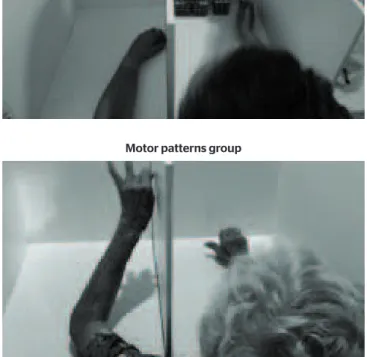
Before mirror therapy, Kinesiotherapy was applied in the irst 10 minutes of the session, with passive muscle stretching and joint mobilizations in upper limbs. Upon mirror therapy, a rectangular platform measuring 40x70 cm was used, where a mirror was put in the sagittal plane and could be removed according to the side of hemiparesis of each patient (Figure 1). he platform extension and sides were closed to avoid patients to be drawn atten-tion by the environment. he platform with the mirror was secured on a table where the patient would be sit-ting by, on a comfortable chair with backrest, with legs leaned on the ground. Patients were oriented to watch the relection of their normal hand on the mirror as it was the afected one, and to perform activities bilaterally. he sessions lasted 30 minutes and the focus of interven-tion was task repetiinterven-tion. To avoid muscle fatigue, patients could rest in an interval of 1-2 minutes between tasks.
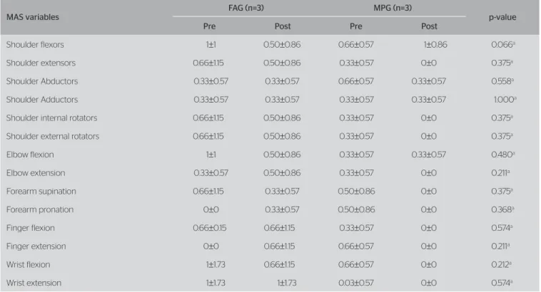
FAG subjects performed activities in the mirror with recreational objects (cups, cubes, balls, toys, bottles) in
varied colors, sizes and shapes. Activities were related to functional range, itting, transferring and stacking objects. MPG subjects performed movements of inger lexion and extension, inger adduction/abduction, fore-arm pronation and supination, elbow extension, without relating them to functional activities (Figure 2). During therapy, subjects were verbally commanded by examin-ers so they were motivated and corrected whenever the activities were performed incorrectly.
he study was approved by the Ethics committee of UFRN (CAAE: 11732712.8.0000.5537), and sub-jects who agreed to participate signed the informed consent form.
Data were descriptively analyzed using the software SPSS 20.0®. Absolute frequency (n) and percentage (%) of categorical variables were calculated, as well as mean and standard deviation of continuous variables. Mann-Whitney, Fisher’s Exact test, chi-square tests were used, and also generalized estimating equation (GEE). Signiicance level was set at 5% in order to minimize type error.
RESULTS
Initially, 20 patients with VCA sequelae were assessed, among which only 6 met all inclusion criteria. hese six patients were randomly and equally divided into two groups: functional activities (FAG) and motor pattern
Figure 1. Dimensions of the mirror used in mirror therapy
40 cm
(MPG). Table 1 shows demographic data of subjects and the characteristics of their lesions in each group.
here was a homogeneous relation between the vari-ables gender, age, side and time of lesion, which means no signiicant diferences between groups.
Results before and after intervention related to the variables of Fugl-Meyer and FIM scales for FAG and MPG, compared to groups of treatment (analysis between groups) and to the junction of both groups are described in Table 2. No signiicant diferences were found before and after MT when GAF and MPG were compared or assessed on a stand-alone basis. However, when the groups of treatment were joined, a signiicance was found in FIM, FIMc (p=0.002) and FIMt (p=0.002), respectively.
Resistance to passive movements of the upper limbs through MAS did not vary signiicantly between groups throughout treatment, as shown in Table 3.
DISCUSSION
MT is beneicial for patients who had stroke, as reported
in many studies3,5,22. he illusion induced by the mirror
improves the training environment, increases somato-sensory information, induces task repetition and boosts
cortical function3. However, the approach of therapy
does not inluence functional and motor gains of the patients in this study.
herapies involving the performance of tasks on the mirror, aimed at functional activities, are more efec-tive when it comes to motor improvements, for they
apply and reinforce the concepts of motor learning23,24.
Functional activities are associated with better motor learning, once the tasks are usually more dynamic, with variations and training aimed at speciic activities, mak-ing assimilation easier. When subjects are trained with simple motor patterns, they can have a good perfor-mance, but also more diiculty to associate them to
ADLs25. Although studies point it out, no diferences
were found between the therapy groups.
There were no significant variations between ther-apy groups in pre and post-treatment assessments by FIM and FM. However, when groups were put together, there was an improvement in cFIM (p=0.002) and tFIM (p=0.002). Performing recreational tasks aimed at functional activities is believed to require more of the cognitive function along with sensory-motor areas for task execution compared to tasks of
Table 1. Sample characteristics
Variables FAG (n=3) Mean
MPG (n=3)
Mean p-value
Gender (n)
Male 0 1
Female 3 2 0.500a
Age (years) 63±9.53 66.66±14.22 0.386b
Marital status (n)
Married 1 2
Widow/er 1 1 –
Single 1 0
Type of lesion (n)
Ischemic 100 100 –
Hemorrhagic 0 0
Side afected (n)
Left 2 2 0.800a
Right 1 1
Time of lesion (years) 4.66±1.52 5.33±1.52 0.386b
FAG: functional activities group; MPG: motor patterns group; a: Fisher’s exact test; b: Mann-Whitney test
Figure 2. Mirror therapy with functional activities and motor patterns Functional activities group
motor patterns, which do not require attention, cog-nition, and activation of brain areas involved. Some studies reported a better response by the mirror-neu-ron system when the movements performed on the mirror are related to specific tasks in comparison to
tasks without definite aims13,23,24.
The most complex movement sequences require more attention and cognition in order to be performed. Therefore, our findings do not support data reported in literature. In a systematic review, the authors reported that MT is beneficial for ADLs and their effects on
motor function are associated with the MT approach10.
Table 3. Comparison of Modified Ashworth Scale between Functional Activities Group and Motor Patterns Group
MAS variables
FAG (n=3) MPG (n=3)
p-value
Pre Post Pre Post
Shoulder flexors 1±1 0.50±0.86 0.66±0.57 1±0.86 0.066a
Shoulder extensors 0.66±1.15 0.50±0.86 0.33±0.57 0±0 0.375a
Shoulder Abductors 0.33±0.57 0.33±0.57 0.66±0.57 0.33±0.57 0.558a
Shoulder Adductors 0.33±0.57 0.33±0.57 0.33±0.57 0.33±0.57 1.000a
Shoulder internal rotators 0.66±1.15 0.50±0.86 0.33±0.57 0±0 0.375a
Shoulder external rotators 0.66±1.15 0.50±0.86 0.33±0.57 0±0 0.375a
Elbow flexion 1±1 0.50±0.86 0.33±0.57 0.33±0.57 0.480a
Elbow extension 0.33±0.57 0.50±0.86 0.33±0.57 0±0 0.211a
Forearm supination 0.66±1.15 0.33±0.57 0.50±0.86 0±0 0.375a
Forearm pronation 0±0 0.33±0.57 0.50±0.86 0±0 0.368a
Finger flexion 0.66±0.15 0.66±1.15 0.33±0.57 0±0 0.574a
Finger extension 0±0 0.66±1.15 0.66±0.57 0±0 0.211a
Wrist flexion 1±1.73 0.66±1.15 0.66±0.57 0±0 0.212a
Wrist extension 1±1.73 1±1.73 0.03±0.57 0±0 0.574a
FAG: functional activities group; MPG: motor patterns group; MAS: Modified Ashworth Scale; Pre: pre-treatment assessment; Post: post-treatment assessment Table 2. Statistical description of clinical and functional measures in chronic hemiparesis patients
Variables mFIM cFIM tFIM tFM
FAG (n=3)
Mean±SD Pre 86.33±3.21 33.33±1.52 119.66±2.51 49.66±19.73
Post 86.33±1.52 34±1 120.33±1.52 51±19.05
χ2 0.66 3.87 3.66 0.001
p-value 0.72a 0.14a 0.99a 0.99a
MPG (n=3)
Mean±SD Pre 84±37 29.33±6.35 113.33±11.01 53.33±1.54
Post 84±6.08 30.33±6.35 114.33±11.01 54±3
χ2 0.57 2.58 3.66 0.001
p-value 0.44a 0.74a 0.99a 0.99a
Analysis between groups
χ2 0.001 1.58 1.36 0.13
p-value 0.99a 0.20a 0.24a 0.13a
Both groups (n=6)
χ2 0.001 9.37 9.37 2.07
p-value 0.99a 0.002a 0.002a 0.15a
Responses of mirror visualization and task perfor-mances aimed at a certain purpose were compared, which promoted a significant activation of the bilat-eral sensorial-motor cortex, including the primary motor, pre-motor, and primary sensorial-motor cortex areas, compared to the group performing tasks without
established aims24. MT and conventional treatment
are more beneficial to the upper limbs motor
func-tion when associate with specific tasks after VCA23.
Many activation ADN adaptation mechanisms take place after VCA, including increased use of the healthy side of the brain, boosted by the corticospinal tract
acti-vation and the mirror-neuron system after MT tasks26.
he cortical reorganization was assessed by magnetic resonance imaging after MT in subjects who had had stroke, and some changes in pattern of primary motor cortex activation were found on the side afected, but without correlation to functional improvements and
balance of activation between brain hemispheres3. he
corticospinal tract is activated by the visualization of movements performed with the normal limb on the
mirror16 and boosted when the mirror is associated
with the virtual environment27.
Motor function of the upper limbs is an impor-tant prognostic factor for functional recovery after VCA. However, we found no signiicant variations in the muscle groups assessed in MAS or in FM values. After six weeks of MT, we did not observed any efects on sensory-motor function assessed by FM in ADLs, but there was a signiicant change in resistance to
passive movements in inger lexor muscles4. Other
studies have reported improvement in motor function after MT using FM and have associated this outcome to the appropriate visual input replacing the reduced
proprioceptive input on the afected limb3,5.
he literature is not consensual as to the minimum time of therapy session and the durations of MT efects. Some authors have applied MT for 30 minutes in lower
limbs after VCA, and showed this time is insuicient28.
Other authors also used 30-minute sessions for 4 weeks and reported motor improvement of wrist and hand, with tasks related to speciic activities with objects23. he small size of our sample, as well as the short period of ther-apy, may have inluenced our indings. hese results can only be applied to subjects classiied as mild to moderate phase of chronic VCA. Assessment scales were used in our study, but more accurate analyses of the movements performed by patients through kinematics were not made.
CONCLUSION
In general terms, functional improvement is achieved by mirror therapy, regardless of the use of functional activi-ties or movement patterns. he literature addressing mir-ror therapy modes of execution is very scarce, so there is a need to perform further studies with larger samples in order to truly assess the eicacy of this therapy.
REFERENCES
1. Costa FA, Silva DLA, Rocha VM. Estado neurológico e cognição de pacientes pós-acidente vascular cerebral. Rev Esc de Enferm USP. 2011;45(5):1083-8.
2. Almeida, SEM. Análise epidemiológica do acidente vascular cerebral no Brasil. Rev de Neurocienc. 2012;20(4):481-2.
3. Michielsen ME, Selles RW, Geest JNV, Eckhardt M, Yavuzer G, Stam HJ,
et al. Motor recovery and cortical reorganization after mirror therapy in chronic stroke patients: A phase II randomized controlled trial. Neurorehabilitation Neural Repair. 2011;24(3):223-33.
4. Thieme H, Bayn M, Wurg M, Zange C, Pohl M, Behrens J. Mirror therapy for patients with severe arm paresis after stroke – A randomized controlled trial. Clin Rehabil. 2012;27(4):314-24.
5. Wu CY, Huang PC, Chen YT, Lin KC, Yang HW. Efects of mirror therapy on motor and sensory recovery in chronic stroke: A randomized controlled trial. Arch Phys Med Rehabil. 2013;94:1023-30.
6. Trevissan CM, Trintinaglia V. Efeito das terapias de imagem motora e de movimento induzido por restrição na hemiparesia crônica: Estudo de caso. Fisioter Pesqui. 2010;17(3):264-9.
7. Teixeira INDO. O envelhecimento cortical e a reorganização neural após o acidente vascular encefálico (AVE): Implicações para reabilitação. Ciênc Saúde Coletiva. 2008;13(2):2171-8.
8. Yavuzer G, Selles R, Sezer N, Sutbeyaz S, Bussmann JB, Koseoglu F,
et al. Mirror therapy improves hand functions in subacute stroke: A
randomized controlled trial. Arch Phys Med Rehabil. 2009;89:393-8.
9. Dogan-Aslan M, Nakipoglu-Yuzer GF, Dogan A, Karaby I, Ozgirgin N. The efect of electromyographic biofeedback treatment in improving upper extremity functioning of patients with hemiplegic stroke. J Stroke Cerebrovasc Dis. 2012;21(3):187-92.
10. Thieme H, Mehrholz J, Pohl M, Behrens J, Dohle C. Mirror therapy for improving motor function after stroke. Stroke. 2013;44:1-2.
11. Ramachandran VS, Rogers-Ramachandran D, Stewart M. Perceptual correlates of massive cortical reorganization. Science. 1992;258:1159-60.
12. Machado S, Velasques B, PaesF, Cunha M, Basile LF, Budde H, et al.
Terapia-espelho aplicada à recuperação funcional de paciente pós-acidente vascular cerebral. Rev Neurocienc. 2011;19(1):171-5.
13. Small SL, Buccino G, Solodkin AS. The mirror neuron system and treatment of stroke. Dev Psychobiol. 2012;54(3):293-310.
15. Yun GJ, Chun MH, Park JY, Kim BR. The synergic efects of mirror therapy and neuromuscular electrical stimulation for hand function in stroke patients. Ann Rehabil Med. 2011;53:316-21.
16. Kang YJ, Ku J, Kim HJ, Park HK. Facilitation of corticospinal excitability according to motor imagery and mirror therapy in healthy subjects and stroke patients. Ann Rebabil Med. 2011;35:747-58.
17. Fugl-Meyer AR, Jaasko L, Olsson S, Steglind S. The post-stroke hemiparetic patients: A method for evaluation of physical performance. Scand J Rehabil Med. 1975;7:13-31.
18. Berg KO, Maki BE, Williams JI, Wood-Dauphinee SL. Clinical and laboratory measures of postural balance in an elderly population. Arch Phys Med Rehabil. 1992;73:1073-80.
19. Almeida OP. Mini Exame do Estado Mental e o diagnóstico de demência no Brasil. Arq Neuropsiquiatr. 1998;56:605-12.
20. Ashworth B. Preliminary trial of carisoprodol in multiple sclerosis. Practitioner. 1964;192:540-42.
21. Riberto M, Miyazaki MH, Juca SSH, Sakamoto H, Pinto PPN, Battistella LR. Validação da versão brasileira da Medida de Independência Funcional. Acta Fisiatr. 2004:11(2):72-6.
22. Lappchen CH, Ringer T, Blessin J, SeidelG, Grieshammer S, Lange R,
et al. Optical illusion alters M1 excitability after mirror therapy: A TMS study. J Neurophysiol. 2012;108:2857-61.
23. Arya KN, Pandian S. Efect of task-based mirror therapy on motor recovery of the upper extremity in chronic stroke patients: A pilot study. Top Stroke Rehabil. 2013;20(3):210-7.
24. Agnew ZK, Wise RJS, Leech R. Dissociating object directed and non-object directed action in the human mirror system; Implications for theories of motor stimulation. Plos One. 2012;7(4):1-9.
25. Kwakkel G. Impact of intensity of practice after stroke: issues for consideration. Disabil Rehabil. 2006;28(13-14):823-30.
26. Byblow WD, Stinear CM, Smith MC, Bjerre L, Flaskager BK, Mccambridge AB. Mirror symmetric bimanual movement priming can increase cortiomotor excitability and enhance motor learning. Plos One. 2012;7(3):1-14.
27. Kang YJ, Park HK, Kim HJ, Lim T, Ku J, Cho S, et al. Upper extremity rehabilitation of stroke: Facilitation of corticospinal excitability using virtual mirror paradigm. J Neuroeng Rehabil. 2012;71(9):2-8.