Correspondence: Dr. Christian R. Gernhardt, Martin-Luther-University Halle-Wittenberg, University School of Dental Medicine Depart-ment of Operative Dentistry and Periodontology, Grosse Steinstrasse, 19 D- 06108 Halle/Saale, Germany. Tel: +49-345-557-3762. Fax: +49-345-557-3773. e-mail: [email protected]
Influence of Pressure Application Before Light-Curing
on the Bond Strength of Adhesive Systems to Dentin
Christian Ralf GERNHARDT1 Katrin BEKES1
Petra HAHN2 Hans-Günter SCHALLER1
1Department of Operative Dentistry and Periodontology, University School of Dental Medicine,
Martin-Luther-University Halle-Wittenberg, Halle, Germany
2Department of Operative Dentistry and Periodontology, University School of Dental Medicine,
Albert-Ludwigs-University, Freiburg, Germany
This study evaluated the influence of defined pressure application before light-curing on the tensile bond strength (TBS) of two adhesive systems to wet and perfused dentin. Bonding sites were analyzed by scanning electron microscopy (SEM) to assess morphological changes. Dentin discs from 120 human third molars were prepared in such a way to allow simulation of intrapulpal pressure. The specimens were randomly assigned to 6 groups (n=20) according to different experimental conditions. The adhesive systems (ScotchbondTM Multi-Purpose and Syntac®) were applied either to wet or perfused dentin. In the latter, compression was
applied before adhesive light-curing . TBS was recorded using an universal testing machine. Qualitative examination of the bonding sites was performed by SEM after dissolving the dentin with 50% nitric acid. Pressure application prior to light-curing resulted in a remarkable decrease of TBS for Syntac®, while no significant change was observed for ScotchbondTM MP. SEM analysis showed no
considerable lengthening of the resin tags after pressure application. These findings suggest that application of adhesive systems to dentin under pressure before light-curing had no positive effect on TBS.
Key Words: bond strength, adhesive systems, dentin, dentin permeability, pressure.
INTRODUCTION
Since the introduction of dentin bonding systems to modern dentistry over 40 years ago, extensive re-search has been undertaken to improve bond strength and clinical properties, and several bonding mecha-nisms have been discussed.
According to proposed concepts, bonding of adhesive systems to human dentin would rely on chemi-cal approach (1) or a micromechanichemi-cal retention to pretreated dentin (1). For this purpose, pretreatment of the smear layer and dentin by using acids, primers or conditioners is recommended. Smear layer can be removed by the total acid-etching technique, which is known to have a positive effect on bond strength of adhesive systems (2) or the smear-covered dentin
surface can be treated with acidic primers or condition-ers. Recently introduced self-etching adhesive systems do not require the phosphoric acid etching step (3).
MATERIAL AND METHODS
One hundred and twenty caries-free freshly extracted third molars were selected and stored in saline (Merck, Darmstadt, Germany) at room temperature for a maximum of 24 h. Ninety teeth were used for TBS evaluation and 30 teeth were used for SEM analysis.
For simulation of intrapulpal pressure and dentin perfusion, the specimens were prepared according to a special procedure previously described (4,5). The teeth were decoronated with a water-cooled diamond-coated band saw (Exakt Trennschleifsystem; PSI Grünwald, Laudenbach, Germany). Pulp was removed and oc-clusal reduction was performed using cylindrical dia-mond burs (Brasseler, Lemgo, Germany) under con-stant water cooling. The distance between the pulp chamber and the occlusal plateau was adjusted to 1.3 mm (± 0.1 mm) and the specimens were apically reduced (parallel to the established flat plane) until specimen thickness reached 3.5 mm (± 0.2 mm). After preparation, the specimens were mounted on an experi-mental apparatus (Fig. 1) and simulation of intrapulpal pressure and physiological dentin perfusion using saline was enabled. The pressure was adjusted to 30 cm H2O
and maintained over throughout the experiment (Fig. 1). A metal ring was then positioned on the occlusal plane and application of the adhesive systems and composite resin into the ring provided a standardized bonding area. Two adhesive systems (Syntac®; Vivadent, Schaan, Liechtenstein and ScotchbondTM Multi-Pur-pose; 3M Dental Products, Loughborough, Great Brit-ain) were used. The materials’ principal ingredients are:
Syntac® - Primer: Tetraethylenglycolmethacrylate, maleic, acid, dimethylketone,water; Adhesive: polyethyleneglycoldimethacrylate, maleic acid, glutaral-dehyde and water; Heliobond: Bis-GMA, dimethacrylate, initiators and stabilizers; ScotchbondTM MP - Etchant: 36% H3PO4; Primer: Polyalkenoic acid, HEMA, water; Adhesive: BisGMA, HEMA.
The specimens were allocated to 6 groups (n=20), according to 3 experimental conditions: SW0 (Syntac®) and SCW0 (ScotchbondTM MP): adhesive system application to wet dentin + light-curing + composite resin application without pressure; SP0 (Syntac®) and SCP0 (ScotchbondTM MP) adhesive system application to perfused dentin + light-curing + composite resin application without pressure; SP1(Syntac®) and SCP1 (ScotchbondTM MP): adhesive system application to perfused dentin + no light-curing + composite resin application under pressure (7 N) + light-curing.
The applied pressure of 7 N was determined in two series of preliminary tests involving 5 dentists and 5 dental students. The pressure applied by the volun-teers while placing composite material in a standardized cavity was measured using a universal testing machine (Zwick, Ulm, Germany). The mean pressure was 7 N. The same light-curing unit (Elipar Visio; Espe, Seefeld, Germany; 400 mW/cm²) was used for adhesive and composite polymerization. The adhesive systems were applied as per manufacturers’ instructions. Syntac®: Syntac® Primer was applied and air-thinned after 15 s,
Syntac® Adhesive was applied and dried after 10 s, and,
finally, Heliobond® (Vivadent, Schaan, Liechtenstein)
was applied and blown to a thin layer. ScotchbondTM
MP: 36% phosphoric acid gel was applied to dentin, rinsed after 15 s, ScotchbondTM MP Primer was applied
for 5 s and blown to a thin layer, and, finally, ScotchbondTM MP Adhesive was applied. Excess was
removed with a mild compressed air stream. Tetric®
hybrid composite resin (Vivadent, Schaan, Liechtenstein; shade A3) was used in all groups. The metal ring of the test apparatus was filled with the composite in 2-mm-thick increments that were light-cured for 60 s.
Fifteen minutes after polymerization, TBS was tested in 15 specimens of each group using a universal testing machine (Zwick, Ulm, Germany) at a crosshead speed of 1 mm/min. Maximum force until fracture was recorded from a personal computer and presented graphically. TBS was expressed as MPa. TBS means and standard deviations were calculated. Repeated
sures ANOVA were used to evaluate the results (SAS Release 6.12). Differences among groups were calcu-lated by Tukey’s Studentized Range test and by the more sensitively closed test procedure (based on Kruskal Wallis test). Significance level was set at 5%.
Twenty in after light-curing, the 5 specimens of each group were prepared for qualitative analysis. They were immersed in 50% nitric acid for a minimum of 48 h to dissolve dentin hard tissue, then were carefully diluted in distilled water, dried, mounted on aluminium stubs using a conductive silver agent (Silver Print; GC, Tokyo, Japan), sputter-coated with gold in a 15-20 nm layer (SCD 040; Balzers, Wiesbaden, Germany) and analyzed with a scanning electron microscope (DSM 950; Zeiss, Oberkochen, Germany).
RESULTS
The influence of the adhesive systems and application conditions on TBS were statistically significant (p≤0.001; ANOVA). TSB means were significantly higher in the groups with wet dentin specimens com-pared to ones with perfused dentin specimens (p<0.05, Tukey’s Studentized Range test).
The mean TBS of SW0 group (9.03 MPa ± 1.94 MPa) was the highest among all groups with statistically significant difference (p<0.05, closed test procedure). The mean TBS for the corresponding group on perfused
dentin (SP0 group) was 3.53 MPa (± 1.07 MPa). When pressure was applied before light-curing (SP1 group), the mean TBS dropped to 0.26 MPa (± 0.22 MPa), which was significantly lower than that of all tested groups (p<0.05, closed test procedure).
TBS of SCW0 and SCP0 groups were 6.57 MPa (± 1.79 MPa) and 4.26 MPa (±1.33 MPa), respectively, and significant difference (p<0.05, closed test procedure) was found between these groups. When pressure was applied before light-curing (SCP1 group), a mean TBS of 4.97 MPa (± 1.14 MPa) was recorded. Closed test procedure showed no statistically significant difference (p>0.05) between SCP0 and SCP1.
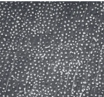
SEM analysis of the specimens treated with Syntac®, showed a homogeneous tag formation on wet
dentin. Tag mean length was approximately 10 µm (Fig. 2). The use of Syntac® on perfused dentin resulted in a
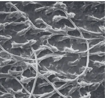
more heterogeneous distribution of tags and in a reduction in length to an average of 5 µm. In SP1 group, the use of compression resulted in nearly no tag formation (Fig. 3). In the groups treated with ScotchbondTM MP, SEM
analysis showed a more extensive and homogeneous tag formation for the wet and perfused dentin specimens (SCW0 and SCP0 groups) compared to the specimens treated with Syntac® (Fig. 4). After pressure application,
(SCP1 group), tag formation did not increase compared to the perfused specimens without pressure. However, occasionally, tags were up to 100 µm in length (Fig. 5).
Figure 3. Nearly no tag formation in a perfused dentin specimen treated with Syntac
after pressure application before light-curing (×500). Figure 2. Characteristic tag formation in a wet dentin specimen
treated with Syntac
DISCUSSION
In the present study, saline was used for storage of the freshly extracted teeth. This is a well-known procedure, as it does not influence the chemical and physical properties of human dentin (6). The prepara-tion of the specimens followed a clinical relevant proce-dure. In contrast to other investigations, preparation was performed using diamond burs under constant water spray, which is known to result in considerably higher bond strength, if compared to other techniques (7-9). It is known that regional differences in dentin anatomy and permeability have a significant influence on dentin bond strength (10). The constant distance between the occlusal plane and the pulp chamber (2.0 mm ± 0.2 mm) resulted in fairly well standardized experimental conditions. To eliminate the influence of light intensity and irradiation time, which are known to affect the bond strength (11), the polymerization time in this study was defined to 60 s using the same light curing unit. In all groups, the same composite material and shade were used in order to avoid composite-related influences on bond strength (12).
Polymerization shrinkage, which occurs during light-curing, has a direct effect on dentinal adhesion and encourages the formation of minifractures. Therefore,
it is known that the adhesion force could be measured immediately after application (12). For this reason, in the present study, TBS was measured 15 min after adhesive application application.
In this study, bond strength means were lower than those reported elsewhere (4). In contrast to other studies, the prepared dentin specimens were located in a defined distance close to the pulp chamber. It is known that the use of dentin specimens located farther from the pulp results in higher bond strength (13). The amount of high mineralized peritubular dentin is greater in deep dentin than in the outer regions. Therefore, demineralization and hybrid layer formation is not as efficient as in coronal dentin (14). Moreover, it should be considered that TBS tests tend to provide lower bond strength if compared to shear bond strength measurements. Here, the different stress distribution is known to be the major factor for increased bond strength values.
In the present investigation, the use of Syntac®
on wet dentin specimens without perfusion (group SW0) produced the highest TBS means of all groups. The mean TBS for Syntac® on perfused dentin (group
SP0: 3.53 MPa) was significantly lower than that recorded on wet dentin (group SW0: 9.03 MPa). These results are comparable to those of previous investigations
Figure 4. Extensive tag formation with the presence of anastomoses in a perfused dentin specimen treated with ScotchbondTM MP (×500).
using the same experimental design (4). Regarding ScotchbondTM MP, a significant decrease of bond
strength was detected between wet (SCW0 group: 6.57 MPa) and perfused (SCP0 group: 4.26 MPa) dentin . In contrast to the present investigation, Swift and Triolo (15) found higher bond strength for wet dentin than for almost dry dentin using ScotchbondTM MP. However,
they did not tested perfused specimens with simulated, intrapulpal pressure.
The application of Syntac® Primer, which contains
maleic acid, and 36% phosphoric acid etching prior to application of ScotchbondTM MP, opens and dilates the
dentinal tubules, thus increasing dentin permeability (1). Therefore, the simulation of pulpal pressure during in vitro tests is described as indispensable in order to achieve the best comparability in relation to in vivo conditions (2).
For this reason, a constant hydrodynamic pres-sure of 30 cm (H2O) was applied in this investigation in
4 groups in order to obtain a perfused tooth substance during the whole experimental period. The simulated intrapulpal pressure (30 cm H2O) used in the present
study is in a physiological range.
In this study, saline was used as perfusion solution. Isotonic solutions have been considered as suitable to produce a perfusion pressure (16). More recent investigations revealed that diluted human serum has a positive effect on tensile strength (5,17). The given reason is the precipitation of serum proteins in the dentinal tubules, which should lead to a reduced liquid outflow. In the above mentioned studies, a significant increase in tensile strength was described when bovine serum was used compared to saline. When saline was used for perfusion, TBS is almost identical to the values determined in the present study (17).
The application of compression before light-curing of the composite resin reduced bond strength of Syntac® to an unexpected low level (0.26 MPa). This
could be due to some kind of dilution of the composite material applied after the use of the dentin bonding agent and, in the case of Syntac®, the additional unfilled
bonding resin Heliobond®. It seems reasonable that
light-curing of the bonding resin will be partially inhibited by the composite material (18). Thus, complete polymerization of the unfilled dentin bonding agent could not necessarily be expected. Another reason for this outcome could be the destruction of the glutaraldehyde fixed collagen mash by compression.
Probably Syntac® can only establish bonding to dentin
if light-cured before application of the composite resin. Other investigators found similar results when comparing bond strength before and after light curing of the dentin adhesive (19).
The SEM analysis could not completely support these findings. The formation of tags decreased in the groups with perfused dentin combined with compression. Syntac® applied to wet dentin showed the
highest bond strength. However, compared to ScotchbondTM MP, tag formation was visibly lower.
It is known that tag formation is only one part of the adhesion mechanism. Obviously, in the case of Syntac®, a greater role must be ascribed to the known
micromechanical retention than to the formation of tags and chemical bond.
In the group treated with ScotchbondTM MP,
compression before polymerization (SCP1 group) had no significant influence on TBS compared to the per-fused group without compression (SCP0 group). Com-pared to wet dentin (SCW0 group), bond strength was lower, but not significantly reduced. A possible explanation might be the absence of an unfilled bonding resin like Heliobond®. Furthermore, hydrophilic monomer
penetration might be enhanced in the case ScotchbondTM
MP (20). Regarding SEM analysis, it was observed an increased occurrence of anastomoses between the dentinal tubules in all groups treated with ScotchbondTM
MP. No significant morphological differences could be observed among the three groups treated with ScotchbondTM MP. This might be due to the improved
bonding properties of this material. Several investiga-tors described a significant role in adhesion to the outflow from lateral tubules and to the formation of resin caps on the tags (20). It seems reasonable that for ScotchbondTM MP, adhesive penetration and tag forma-tion is improved and plays a greater role in the adhesion mechanism compared to Syntac®. Furthermore, the
results of the present investigation fully agree with previous reports, stressing that pre-curing of the adhe-sive system causes a significant increase in the dentin bond strength, if compared to curing together with composite placement (19).
com-pression before light-curing could not be confirmed in the present study.
In the present investigation, the effect of pres-sure application on the adhesive systems during poly-merization seemed to material-dependent. The results showed that compression of composite filling after Syntac® application was deleterious to the adhesive
mechanism and resulted in an unacceptable decrease of TBS. The decrease of bond strength of ScotchbondTM
MP to dentin, though noticeable, was not significant. Keeping this results in mind, it is recommendable to follow the manufacturers’ instructions and pre-cure dentin bonding systems before placement of composite. It remains to be clarified how other systems would react to compression before light-curing.
RESUMO
Este estudo avaliou a influência da aplicação de uma pressão definida antes da polimerização sobre a resistência à tração de dois sistemas adesivos à dentina molhada e perfundida. Os sítios de adesão foram analisados em microscopia eletrônica de varredura (MEV) a fim de determinar a ocorrência de alterações morfológicas. Discos de dentina de 120 terceiros molares humanos foram preparados de tal forma a simular a pressão intrapulpar. Os espécimes foram alocados aleatoriamente em 6 grupos (n=20), de acordo com as diferentes condições experimentais propostas. Os sistemas adesivos (Syntac® e ScotchbondTM Multi-Purpose) foram aplicados sobre a dentina molhada ou perfundida. Nesta última, compressão (7 N) foi aplicada antes da polimerização do sistema adesivo. A resistência à tração foi registrada em máquina universal de ensaios. A análise qualitativa dos sítios de adesão foi realizada em MEV após dissolução da dentina em 50% de ácido nítrico. A aplicação da pressão antes da polimerização do sistema adesivo resultou em diminuição significativa da resistência à tração do Syntac®, ao passo que para o ScotchbondTM MP nenhuma alteração significativa foi observada. A análise em MEV não mostrou aumento considerável do tamanho dos tags de resina após a aplicação da pressão. Esses achados sugerem que a aplicação de sistemas adesivos à dentina sob pressão previamente à polimerização não teve efeito positivo sobre a resistência à tração.
REFERENCES
1 . Van Meerbeek B, De Munck J, Yoshida Y, Inoue S, Vargas M, Vijay P et al. Buonocore memorial lecture. Adhesion to enamel and dentin: current status and future challenges. Oper Dent 2003;28:215-235.
2 . Breschi L, Mazzoni A, Ruggeri A, Cadenaro M, Di Lenarda R, De Stefano-Dorigo E. Dental adhesion review: aging and stabil-ity of the bonded interface. Dent Mater 2008;24:90-101. 3 . Chan KM, Tay FR, King NM, Imazato S, Pashley DH.
Bond-ing of mild self-etchBond-ing primers/adhesives to dentin with
thick smear layers. Am J Dent 2003;16:340-346.
4 . Gernhardt CR, Kielbassa AM, Hahn P, Schaller HG. Tensile bond strengths of four different adhesive systems on irradi-ated and non-irradiirradi-ated human dentin in vitro. J Oral Rehabil 2001;28:814-820.
5 . Gernhardt CR, Bekes K, Fechner K, Schaller HG. The influ-ence of human plasma used for in vitro dentin perfusion on microtensile bond strength of 5 self-conditioning adhesive systems. Quintessence Int 2006;37:429-435.
6 . Lee JJ, Nettey-Marbell A, Cook A, Jr., Pimenta LA, Leonard R, Ritter AV. Using extracted teeth for research: the effect of storage medium and sterilization on dentin bond strengths. J Am Dent Assoc 2007;138:1599-1603.
7 . Sadek FT, Cury AH, Monticelli F, Ferrari M, Cardoso PE. The influence of the cutting speed on bond strength and integrity of microtensile specimens. Dent Mater 2005;21:1144-1149. 8 . Gupta R, Tewari S. Effect of rotary instrumentation on composite bond strength with simulated pulpal pressure. Oper Dent 2006;31:188-196.
9 . Reis A, Rocha de Oliveira Carrilho M, Schroeder M, Tancredo LL, Loguercio AD. The influence of storage time and cutting speed on microtensile bond strength. J Adhes Dent 2004;6:7-11. 10. Inoue S, Pereira PN, Kawamoto C, Nakajima M, Koshiro K, Tagami J et al.. Effect of depth and tubule direction on ultimate tensile strength of human coronal dentin. Dent Mater J 2003;22:39-47.
11. Yamamoto A, Tsubota K, Takamizawa T, Kurokawa H, Rikuta A, Ando S et al.. Influence of light intensity on dentin bond strength of self-etch systems. J Oral Sci 2006;48:21-26. 12. Ilie N, Kunzelmann KH, Hickel R. Evaluation of micro-tensile bond strengths of composite materials in comparison to their polymerization shrinkage. Dent Mater 2006;22:593-601. 13. Kaaden C, Powers JM, Friedl KH, Schmalz G. Bond strength
of self-etching adhesives to dental hard tissues. Clin Oral Investig 2002;6:155-160.
14. Yesilyurt C, Bulucu B. Bond strength of total-etch and self-etch adhesive systems on peripheral and central dentinal tissue: a microtensile bond strength test. J Contemp Dent Pract 2006;7:26-36.
15. Swift EJ, Jr., Triolo PT, Jr. Bond strengths of Scotchbond Multi-Purpose to moist dentin and enamel. Am J Dent 1992;5:318-320.
16. Pioch T, Staehle HJ, Schneider H, Duschner H, Dorfer CE. Effect of intrapulpal pressure simulation in vitro on shear bond strengths and hybrid layer formation. Am J Dent 2001;14:319-323.
17. Gernhardt CR, Schaller HG, Kielbassa AM. The influence of human plasma used for dentin perfusion on tensile bond strength of different light-curing materials. Am J Dent 2005;18:318-322.
18. Hickel R, Heid M, Kunzelmann KH, Petschelt A. Curing depth of light curing composites under ceramic material. Dtsch Zahnärztl Z 1992;47:182-185.
19. Coelho Santos MJ, Navarro MF, Tam L, McComb D. The effect of dentin adhesive and cure mode on film thickness and microtensile bond strength to dentin in indirect restorations. Oper Dent 2005;30:50-57.
20. Chappell RP, Eick JD. Shear bond strength and scanning electron microscopic observation of six current dentinal ad-hesives. Quintessence Int 1994;25:359-368.