Morphology and thickness of the diffusion of resin through
demineralized or unconditioned dentinal matrix
Morfologia e espessura da difusão de resina através da matriz de
dentina desmineralizada ou sem condicionamento
César Augusto Galvão Arrais* Marcelo Giannini**
ABSTRACT:The formation of a hybrid layer is the main bonding mechanism of current dentin-bonding systems. This study evaluated the morphology and thickness of the resin-infiltrated dentinal layer after the application of adhesive systems. The dentin-bonding agents were evaluated on flat dentinal preparations confected on the occlusal surfaces of human teeth. The test specimens were prepared and inspected under scanning electron microscopy at a magnification of X 2,000. The adhesive systems were responsible for different hybrid layer thicknesses (p < 0.05), and the mean valu-es were: for Scotchbond MP Plus (SM), 7.41±1.24µm; for Single Bond (SB), 5.55±0.82µm; for Etch & Prime 3.0 (EP), 3.86±1.17µm; and for Clearfil SE Bond (CB), 1.22±0.45µm. The results suggest that the conventional three-step ad-hesive system (SM) was responsible for the thickest hybrid layer, followed by the one-bottle adad-hesive (SB). The self-et-ching adhesives, EP and CB, produced the formation of the thinnest hybrid layers.
UNITERMS:Dentin; Dentin-bonding agents.
RESUMO:A formação da camada híbrida representa o principal mecanismo de união dos sistemas adesivos odontoló-gicos. Este estudo avaliou a micromorfologia e a espessura da camada híbrida formada por adesivos dentinários. Os adesivos foram avaliados em preparos planificados de dentina na região oclusal de dentes humanos. Os espécimes fo-ram preparados e analisados em MEV sob 2.000 X de magnitude. Os sistemas adesivos mostrafo-ram formação de dife-rentes espessuras de camada híbrida (p < 0,05) e suas médias foram (µm): para o Scotchbond MP Plus (SM), 7,41±1,24; para o Single Bond (SB), 5,55±0,82; para o Etch & Prime 3.0 (EP), 3,86±1,17 e, para o Clearfil SE Bond (CB), 1,22±0,45. Os resultados sugerem que o adesivo convencional (SM) seguido do adesivo de frasco único (SB) mostraram a formação das camadas híbridas com maior espessura, enquanto os adesivos autocondicionantes EP e CB, formaram respectivamente as mais delgadas.
UNITERMOS:Dentina; Adesivos dentinários.
INTRODUCTION
Adhesive restorative techniques have become more acceptable as to their their clinical perfor-mance due to the improvement of the bonding of
resin to enamel and dentin5
. Several authors have reported that to obtain proper bonding it is essen-tial to form a hybrid layer in the composite re-sin-dentin interface7,5,13,19
. The formation of the hybrid layer was firstly described by Nakabayashi
et al.9
(1982). It results in a micromechanically in-terlocked entanglement that originates a mixed structure composed of a demineralized collagen network surrounded by resin monomers.
The conventional adhesive systems are utilized in three steps, which include etching of dentin and enamel with phosphoric acid, application of hydro-philic primers and application of an adhesive
re-sin. Acid etching removes the smear layer, opens dentinal tubules, increases dentinal permeability and demineralizes peritubular and intertubular dentin. After rinsing with water, approximately 70% of the volume of demineralized dentin, or 50% of the intertubular area, is filled by water, which
replaces the removed minerals3,11,13
.
Most primers are composed of one or more resin monomers, which contain two functional groups (hydrophilic and hydrophobic groups) dissolved in acetone, water, ethanol, or some combination of these solvents. The infiltration of the adhesive into the wet demineralized dentin is possible due to the hydrophilic character of the primer. The den-tin-bonding agent must physically replace water in order to infiltrate the spaces between the exposed collagen fibrils. Organic solvents have the ability to
* Graduate Student (Master Degree).
carry resin monomers, thereby facilitating infiltra-tion of the adhesive into the wet dentinal substra-te. The hydrophobic group has an affinity for the adhesive resin, which finalizes the application of
conventional adhesive systems5
.
In order to simplify the bonding procedure by reducing the number of bottles and steps, manu-facturers have attempted to combine either et-chant and primer or primer and bonding resin. One-bottle adhesive systems combine the func-tions of the primer and adhesive components of conventional three-step adhesives. However, these systems still require a separate conditioning step7,13,19.
Self-etching primers are generally less techni-que-sensitive, when compared with systems that require a separate acid-etching step and the
“wet-bonding” protocol11
. Regarding the bonding mechanism, the acidic primer allows for the incor-poration of the smear layer into the hybrid layer. Moreover, the acidic resin monomer must still be able to penetrate beyond the smear layer into the underlying mineralized dentin after its early
reac-tion with mineral components of the smear layer18
. The formation, quality and morphology of the resin-dentin interdiffusion zone have been an im-portant topic of the researches on adhesives systems6,13,14,16,18,19
. This study investigated the mi-cromorphology and thickness of the hybrid layer formed at the interface between the composite re-sin and the dentinal surface.
MATERIAL AND METHODS
The teeth utilized in this study were obtained accordingly to the protocol (25/2001) analyzed and approved by the Ethical Committee in Rese-arch, Piracicaba Dentistry School, UNICAMP, and with the informed consent of donors. Sixteen sound human third molars, refrigerated in a solu-tion of 2% formalin for up to two weeks after ex-traction, were utilized in this study. The teeth were cleaned of gross debris and stored in distilled wa-ter for twenty-four hours before the beginning of the experiment.
Roots were removed using a diamond disk (KG Sorensen) mounted in a low-speed handpiece. The crowns were mesiodistally sectioned with a dia-mond saw (650 - SBT), under water lubrication, in order to obtain thirty-two half-crowns. The thick-ness of the dentinal substrate has been considered
an important experimental variable15,23
. Therefore, the occlusal enamel perpendicular to the long axis
of the crown was removed, and 2.0 mm of dentin were left from the flat dentinal surface to the roof of the pulp chamber of each specimen – the thick-ness of the remaining dentin was measured by means of a digital micrometer (Starrett).
Flat dentinal surfaces were wet-abraded with 600-grit silicon carbide paper (3M), for twenty se-conds, in order to create a standardized smear la-yer10,16,17,20,21
. The test specimens were randomly di-vided into four groups (n = 8), which underwent the application of a conventional three-step adhe-sive system (Scotchbond MP Plus), a one-bottle system (Single Bond) and two self-etching systems (Etch & Prime 3.0 and Clearfil SE Bond). After each adhesive system was utilized according to the ma-nufacturers’ instructions, the composite resin Z100 (3M) was applied and cured for forty seconds. The bonded test specimens were then stored in distilled water for twenty-four hours at 37ºC. The composition and pH values of the dentin-bonding agents are shown in Table 1.
The axial surfaces of the test specimens, crea-ted by means of mesiodistal sectioning, were polis-hed using wet aluminum oxide paper (600, 1000 and 1200 grit) and diamond pastes (6, 3, 1 and
¼ mm) on a polishing machine (APL-4 - Arotec).
Each specimen was etched with 37% phosphoric acid for five seconds to facilitate the observation of the hybrid layer on the resin-dentin interface.
For scanning electron microscopy (SEM), the specimens were fixed in Karnovsky solution, post-fixed in osmium tetroxide solution, dehydrated in ascending acetone concentration (30, 50, 70, 90 and 100%), critical-point dried (CPD 030 - Balzers) and sputter-coated with gold (MED 010 - Balzers). Each specimen was examined at a magnification of X 2,000 magnification (DSM 940A – Zeiss) so that the thickness of the hybrid layer could be measu-red. The measurements and the micromorphologi-cal study were carried out on the area above the pulp chamber. A tool of the scanning electron mi-crocope allowed to measure the thickness of the
hybrid layer and the results are expressed inmm.
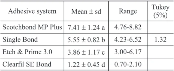
The mean values of thickness of the hybrid layer were statistically analyzed by means of the one-way analysis of variance (ANOVA) and Tukey’s test, at a level of significance of 5%.
RESULTS
means of the Tukey’s test, which revealed that ad-hesives formed hybrid layers with different thick-nesses (p < 0.05). Table 3 displays the mean values of thickness of the hybrid layer and standard de-viations for each adhesive system. The conventio-nal three-step adhesive Scotchbond MP Plus
for-med the thickest hybrid layer (7.41 ± 1.24 µm),
while the self-etching system Clearfil SE Bond
for-med the thinnest hybrid layer (1.22 ± 0.45 µm).
When the bonding systems were applied on dentin conditioned with phosphoric acid, the thickness of the resin-infiltrated layers was greater than that observed after the utilization of self-etching den-tin-bonding systems, which were applied directly to the smear layer.
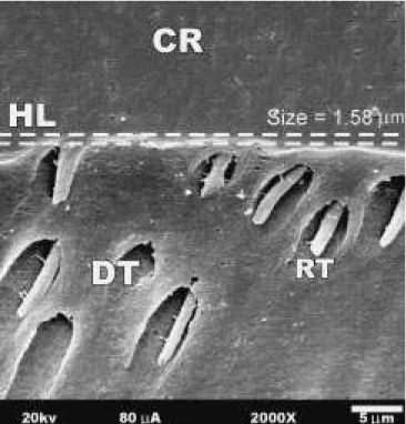
The penetration of resin monomers into denti-nal tubules and the formation of a resdentin in-terdiffusion zone, or hybrid layer, were observed in all groups. Figures 1, 2, 3 and 4 show the interfa-ces formed by the adhesives systems. The resin
pe-netrated into dentinal tubules up to 40 mm,
for-ming a funnel-shaped configuration of the tag neck. Some lateral branches of adhesive were ob-served for all dentin-bonding agents – they are the result of the penetration of monomer into lateral canals, which communicate with adjacent tubu-les.
DISCUSSION
Although self-etching primers present less
aci-dity than 35% phosphoric acid gels12
, they have been able to etch through the smear layer into the underlying mineralized dentin and form a hybridi-zed complex. For self-etching adhesives, the hybri-dized complex comprises two portions: the zone of authentic hybrid layer and the zone of hybridized smear layer. The authentic hybrid layer is that portion where collagen fibrils remain attached to
TABLE 1 -Composition of the adhesive systems utilized in this study.
Adhesive systems Composition
Scotchbond MP Plus (conventional three-step - 3M Dental Products)
Conditioner12
: 35% phosphoric acid (pH 0.02).
Primer15
: HEMA, PAA and water (pH 3.5).
Adhesive: Bis-GMA and HEMA.
Single Bond (one-bottle - 3M Den-tal Products)
Conditioner12
: 35% phosphoric acid (pH 0.02).
Adhesive: Bis-GMA, HEMA, PAA, ethanol and water (pH 5*).
Etch & Prime 3.0 (selfetching -Degussa Hüls)
Universal: HEMA, ethanol and water.
Catalyst2: Tetra-methacryloxyethilpyrophosphate and HEMA (pH of mixture = 1,4).
Clearfil SE Bond (selfetching -Kuraray Co.)
SE-Primer19
: MDP, HEMA, CQ, N,N-Diethanol p-toluidine, hydrophilic dimethacrylate and water (pH 2).
SE-Bond: MDP, Bis-GMA, HEMA, hydrophobic dimethacrylate, CQ, N,N-Diethanol p-toluidine e silanated colloidal silica.
*pH as reported by the manufacturer. HEMA = 2-hydroxyethyl methacrylate; PAA = polyalkenoic acid copolymer; Bis-GMA = bisphenol-glycidyl methacrylate; MDP = 10-methacryloxydecil-dihydrogen phosphate; CQ = dil-camphor-quinone.
TABLE 2 -One-way analysis of variance.
Source Sum of squares df
Mean
square F-value
Adhesive system 165.74 3 55.25 58.78*
Residual 26.40 28 0.94
Total 192.14 31
*Statistically significant at the level of 5%.
TABLE 3 - Mean values of thickness (µm) of the re-sin-dentin interdiffusion zone.
Adhesive system Mean±sd Range Tukey (5%)
Scotchbond MP Plus 7.41±1.24 a 4.76-8.82
Single Bond 5.55±0.82 b 4.23-6.52 1.32
Etch & Prime 3.0 3.86±1.17 c 3.00-6.17
Clearfil SE Bond 1.22±0.45 d 0.70-2.10
FIGURE 1 -Photomicrograph of the resin-dentin interfa-ce formed by Scotchbond MP Plus. CR = composite resin, HL = hybrid layer, RT = resin tag, DT = dentinal tubule.
FIGURE 2 -Photomicrograph of the resin-dentin inter-face formed by Single Bond. CR = composite resin, HL = hybrid layer, RT = resin tag, DT = dentinal tubule.
FIGURE 3 -Photomicrograph of the resin-dentin inter-face formed by Etch & Prime 3.0. CR = composite resin, HL = hybrid layer, RT = resin tag, DT = dentinal tubule.
the underlying dentin, and the hybridized smear layer is the portion above it, where the smear layer
is incorporated into a hybridized complex18.
The early versions of self-etching adhesives4,20
contained Phenyl-P, as the acidic monomer, and HEMA, which resulted in a solution with a pH va-lue of 1.4-0.84. Clearfil Liner Bond II etched be-yond the smear layer and demineralized the
un-derlying dentin to a depth of 1.2-1.4µm18
. Studies have reported that these self-etching adhesives
formed a 1-2-µm-thick hybrid layer6,8,14,18,21
, which is rarely thicker than 2.5µm10,24.
Clearfil SE Bond replaced Phenyl-P by another acidic phosphate resin monomer (MDP) and its
pri-mer showed pH = 2.0. Tayet al.18
(2000) analyzed the effect of the acidity of self-etching primers and the thickness of the smear layer. They reported an underlying authentic hybrid layer with the
thick-ness of 0.4 to 0.5µm and a hybridized smear layer
with the thickness of 0.4 to 0.7µm for Clearfil SE
Bond. The aforementioned authors prepared the specimens for transmission electron microscopy and, regardless of the different methods of prepa-ration16
, the range of thickness of the hybridized
complex (0.8-1.2 µm) observed in their study
in-cludes the mean value obtained in our experiment. Studies reported that the depth of deminerali-zed dentin is related to the concentration and pH of
the acidic monomer2,4,20
. Thus, a minimum pH va-lue of 2.8 is required so that the primer solution can effectively demineralize the dentin within
thirty seconds18
. Acidic pyrophosphate resin mo-nomer, present in the catalyst bottle, ionizes when in contact with the water from the catalyst bottle,
originating a solution2
with pH = 1.4. The acidity of the Etch & Prime 3.0 adhesive is higher than that of the other self-etching primer, therefore, the depth of demineralized intact dentin and the thick-ness of the authentic hybrid layer were also greater. Although Etch & Prime 3.0 produced the thickest hybrid layer, when compared to the other tested self-etching adhesive, studies have shown that it is less effective, on dentinal substrate, as
to marginal microleakage and shear bond
strength1,17
.
Similarly to what was observed for Clearfil SE Bond, the demineralization of dentin and the pene-tration of HEMA occur simultaneously for Etch & Prime 3.0. The formation of a hybrid layer after the application of the Scotchbond MP Plus and Single
Bond adhesive systems depends on previous acid-ethching and on the uniform infiltration of re-sin monomers into the spaces between the colla-gen fibrils of the demineralized dentinal subsurfa-ce9
. The etching agent (32-37% phosphoric acid) demineralizes dentin by removing hydroxyapatite and exposing collagen fibrils in the few microns
(3-5 µm) of the most superficial layer of dentin3,12.
Therefore, the depth of demineralized dentin duced by phosphoric acid is greater than that pro-duced by self-etching primers, which results in a thicker hybrid layer.
The Scotchbond MP Plus bonding system was
able to produce the thickest hybrid layer (7.41 ±
1.24 µm). Since the etching time and the etching
agent were the same, other mechanisms must be responsible for the thicker resin-infiltrated denti-nal layer seen after the application of Scotchbond MP Plus, when compared to Single Bond. A possi-ble explanation is that this primer is more acidic (pH 3.5) than the Single Bond one-bottle adhesive (pH 5). The acidity of the primer of Scotchbond MP may have contributed to a second demineralizati-on of the underlying dentin and, thus, allowed for deeper penetration of monomer into the deminera-lized dentinal matrix. Similar values of thickness of the hybrid layer have been reported for Scot-chbond MP Plus6,8,14,15,19,22
and Single Bond7,13,15,19
. That confirms the validity of preparing specimens for SEM and measuring the thickness of the hybrid layer by means of the techniques employed in the present study.
CONCLUSION
According to the methodology employed and ba-sed on the obtained results and on the statistical analyses, it can be concluded that:
1. all tested adhesive systems formed a hybrid la-yer, although with significantly different thick-nesses;
2. the conventional three-step adhesive, followed by the one-bottle adhesive, exhibited the thic-kest hybrid layer;
3. self-etching adhesives exhibited the thinnest hybrid layer.
ACKNOWLEDGMENTS
REFERENCES
1. Burmann P, Cardoso PEC, Silveira B,et al.The influence of composite resin polymerization techniques on microleaka-ge [abstract 315]. J Dent Res 2000;79:183.
2. Cardoso PEC, Braga RR, Carrilho MRO. Evaluation of mi-cro-tensile, shear and tensile tests determining the bond strength of three adhesive systems. Dent Mater 1998; 14:394-8.
3. Carvalho RM, Yoshiyama M, Pashley EL, et al. In vitro
study on the dimensional changes of dentine after demine-ralization. Arch Oral Biol 1996;41:369-77.
4. Chigira H, Yukitani W, Hasegawa T,et al. Self-etching den-tin primers containing phenyl-P. J Dent Res 1994; 73:1088-95.
5. Eick JD, Gwinnett AJ, Pashley DH,et al. Current concepts on adhesion to dentin. Crit Rev Oral Biol Med 1997; 8:306-35.
6. Ferrari M, Cagidiaco CM, Kugel G,et al. Dentin infiltration by three adhesive systems in clinical and laboratorial con-ditions. Am J Dent 1996;9:240-4.
7. Ferrari M, Goracci G, Garcia-Godoy F. Bonding mecha-nism of three one-bottle systems to conditioned and unconditioned enamel and dentin. Am J Dent 1997; 10:224-30.
8. Marshall SJ, Tomsia AP, Marshall GW. Resin-dentin inter-diffusion zone thickness [abstract 2415]. J Dent Res 1997;76:315.
9. Nakabayashi N, Kojima K, Masuhara E. The promotion of adhesion by the infiltration of monomers into tooth subs-trates. J Biomed Mater Res 1982;16:265-73.
10. Nakajima M, Sano H, Burrow MF, et al. Tensile bond strength and SEM evaluation of caries-affected dentin using dentin adhesives. J Dent Res 1995;74:1679-88. 11. Pashley DH, Carvalho RM. Dentine permeability and
denti-ne adhesion. J Dent 1997;25:355-72.
12. Perdigão J, Lambrechts P, Van Meerbeek B,et al. A mor-phological field emission SEM study of the effect of six phosphoric acid-etching agents on human dentin. Dent Mater 1996;12:262-71.
13. Perdigão J, Ramos JC, Lambrechts P.In vitrointerfacial re-lationship between human dentin and one-bottle dental adhesives. Dent Mater 1997;13:218-27.
14. Prati C, Chersoni S, Mongiorgi R, et al. Resin infiltrated dentin layer formation of new bonding systems. Oper Dent 1998;23:185-94.
15. Prati C, Chersoni S, Mongiorgi R, et al. Thickness and morphology of resin-infiltrated dentin layer in young, old and sclerotic dentin. Oper Dent 1999;24:66-72.
16. Sano H, Yoshiyama M, Ebisu S,et al. Comparative SEM and TEM observations of nanoleakage within the hybrid la-yer. Oper Dent 1995;20:160-67.
17. Segre INW, Giannini M, Pimenta LAF. Shear bond strength evaluation of different hydrophilic adhesive system [abs-tract 1850]. J Dent Res 2000;79:375.
18. Tay FR, Sano H, Carvalho RM, et al. An ultrastructural study of the influence of acidity of self-etching primers and smear layer thickness on bonding to intact dentin. J Adhes Dent 2000;2:83-98.
19. Vargas MA, Cobb DS, Denehy GE. Interfacial micromor-phology and shear bond strength of single-bottle pri-mer/adhesives. Dent Mater 1997;13:316-24.
20. Watanabe I, Nakabayashi N, Pashley DH. Bonding to ground dentin by a phenyl-P self-etching primer. J Dent Res 1994;73:1212-20.
21. Yoshiyama M, Sano H, Ebisu S,et al. Regional strengths of bonding agents to cervical sclerotic root dentin. J Dent Res 1996;75:1404-13.
22. Youssef MN, Guaraldi E, Sato CT,et al. Estudo comparati-vo de quatro filosofias adesivas quanto à penetração na dentina. Rev Assoc Paul Cir Dent 1998;52:236-9.