Milad ALIKHANI(a)
Parichehr GHALAIANI(a)
Elham ASKARIYAN(b)
Zahra Ahmadi KHUNSARAKI(c)
Atefeh TAVANGAR(a)
Aliasghar NADERI(a)
(a) Isfahan University of Medical Sciences, Department of Oral and Maxillofacial Medicine, Isfahan, Iran.
(b) Islamic Azad University, Department of psychology, Najafabad Branch, Najafabad, Iran.
(c) Isfahan University of Medical Sciences, Dental School, Isfahan, Iran.
Association between the clinical severity
of oral lichen planus and anti-TPO level
in thyroid patients
Abstract: This study considered a possible relationship between the severity of oral lichen planus (OLP), serum anti-TPO autoantibodies (TPOAb) titer and thyroid disease in OLP patients. Forty-six OLP patients with positive TPOAb results (> 35 IU/ml) who had also been diagnosed with thyroid disease were included in the study group. The control group consisted of 46OLP patients with no thyroid disease. The study and control groups (92) were divided to two subgroups of erosive OLP (EOLP) and non-erosive OLP (NEOLP). Serum TPOAb levels and IL-8 (to measure OLP severity) were evaluated using the independent t-test, chi-square and conditional logistic regression
analysis (α = 0.05). A signiicant positive correlation was found between serum IL-8 and TPOAb levels in the study group (r = 0.783; p = 0.001). The positive blood levels of TPOAb were signiicantly associated with an increased risk of EOLP (OR = 4.02 at 95%CI; 1.21–13.4; p = 0.023). It is possible to used positive serum TPOAb levels
in patients with OLP as in indicator of possible undetected thyroid disorders in those patients. Because erosive OLP has been associated with TPOAb in thyroid patients, it may be useful to determine TPOAb levels of such patients to diagnose a possible undetected thyroid disorders and follow-up for malignancy.
Keywords: Lichen Planus, Oral; Autoimmunity; Thyroid Diseases; Cytokines.
Introduction
Oral lichen planus (OLP) is a chronic inlammatory disease affecting
the oral mucosa that may also involve the skin and the genitalia.1 OLP may appear clinically as a reticular, erythematous or erosive type of lesion.2 Although the etiopathology of OLP remains unidentiied, it is believed
that immune dysregulation plays a crucial role.2,3 Immune dysregulation in OLP includes unusual production of inlammatory mediators,4 of which
cytokines are the most prominent.5
The abnormal expression pattern of cytokines such as IL-1, -2, -4, -5, -6, -8, -10, -18 and TNF-α has been found in the lesions, serum and saliva of patients
with OLP.5 Elevated levels of IL-8 in the serum of OLP patients have been
recorded and reported in several studies.6,7,8,9 It has also been reported that the clinical severity of OLP is directly associated with the IL-8 level in the serum.9
Declaration of Interests: The authors certify that they have no commercial or associative interest that represents a conflict of interest in connection with the manuscript.
Corresponding Author:
Aliasghar Naderi
E-mail: [email protected]
DOI: 10.1590/1807-3107BOR-2017.vol31.0010
Submitted: Apr 15, 2016
A high frequency of circulating anti-nuclear antibodies (ANA), anti-thyroglobulin antibodies (TGA) and anti-thyroid microsomal autoantibodies (TMA) has been reported in OLP patients.10,11 These findings suggest a possible association between autoimmune disease, including thyroid disorders, and OLP.10 It is known that autoantibodies against thyroid gland antigenic components such as thyroid peroxidase (TPO) and thyroglobulin (TG) can induce epithelial cell damage12,13 and that these autoantibodies are closely linked to Hashimoto’s thyroiditis and Graves’ disease.14,15
Although a causal relationship between OLP and thyroid gland disorders has not been discovered, the association of OLP and thyroid disease has been studied.16 It has been suggested that patients with a history of thyroid gland disorders, especially hypothyroidism, are more likely to have OLP than healthy subjects.17,18 No studies were found in English-language publications, however, that had found a possible association between the severity of clinical signs of OLP and the serum titer of anti-TPO antibodies (TPOAb) in OLP patients who have also been diagnosed with thyroid disease. The current study examined a possible association between the clinical severity of OLP and the titer of serum TPOAb in OLP patients with thyroid disease.
Methodology
Because the clinical severity of OLP is associated with elevated serum IL-8 levels,9 IL-8 serves as a
reliable tool to assess the clinical severity of this disease. A possible association between the clinical severity of OLP and the titer of serum TPOAb in OLP patients with thyroid disease was determined using serum IL-8 level as an indicator in the present study.
Between November 2010 and December 2013, 340 OLP patients visited the Institute of
Dentistry, Isfahan University of Medical Science,
Iran. A total of 216 individuals who denied a
history of thyroid disease or thyroid medication consumption were excluded by evaluating their self-assessed general health status forms. Also excluded were patients suspected of having
drug or dental restoration-related oral lichenoid lesions. An additional 62 OLP patients with areca quid chewing habits, hypertension and autoimmune diseases such as systemic lupus erythematosus, rheumatoid arthritis, Sjögren’s syndrome, pemphigus vulgaris and cicatricial
pemphigoid were also excluded. In total, 278 (81.5%)
individuals were excluded from the present study.
A total of 62 patients (18.5%) were included in the
initial study group. All participants had a history of taking thyroxin or suffering from thyroid gland disorders according to their self-assessed general health status forms. Diagnosis of OLP in the 62 patients was initially done by clinical impression, which was
later conirmed histologically using the most recent
diagnostic criteria.19
Blood samples were collected from all OLP
patients with self-reported thyroid disease (n = 62).
TSH, thyroxine (T4) and tri-iodothyronine (T3) thyroid function tests were carried and TPOAb and serum IL-8 levels were measured. TPOAb values higher than 35 IU/ml were considered positive.20
Of the 62 patients assessed, 46 (74%) were TPOAb positive and were included in the inal study group.
The patients were referred to an endocrinologist
to conirm the diagnosis of thyroid gland disorder based on the indings of the thyroid gland function
tests. Of the 46 patients, 34 were diagnosed with Hashimoto’s thyroiditis, 8 with Graves’s disease and 4 with hyperthyroidism.
The 46 OLP patients were matched for gender and age with 46 patients having no reported history of thyroid disease to serve as the control group. Because
10% to 15% of clinically healthy individuals may have
high TPOAb titer,21 blood samples were taken from all 46 controls to rule out thyroid malfunction and their TSH, T3, T4, TPOAb and serum IL-8 levels were measured. None recorded positive TPOAb levels (>35 IU/ml) and all were included in the control
group. The Ethics Committee of Isfahan University
of Medical Sciences approved the protocol of this study and an informed consent was obtained from each participant.
Non-erosive OLP (NEOLP) was deined based on
clinical presentation of radiating grayish-white striae and/or plaque separately or in combination. Any sign of non-traumatic ulceration in the oral mucosa associated with typical lichen signs indicated erosive OLP (EOLP).22 The participants underwent
evaluations of for TPOAb level using IMMULITE
2000 anti-TPO (EURO/DPC; United Kingdom). IL-8
levels were measured using an Interleukin-8 Elisa
kit (Bender MedSystems; Austria).
SPSS (v. 18) was used to analyze the data. The
distribution of the variables was examined for
normality using the Kolmogorov-Smirnov test. Because
the data was normally distributed, a parametric independent t-test was carried out. The difference in the presence of TPOAb was compared between the EOLP and NEOLP subjects using a chi-square test.
The odds ratios (OR) at a 95% conidence interval (CI)
was calculated using conditional logistic regression adjusted for confounders. The threshold of statistical
signiicance was set at р < 0.05.
Results
Most of the OLP patients (n = 34; 74%) in each
group (study and control) were women. The mean age of the study and control subjects at admission
to the university dental clinic was 45 years (17–68) and 45 years (18–72), respectively.
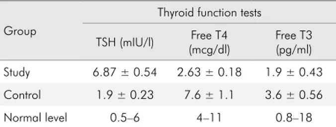
The regular blood levels of TSH were 0.4–4 mIU/l, T4 were 4.5–11.2 mcg/dl and T3 were 2.3-4.2 pg/ml.20
The mean results of the thyroid function tests for each
group conirmed that the mean blood levels of the three
hormones in the control group were within normal
limits (Table 1). The indings were also normal in the
individuals of the control group, further verifying that the control group was free of thyroid gland malfunction.
Most (n =30, 65.4%) of the 46 OLP patients with
positive TPOAb levels (study group) were diagnosed
with EOLP; only 16 (34.6%) were diagnosed with
NEOLP. The majority of individuals (n=32, 69.5%)
in the control group were diagnosed with NEOLP; only 14 (30.5%) were identiied with EOLP. The
mean TPOAb level in the total study group was
90.3 ± 18.8 (IU/ml); this value was 102 ± 0.23 for the NEOLP patients compared to 11 ± 4.8 (IU/ml)
in the control group. There was a large difference between the study and control groups for serum IL-8 versus TPOAb levels and this difference
was statistically signiicant (p = 0.001) (Table 2). A signiicant positive correlation was also found
between serum IL-8 and TPOAb levels in the study
group (r = 0.783; p = 0.001). There was no signiicant
correlation between IL-8 and TPOAb levels in the
control group (p = 0.65).
All OLP patients, regardless of TPOAb status
(n = 92) were divided into EOLP (n = 44) and NEOLP (n = 48) groups (Table 3). A statistically signiicant
correlation was found between the occurrence of EOLP and positive TPOAb (> 35 IU/ml) that indicates
that TPOAb positivity is signiicantly associated with an increased chance of EOLP (OR = 4.02 at 95%CI; 1.21–13.4; p = 0.023). A signiicant difference was
also found between groups for serum IL-8 level.
Increased IL-8 levels were signiicantly associated with an increased chance of EOLP (OR = 1.276 at 95%CI; 1.092–1.49; p = 0.002).
Table 1. Mean level (±SD) of thyroid gland function tests in study and control groups.
Group
Thyroid function tests
TSH (mIU/l) Free T4 (mcg/dl)
Free T3 (pg/ml)
Study 6.87 ± 0.54 2.63 ± 0.18 1.9 ± 0.43
Control 1.9 ± 0.23 7.6 ± 1.1 3.6 ± 0.56
Normal level 0.5–6 4–11 0.8–18
Table 2. Prevalence of clinical presentations of oral lichen planus (OLP) and the mean anti-TPO autoantibodies (TPOAb) serum levels (IU/ml) and IL-8 (pg/ml) in study and control groups.
Group
Type of OLP Serum markers
Marker correlation EOLP
(%)
NEOLP
(%) TPOAb IL-8
Study 30 (65.4)
16
(34.6) 90.3 ± 18.8 12.39 ± 2.65
r = 0.783; p = 0.001
Control 14 (30.5)
32
(69.5) 11 ± 4.8 3.9 ± 1.7 p = 0.65
Discussion
The current study examined the association between the clinical severity of OLP and TPOAb titer in patients with thyroid disease. The results indicate that the severity of the clinical expression of OLP lesions were directly linked to the level of serum IL-8, as previously reported,9 and also to the
level of TPOAb.
Cytokines play an important role in controlling
the direction, extent and duration of immune response.23 Abnormal expression of cytokines may
lead to the onset of autoimmunity.23,24 IL-8 plays affects pathogenesis of inlammation25 and has the
capacity to recruit T cells as well as non-specific
inlammatory cells to the sites of inlammation by
activating neutrophils.26 Serum IL-8 levels are sensitive
markers that can be used to monitor disease activity and the severity of OLP.9 The level of serum IL-8 also
appears to signify an association with reticular and erosive forms of OLP.9 Because IL-8 is a pluripotent
pro-tumorigenic cytokine that is known to induce angiogenesis, tumor cell proliferation and tumor cell migration,27,28,29 evaluation of IL-8 serum level may also serve as an indicator of the severity of OLP and the risk of the developing malignancies.
The results of the current study conirm the initial
hypothesis that there is a significant association between the clinical severity of OLP and the level
of TPOAb in patients with thyroid disease; however,
logical bias may exist in the extent of the differences because the initial inclusion criteria stipulated that TPOAb levels should be higher in the study group
than in the control group. The present study showed
that IL-8 serum levels were signiicantly higher in
OLP patients with high TPOAb levels who had also been diagnosed with thyroid disease compared to OLP patients with no thyroid disease. The current
indings suggest that OLP patients who have positive
levels of TPOAb are about four times more likely to develop EOLP than patients with negative TPOAb levels, although this assumption requires further study combined with long-term patient follow-ups. The finding of a correlation between EOLP and
TPOAb further supports the conclusion of Chang
et al.10 who reported that patients with severe clinical
EOLP have a signiicant probability of development
of autoimmune markers for thyroid disease such as abnormal TGA and TMA.
There was a substantial difference between the study and control groups for serum IL-8 level.
Karanikas et al.30 showed that TPOAb titer correlates
with increased presence of T cells that produce cytokines, which are responsible for cell damage.
They also found increased production of TNF-α by CD8+ cytotoxic T lymphocytes in patients with high
TPOAb titer.30 Because, in many cell types, synthesis
of IL-8 is strongly stimulated by TNF-α,31 it appears
reasonable for IL-8 levels to be signiicantly higher
in OLP patients with positive TPOAb values and thyroid disease. This may also explain the increased prevalence of EOLP in patients with thyroid disease than in OLP patients free of thyroid disease. The
histological indings show that there was very high neutrophilic iniltration of the epithelium in EOLP
than in other forms of the disease.32 This iniltration
Table 3. Mean levels of serum positive* anti-TPO autoantibodies (TPOAb) and IL-8 (pg/ml) serum levels in patients with EOLP and NEOLP.
OLP (n = 92)*
TPOAb Pos. TPOAb/EOLP
Serum IL-8 level (pg/ml) ± SD
IL-8 levels/EOLP
pos./neg. **
OR (% CI) OR (95%CI)
(%)
EOLP (n = 44) 30 /14 4.02 10.45 ± 4.1*
1.276 [1.092–1.49] (p = 0.002\0
(68/31) [1.21–13.4]
NEOLP (n = 48) 16/32 (p = 0.023) 6.04 ± 3.93 *
(33/66)
can, at least partially, be attributed to the higher level of IL-8 in EOLP patients.
The present study showed that 46 of the 340 initial patients (14%) had thyroid disease with positive TPOAb values. Similar indings were reported by Lo Muzio et al.16 and Siponen et al.,18 who found
a history of thyroid gland pathosis in 14% and 15% of OLP cases, respectively. Although a causal
relationship between OLP and thyroid disease was not investigated in the current study, it can be speculated that circulating thyroid antibodies may trigger an autoimmune response in the oral mucosa and cause development of OLP. This immune response could
also occur in cases in which OLP precedes the onset
of thyroid dysfunction;16 therefore further studies on
the association between thyroid disease and OLP are required.
In conclusion, because the erosive pattern of OLP is
associated with positive indings of TPOAb in patients
with thyroid disease, it could be a clinically useful method of determining TPOAb levels in patients with EOLP to allow diagnosis of hidden thyroid disorders. The high levels of TPOAb found may indicate high levels of IL-8 and point to increased risk of development of malignancies and warrants much closer follow-ups.
1. Eisen D, Carrozzo M, Bagan Sebastian JV,
Thongprasom K. Oral lichen planus: clinical features and management. Oral Dis. 2005;11(6):338-49. doi: 10.1111/j.1601-0825.2005.01142.x
2. Lodi G, Scully C, Carrozzo M, Griffiths M, Sugerman
PB, Thongprasom K. Current controversies in oral lichen planus: report of an international consensus meeting. Part 1. Viral infections and etiopathogenesis. Oral Surg Oral Med Oral Pathol Oral Radiol Endod. 2005;100(1):40-51. doi:10.1016/j.tripleo.2004.06.077
3. Sugerman PB, Savage NW, Walsh LJ, Zhao ZZ, Zhou XJ,
Khan A et al. The pathogenesis of oral lichen planus. Crit Rev Oral Biol Med. 2002;13(4):350-65.
doi:10.1177/154411130201300405
4. Tao XA, Li CY, Rhodus NL, Xia J, Yang XP, Cheng B. Simultaneous detection of IFN-gamma and IL-4 in lesional tissues and whole unstimulated saliva from patients with
oral lichen planus. J Oral Pathol Med. 2008;37(2):83-7. doi:10.1111/j.1600-0714.2007.00593.x
5. Lu R, Zhang J, Sun W, Du G, Zhou G. Inflammation-related
cytokines in oral lichen planus: an overview. J Oral Pathol Med. 2015;44(1):1-14.
doi:10.1111/jop.12142
6. Rhodus NL, Cheng B, Bowles W, Myers S, Miller L, Ondrey F. Proinflammatory cytokine levels in saliva before and after treatment of (erosive) oral lichen
planus with dexamethasone. Oral Dis. 2006;12(2):112-6. doi:10.1111/j.1601-0825.2005.01165.x
7. Rhodus NL, Cheng B, Myers S, Miller L, Ho V, Ondrey
F. The feasibility of monitoring NF-kappaB associated
cytokines: TNF-alpha, IL-1alpha, IL-6, and IL-8 in
whole saliva for the malignant transformation of
oral lichen planus. Mol Carcinog. 2005;44(2):77-82. doi:10.1002/mc.20113
8. Zhang Y, Lin M, Zhang S, Wang Z, Jiang L, Shen J et al. NF-kappaB-dependent cytokines in saliva and serum
from patients with oral lichen planus: a study in an ethnic Chinese population. Cytokine. 2008;41(2):144-9. doi:10.1016/j.cyto.2007.11.004
9. Sun A, Wang JT, Chia JS, Chiang CP. Serum interleukin-8 level is a more sensitive marker than serum interleukin-6 level in monitoring the disease activity of oral lichen
planus. Br J Dermatol. 2005;152(6):1187-92. doi:10.1111/j.1365-2133.2005.06497.x
10. Chang JY, Chiang CP, Hsiao CK, Sun A. Significantly
higher frequencies of presence of serum autoantibodies
in Chinese patients with oral lichen planus. J Oral Pathol Med. 2009;38(1):48-54. doi:10.1111/j.1600-0714.2008.00686.x 11. Lin HP, Wang YP, Chia JS, Sun A. Modulation of serum
anti-thyroglobulin and anti-thyroid microsomal autoantibody levels by levamisole in patients with oral
lichen planus. J Formos Med Assoc. 2011;110(3):169-74. doi:10.1016/S0929-6646(11)60027-2
12. Rodien P, Madec AM, Ruf J, Rajas F, Bornet H, Carayon
P et al. Antibody-dependent cell-mediated cytotoxicity
in autoimmune thyroid disease: relationship to
antithyroperoxidase antibodies. J Clin Endocrinol Metab. 1996;81(7):2595-600. doi:10.1210/jcem.81.7.8675583
13. Chiovato L, Bassi P, Santini F, Mammoli C, Lapi P, Carayon
P et al. Antibodies producing complement-mediated thyroid cytotoxicity in patients with atrophic or goitrous
autoimmune thyroiditis. J Clin Endocrinol Metab. 1993;77(6):1700-5. doi:10.1210/jcem.77.6.7903315 14. Bjoro T, Holmen J, Krüger O, Midthjell K, Hunstad K,
Schreiner T et al. Prevalence of thyroid disease, thyroid dysfunction and thyroid peroxidase antibodies in
a large, unselected population. Eur J Endocrinol. 2000;143(5):639-47. doi:10.1530/eje.0.1430639
15. Prummel MF, Wiersinga WM. Thyroid peroxidase autoantibodies in euthyroid subjects. Best Pract
Res Clin Endocrinol Metab. 2005;19(1):1-15. doi:10.1016/j.beem.2004.11.003
16. Lo Muzio L, Santarelli A, Campisi G, Lacaita M, Favia
G. Possible link between Hashimoto’s thyroiditis and
oral lichen planus: a novel association found. Clin Oral Investig. 2013;17(1):333-6. doi:10.1007/s00784-012-0767-4 17. Garcia-Pola MJ, Llorente-Pendás S, Seoane-Romero JM,
Berasaluce MJ, García-Martín JM. Thyroid disease and oral lichen planus as comorbidity: a prospective case-control study. Dermatology. 2016;232(2):214-9. doi:10.1159/000442438 18. Siponen M, Huuskonen L, Läärä E, Salo T. Association
of oral lichen planus with thyroid disease in a Finnish
population: a retrospective case-control study. Oral
Surg Oral Med Oral Pathol Oral Radiol Endod.
2010;110(3):319-24. Doi:10.1016/j.tripleo.2010.04.001 19. Meij EH, Waal I. Lack of clinicopathologic correlation
in the diagnosis of oral lichen planus based on the presently available diagnostic criteria and suggestions
for modifications. J Oral Pathol Med. 2003;32(9):507-12. Doi:10.1034/j.1600-0714.2003.00125.x
20. Gardner D. Normal hormone reference ranges.
In: Gardner DG, Shoback D. Greenspan’s basic & clinical
endocrinology. 9th ed. McGraw-Hill; 2011. Appendix.
21. Chardès T, Chapal N, Bresson D, Bès C, Giudicelli V,
Lefranc MP et al. The human anti-thyroid peroxidase autoantibody repertoire in Graves’ and Hashimoto’s autoimmune thyroid diseases. Immunogenetics.
2002;54(3):141-57. Doi:10.1007/s00251-002-0453-9
22. DeRossi SS1, Ciarrocca KN. Lichen planus, lichenoid drug
reactions, and lichenoid mucositis. Dent Clin North Am. 2005;49(1):77-89. Doi:10.1016/j.cden.2004.08.004
23. Roescher N, Tak PP, Illei GG. Cytokines in Sjogren’s
syndrome. Oral Dis. 2009;15(8):519-26. Doi:10.1111/j.1601-0825.2009.01582.x
24. Moudgil KD, Choubey D. Cytokines in autoimmunity:
role in induction, regulation, and treatment. J Interferon Cytokine Res. 2011;31(10):695-703. Doi:10.1089/jir.2011.0065
25. Dobreva I, Waeber G, James RW, Widmann C. Interleukin-8 secretion by fibroblasts induced by low
density lipoproteins is p38 MAPK-dependent and leads to cell spreading and wound closure. J Biol Chem. 2006;281(1):199-205. Doi:10.1074/jbc.M508857200
26. Schröder JM. The neutrophil-activating peptide 1/ interleukin 8, a novel neutrophil chemotactic cytokine.
Arch Immunol Ther Exp (Warsz). 1992;40(1):23-31. 27. Silverman S Jr, Gorsky M, Lozada-Nur F. A prospective
follow-up study of 570 patients with oral lichen planus:
persistence, remission and malignant association.
Oral Surg Oral Med Oral Pathol. 1985;60(1):30-4. Doi:10.1016/0030-4220(85)90210-5
28. Arihiro K, Oda H, Kaneko M, Inai K: Cytokines facilitate chemotactic motility of breast carcinoma cells. Breast
Cancer. 2000;7(3):221-30. Doi:10.1007/BF02967464
29. Singh RK, Gutman M, Radinsky R, Bucana CD, Fidler IJ. Expression of interleukin 8 correlates with the metastatic
potential of human melanoma cells in nude mice. Cancer Res. 1994;54(12):3242-7
30. Karanikas G, Schuetz M, Wahl K, Paul M,
Kontur S, Pietschmann P et al. Relation of anti-TPO
autoantibody titre and T-lymphocyte cytokine production
patterns in Hashimoto’s thyroiditis. Clin Endocrinol (Oxf). 2005;63(2):191-6.
doi:10.1111/j.1365-2265.2005.02324.x
31. Osawa Y, Nagaki M, Banno Y, Brenner DA, Asano T, Nozawa Y et al. Tumor necrosis factor alpha-induced
interleukin-8 production via NF-kappaB and phosphatidylinositol 3-kinase/Akt pathways inhibits cell apoptosis in human hepatocytes. Infect Immun.
2002;70(11):6294-301.
doi:10.1128/IAI.70.11.6294-6301.2002
32. Fernández-González F, Vázquez-Álvarez R,
Reboiras-López D, Gándara-Vila P, García-García A, Gándara-Rey JM. Histopathological findings in oral
lichen planus and their correlation with the clinical