Pseudomonas aeruginosa
: Study of Antibiotic Resistance and
Molecular Typing in Hospital Infection Cases in a Neonatal
Intensive Care Unit from Rio de Janeiro City, Brazil
MM Loureiro, BA de Moraes, VLF Mendonça, MRR Quadra*, GS Pinheiro*, MD Asensi/
+Laboratório de Enterobactérias, Departamento de Bacteriologia, Instituto Oswaldo Cruz-Fiocruz, Av. Brasil 4365, 21045-900 Rio de Janeiro, RJ, Brasil *Hospital Maternidade Alexander Fleming II, Sistema Único de Saúde, Rio de Janeiro, RJ, Brasil
This study had the objective of to analyze the demographic and bacteriologic data of 32 hospitalized newborns in an neonatal intensive care unit of a public maternity hospital in Rio de Janeiro city, Brazil, seized by
Pseudomo-nas aeruginosa sepsis during a period ranged from July 1997 to July 1999, and to determine the antimicrobial resistance percentage, serotypes and pulsed field gel electrophoresis (PFGE) patterns of 32 strains isolated during this period. The study group presented mean age of 12.5 days, with higher prevalence of hospital infection in males (59.4%) and vaginal delivery (81.2%), than females (40.6%) and cesarean delivery (18.8%), respectively. In this group, 20 (62.5%) patients received antimicrobials before positive blood cultures presentation. A total of 87.5% of the patients were premature, 62.5% presented very low birth weight and 40.6% had asphyxia. We detected high antimicrobial resistance percentage to β-lactams, chloramphenicol, trimethoprim/sulfamethoxazole and tetracy-cline among the isolated strains. All isolated strains were classified as multi-drug resistant. Most strains presented serotype O11 while PFGE analysis revealed seven distinct clones with isolation predominance of a single clone (75%) isolated from July 1997 to June 1998.
Key words: Pseudomonas aeruginosa - neonatal intensive care unit - hospital infection - sepsis - Rio de Janeiro - Brazil
Pseudomonas aeruginosa is one of the most common
pathogens involved in hospital infection (HI) causing opportunistic infections in humans, particularly among immunocompromised patients (Bert & Lambert-Zechovsky 1996, Kinoshita et al. 1997, Tsakris et al. 2000), and be-cause of its ubiquitous nature, ability to survive in ad-verse conditions, and affinity for moist environments, re-mains a common pathogen in intensive care units (ICU) (Grundmann et al. 1995, Moolenaar et al. 2000)
The HI causes complication of medical care in ICUs, principally in neonatal intensive care units (NICU), due to the natural immunodeficiency of newborns, submission to invasive procedures of therapy and diagnostic, pres-ence of several underlying risk conditions and indiscriminated antimicrobial use, and can causes signifi-cant morbidity and mortality (Sader et al. 1993, Kettner et al. 1995, Moolenaar et al. 2000). Mortality upon infected patients with P. aeruginosa sepsis was 46.7%, compared
to only 21% for other bacteria in a study that collected blood stream isolates from nearly 50 medical center in the USA (Jones et al. 1997).
The worldwide emergence of multi-resistant bacterial strains is a growing concern, especially in HI cases caused by P. aeruginosa. Among nosocomial bacterial infections,
those caused by P. aeruginosa are associated with
high-est mortality rate, and are difficult to eradicate from
in-fected tissues or blood because those microorganisms are virulent and have a limited susceptibility to antimicro-bials (Kettner et al. 1995, Harris et al. 1999).
The epidemiology of P. aeruginosa infections are
usu-ally studied by the analysis of phenotypic markers, in-cluding biotype, serovar, pyocin production, phage type, and antimicrobial susceptibility pattern (Pitt 1988, Kinoshita et al. 1997). Typing of strains is important for eradication of environmental sources as well as preven-tion of cross-infecpreven-tions and monitoring of antimicrobial therapy efficacy (Poh et al. 1992). Recently, nucleic acid-based methods have been used to assist identification of bacteria to subspecies level in epidemiological studies, providing a higher discriminatory power than phenotypic parameters (Severino et al. 1999)
Chromosomal DNA restriction analysis by pulsed field gel electrophoresis (PFGE) is considered worldwide the most powerful tool to perform hospital epidemiologic stud-ies of P. aeruginosa because of its high discriminatory
capacity. These technique facilitate the elucidation of transmission routes, genetic variability and phylogenetic distances between strains (Sader et al. 1993, Grundmann et al. 1995, Renders et al. 1996, Müller-Premru & Gubina 1999)
This study reports the demographics characteristics of hospitalized newborns seized by P. aeruginosa sepsis
associated with HI, and antimicrobial resistance percent-age, serotypes and PFGE patterns of the isolated strains in a NICU from a public maternity hospital in Rio de Janeiro city, Brazil.
MATERIALS AND METHODS
Hospital and patients -Between July 1997 and July
1999, 32 P. aeruginosa strains were isolated from blood
cultures of different NICU newborns involved in HI cases,
Financial suppor: CNPq and Papes (Fiocruz)
+Corresponding author. Fax: +55-21-2270.6565. E-mail:
at the Hospital Maternidade Alexander Fleming II (HMAF), Rio de Janeiro city, Brazil. It is a maternity hospital provid-ing assistance and perinatal care includprovid-ing a neonatol-ogy intermediate care unity (NIU) with 40 beds and a NICU with 15 beds.
HI cases were defined according to Centers for Dis-eases Control and Prevention (CDC) (Garner et al. 1988). In general, infections that occurred after 48 h of permanence at the hospital were assumed to be hospital acquired.
Blood cultures, strains identification and suscepti-bility testing - 0.2 ml of venous blood obtained from
new-borns were drawn into bottles with 10 ml of Tripticase Soy Broth supplemented (Roche) and incubated to 37°C. After 24 h, the blood cultures were inoculated into Thioglycolate Broth (DIFCO) and plated on Blood Agar and Eosin-Methylene Blue Agar (EMB, DIFCO). The Thioglycolate Broth, and plates of Blood Agar and EMB Agar were incubated at 37°C during a period ranged from 18 to 24 h. When the blood cultures were negative after incubation of 24 h, the inoculation into broth and plates above cited were repeated during a week. Identification of P. aeruginosa strains were performed using the Crystal
System of identification for fermenters and non-ferment-ers (BBL/Becton-Dickinson).
P. aeruginosa sepsis were defined by a single
posi-tive blood culture associated with appropriate clinical manifestations (one of the following clinical signs or symp-toms: fever > 38°C, hypothermia < 36.5°, apnoea, brady-cardia or tachybrady-cardia) according to CDC definitions (Gar-ner et al. 1988)
The antimicrobial susceptibility test was carried out through of disk diffusion method according to National Committee for Clinical Laboratory Standards, NCCLS (1997) recommendations. Quality control was carried out using standard strains of Escherichia coli (ATCC 25922), P. aeruginosa (ATCC 27953) and Staphylococcus aureus
(ATCC 25923). The following concentrations of antimi-crobials drugs (CECON) were used: cephalothin (30 µg), cefoxitin (30 µg), ceftriaxone (30 µg), cefuroxime (30 µg), carbenicillin (100 µg), cefepime (30 µg), ceftazidime (30 µg), piperacillin/tazobactam (100/10 µg), ticarcillin/ clavulanic acid (75/10 µg), imipenem (10 µg), gentamicin (10 µg), amikacin (30 µg), ciprofloxacin (5 µg), trimetho-prim/sulfamethoxazole (1.25/23.75 µg), chloramphenicol (30 µg), tetracycline (30 µg).
Serotyping method - An 18h Nutrient Agar culture (at
37°C) of each strain was used as antigen. One ml of sterile saline solution (0.85 % NaCl) was drawn into tubes con-taining the Nutrient Agar cultures and mixed to produce a suspension containing the bacterial growth. The O-group was identified by slide agglutination with P. aeruginosa
Antisera Kit (Denka Seiken Co. Ltd., Tokyo, Japan), ac-cording to Homma (1982) and Liu et al. (1983).
PFGE method - The strains were submitted to
chro-mosomal DNA extraction and processing according to the procedures previously described by Sader et al.(1994). Nucleic acids present in agarose plugs were digested with 10U of SpeI restriction enzyme at 37ºC during 20 h, and
the electrophoresis procedure was carried out in 1% aga-rose gels and running buffer containing 0.5X TBE with a CHEF DR III pulsed field electrophoresis systems
(Bio-Rad, California). Running conditions consisted of two ramps in sequence (ramp A consisted of an initial switch time of 0.5 sec, a final switch time of 25 sec, and a run time of 20 h; ramp B consisted of an initial switch time of 30 sec, a final switch time of 60 sec, and run time of 4 h). The voltage was 6V/cm for both ramps, and the temperature was kept constant at 13ºC. Fragments were stained with ethidium bromide and photographed. All agarose gels were run twice to verify the reproducibility of the tests.
Band patterns were analyzed using GelCompar II (Ap-plied Maths, Belgium), without using internal markers. The similarities between fingerprints were determined by con-struction of a similarity matrix using the Dice’s coefficient with 1.5% position tolerance and optimization of 1%, and a dendrogram generated using the UPGMA clustering al-gorithm. Definition of clonal structures of P. aeruginosa
strains were made according to Tenover et al.(1995)
Epidemiological analysis of patients - For each
pa-tient, an epidemiological record with demographic and microbiologic data was done. These data were registered in an EXCEL 7.0 program (Microsoft) and analyzed later in Epi-Info program (version 6.04b; CDC, Atlanta, USA). A control group of 70 newborns which did not acquire sepsis during the hospitalization in the NICU-HMAF from July 1997 to July 1998 was created. These control cases were randomly selected from a group of 720 patients that presented negative blood cultures, to evaluate the statis-tical significance of the demographics data. Most vari-ables were compared using the odds ratios (ORs), 95% confidence intervals (CI95), chi-square (χ2) and P values,
except, mean age, mean of antimicrobial drugs used by patients before positive blood presentation and mean of antimicrobials days use before positive blood culture pre-sentation, that were compared using the t-student test.
RESULTS
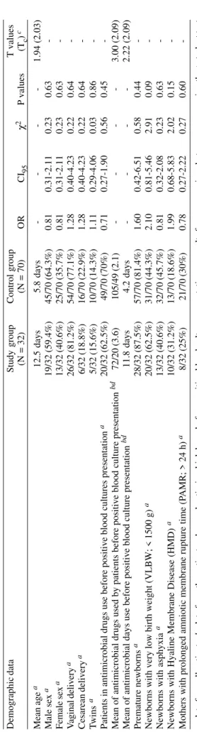
Table I shows the general demographic characteris-tics of the hospitalized newborns in NICU-HMAF, Rio de Janeiro city, Brazil, seized by P. aeruginosa sepsis and
classified as HI cases. The mean age of these newborns was 12.5 days, and the major frequencies of HI were de-tected in male sex (59.4%) and vaginal delivery (81.2%). Twenty patients (62.5%) received antimicrobial drugs be-fore positive blood cultures presentation with mean use of 3.6 antimicrobials per patient and mean of 11.8 days use. A total of 87.5% of the patients were premature, 62.5% presented very low birth weight (VLBW), 40.6% had as-phyxia and 31.2% presented Hyaline Membrane Disease (HMD).
The statistical results of the comparison between study and control groups (Table I) using OR, CI95, χ2, P
values and t-student test, showed statistical significance
in mean of antimicrobial drugs used by patients before positive blood culture presentation (T = 3.00) and mean of antimicrobials days use before positive blood cultures presentation (T = 2.22).
The antimicrobial resistance percentage of the isolated
P. aeruginosa strains (Table III) demonstrated high
anti-microbial resistance percentage (75 to 100% of resistance) to cephalotin, cefoxitin, ceftriaxone, cefuroxime, chloram-phenicol, trimethoprim/sulfamethoxazole and tetracycline in two analyzed periods (July 1997 to July 1998 and Au-gust 1998 to July 1999); high antimicrobial resistance per-centage to ceftazidime, cefepime, carbenicillin and ticarcillin/clavulanic acid during the first analyzed period and no resistant strain in the second; low antimicrobial resistance percentage (0 to 35% of resistance) to piperacillin/tazobactam in the first period and no resistant strain in second; low antimicrobial resistance percentage to gentamicin and amikacin (10.7% each) during the first period and were detected increase in antimicrobial resis-tance percentage in second (50 and 25%, respectively). In the first period there was no isolation of resistant strains to imipenem and ciprofloxacin, but in the second period an emergence of resistant strains was detected.
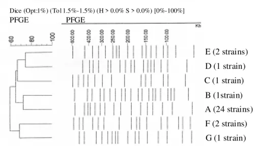
Fig. 1 shows seven distinct PFGE patterns (A-G) ob-served in the 32 P. aeruginosa strains, where the clone A
was the most frequent (epidemic clone; 75% of the strains), while the patterns C and E presented two strains, and the patterns B, D, F and G only one strain, corresponding to 25% of the isolated strains in total.
The dendrogram generated by GelCompar II (Fig. 2), revealed that the seven clones presented low similarity percentage (< 80%) under stringency conditions above cited, indicating that these clones are not genetically re-lated.
The serotyping results (Table IV) demonstrated four serotypes, with isolation predominance of the serotype E (29 strains; 90.7%), followed by serotypes B, D and I (one strain in each; 3.1%). The correspondence nomenclature of the serotypes B, D, E and I with the International Anti-genic Typing Scheme (IATS) serotypes designation are respectively: O2/O5/O16, O9, O11 and O1 (Homma 1982, Liu et al. 1983).
The distribution of serotypes according to the chro-mosomal profiles (PFGE) and isolation periods is shown
TABLE I
Demographic characteristics of newborns with
Pseudomonas aeruginosa
sepsis associated with hospital infection in two years period (July 1997 to July 1999)
Demographic data Study group Control group T values
(N = 32)
(N = 70)
O R C I95 χ 2 P values (Tc ) c Mean age a 12.5 days 5.8 days -1.94 (2.03) Male sex a 19/32 (59.4%) 45/70 (64.3%) 0.81 0.31-2.11 0.23 0.63 -Female sex a 13/32 (40.6%) 25/70 (35.7%) 0.81 0.31-2.11 0.23 0.63 -V aginal delivery a 26/32 (81.2%) 54/70 (77.1%) 1.28 0.40-4.23 0.22 0.64 -Cesarean delivery a 6/32 (18.8%) 16/70 (22.9%) 1.28 0.40-4.23 0.22 0.64 -Twins a 5/32 (15.6%) 10/70 (14.3%) 1.11 0.29-4.06 0.03 0.86
-Patients in antimicrobial drugs use before positive blood cultures presentation
a 20/32 (62.5%) 49/70 (70%) 0.71 0.27-1.90 0.56 0.45
-Mean of antimicrobial drugs used by patients before positive blood culture presentation
bd 72/20 (3.6) 105/49 (2.1) -3.00 (2.09)
Mean of antimicrobial days use before positive blood culture presentation
bd 11.8 days 4.2 days -2.22 (2.09) Premature newborns a 28/32 (87.5%) 57/70 (81.4%) 1.60 0.42-6.51 0.58 0.44
-Newborns with very low birth weight (VLBW; < 1500 g)
a 20/32 (62.5%) 31/70 (44.3%) 2.10 0.81-5.46 2.91 0.09
-Newborns with asphyxia
a 13/32 (40.6%) 32/70 (45.7%) 0.81 0.32-2.08 0.23 0.63
-Newborns with Hyaline Membrane Disease (HMD)
a 10/32 (31.2%) 13/70 (18.6%) 1.99 0.68-5.83 2.02 0.15
-Mothers with prolonged amniotic membrane rupture time (P
AMR; > 24 h)
a 8/32 (25%) 21/70 (30%) 0.78 0.27-2.22 0.27 0.60 -a
: data from all patients;
b
: data from the patients who used antimicrobial drugs before positive blood culture presentation;
c
: results from comparison between means using the
t
-student test
(Tc
= critical value of the
t
-student test); OR: Odds ratio; CI
95
: 95% confidence interval;
χ
2: Chi-square;
d
: variables with statistical significance
TABLE II
Antimicrobial drugs used by 20 newborns before positive blood culture presentation in two years period
(July 1997 to July 1999)
Antimicrobials Frequency %
Ampicillin 18 90
Amikacin 17 85
Cefotaxime 9 45
Oxacillin 9 45
Imipenem 7 35
Ceftazidime 4 20
Vancomycin 4 20
Amphotericin 1 5
Ceftriaxone 1 5
Ciprofloxacin 1 5
Gentamicin 1 5
-15), with antimicrobial resistance varying from 6 to 12 an-timicrobials.
DISCUSSION
P. aeruginosa is the fourth most frequent pathogen
isolated from nosocomial sepsis cases in the NICU-HMAF, accounting for 12.5% (32/255) of the infection cases, dur-ing the study period (data not shown). From the 32 sepsis cases detected in two years, 28 (87.5%) were detected in the first year of the study period. Sader (2000) related similar results notifying that P. aeruginosa was the third
most frequent pathogen isolated from Gram-negative sep-sis cases in a multi-centric study performed in Brazil, ac-counting 14.6% (125/855) of the cases. In another study on bloodstream infection cases in Latin America (Sader et al. 1999), 736 isolates were analyzed and P. aeruginosa
was the fourth more frequent pathogen isolated in these cases, accounting 6.9% (51/736) of the isolates.
The newborns seized by P. aeruginosa sepsis in the
HMAF, when compared with the control group presented as risk conditions for HI acquisition, a more elevated mean of antimicrobials drugs used before positive blood cul-ture presentation (3.6 vs 2.1) and a major time use (11.8 daysvs 4.2 days) of these drugs, during the study period. These observations agree with other authorswho de-scribed the extensive antimicrobial drugs use as predis-posing risk factor to HI acquisition in NICUs, because these extensive use can select multi-drug resistant micro-organisms (Jones et al. 1997, Leroyer et al. 1997, Cordero et al. 1999, Brodie et al. 2000).
Due the clinical picture suggestive of sepsis showed by infected newborns, a large number of patients (62.5%) received antimicrobials drugs before positive blood cul-ture presentation with means use of 3.6 antimicrobials per patient and 11.8 days. In these cases, co-administration of ampicillin plus amikacin was the first empirical thera-peutic scheme adopted in our institution, followed by oxacillin plus cefotaxime (second scheme). These results associated with high levels of antimicrobial resistance detected in the isolated strains, reinforce the idea that the empirical treatment adopted in hospital routine induced selective pressure of multi-drug resistant strains. Several authors described that prolonged use of antimicrobials
TABLE III
Antimicrobial resistance percentage detected in 32 Pseudomonas aeruginosa strains isolated in a two years period
(July 1997 to July 1999)
Antimicrobials 07/97 to 07/98a 08/98 to 07/99b Antimicrobials 07/97 to 07/98 a 08/98 to 07/99 b
(N/%) c (N/%) c (N/%) c (N/%) c
Cephalothin 28/100 4/100 Ticarcillin/clavulanic acid 22/78.6 0/0
Cefoxitin 28/100 4/100 Imipenem 0/0 2/50
Ceftriaxone 27/96.4 4/100 Gentamicin 3/10.7 2/50
Cefuroxime 28/100 4/100 Amikacin 3/10.7 1/25
Ceftazidime 26/92.9 0/0 Ciprofloxacin 0/0 1/25
Cefepime 22/78.6 0/0 Chloramphenicol 26/92.9 4/100
Carbenicillin 24/85.7 0/0 Trimethoprim/sulfamethoxazole 25/89.3 4/100
Piperacillin/tazobactam 9/32.1 0/0 Tetracycline 21/75 4/100
a: 28 strains were isolated during this period; b: 4 strains were isolated during this period; c: number/percentage of isolated resistant strains in period
Kb
ë 1 2 3 4 5 6 7 ë
50 50
100 100
150 150
200 200
250 250
300 300
350 350
Kb Kb
ë 1 2 3 4 5 6 7 ë
50 50
100 100
150 150
200 200
250 250
300 300
350 350
Kb
Fig. 1: λ - pulsed field gel electrophoresis marker (lambda ladder 50 to 1000 Kb); Lanes 1-7: pulsed field gel electrophoresis patterns detected in 32 strains of Pseudomonas aeruginosa isolated from blood culture of neonatal intensive care unit newborns. 1: A (24 strains); 2: B (1 strain); 3: C (1 strain); 4: F (2 strains); 5: G (1 strain); 6: E (2 strains); 7: D (1 strain)
in Table IV. Strains with serotype O11 and chromosomal profile A (23 strains; 71.9%) were the more prevalent and have been isolated from July 1997 to June 1998. The oth-ers serotypes and chromosomal profiles were detected in small number distributed along the study period.
12-before and during NICU stay can lead to selection of multi-resistant P. aeruginosa strains with different resistance
forms, resulting in treatment failure, due selective pres-sure promoted by indiscriminated use of antimicrobials, principally broad-spectrum antimicrobials (Kettner et al. 1995, Bert & Lambert-Zechovsky 1996, Cailleaux et al. 1997, Cordero et al. 1999).
In relation to antimicrobial resistance, cephalothin, cefoxitin, ceftriaxone, cefuroxime, chloramphenicol, tetra-cycline and trimethoprim/sulfamethoxazole, should not be considered effective agents for P. aeruginosa sepsis
treat-ment in our hospital unit, due the high resistance rates detected during the study period. Others authorsdetected high resistance levels to β-lactams and trimethoprim/ sulfamethoxazole in hospital units (Sader et al. 1993, Kettner et al. 1995).
We detected high resistance percentage for ceftazidime, cefepime, carbenicillin and ticarcillin/ clavulanic acid during the first year of analysis and only sensible strains during the second year. These results indicate that resistance to these drugs were most associ-ated with the epidemic strains isolassoci-ated during the first year, in second year were no isolated these strains, and consequently these drugs can be newly considered ef-fective in treatment of P. aeruginosa sepsis at HMAF
routine. Others studies recommend the use of ceftazidime, carbenicillin and cefepime for P. aeruginosa sepsis
treat-ment (Ismaeel 1993, Sader 2000). For ticarcillin/clavulanic acid, some studies related that this antimicrobial was little active against P. aeruginosa strains (Ismaeel 1993, Tassios
et al. 1998).
In a multi-centric study, performed by Sader (2000) in Brazil, were detected that imipenem is the most active com-pound against P. aeruginosa infections followed by
ciprofloxacin. These drugs are considered the most effec-tive agents for treatment of P. aeruginosa sepsis at HMAF,
but some resistant strains were isolated in the second period of analysis (August 1998 to July 1999). Several
B (1strain)
A (24 strains)
C (1 strain)
D (1 strain)
E (2 strains)
F (2 strains)
G (1 strain)
PFGE PFGE
Dice (Opt:1%) (Tol 1.5%-1.5%) (H > 0.0% S > 0.0%) [0%-100%]
B (1strain)
A (24 strains)
C (1 strain)
D (1 strain)
E (2 strains)
F (2 strains)
G (1 strain)
B (1strain)
A (24 strains)
C (1 strain)
D (1 strain)
E (2 strains)
F (2 strains)
G (1 strain)
PFGE PFGE
Dice (Opt:1%) (Tol 1.5%-1.5%) (H > 0.0% S > 0.0%) [0%-100%]
authors consider imipenem and ciprofloxacin as potent agents in treatment of infections caused by multi-resis-tant P. aeruginosa and alerted the emergence of mutants
for these drugs in the last years, due the indiscriminate use of these antimicrobials (Jones et al. 1997, Sader 2000, Tsakris et al. 2000).
Gentamicin and amikacin are considered effective an-timicrobial agents and are largely used in the HMAF rou-tine, but were detected a low number of resistant strains at HMAF during two years of study. This fact reflect the importance of to control the use of these antimicrobials in the hospital unit, for preventing the emergence of aminoglycosides-resistant strains. In addition, Müller-Premru and Gubina (1999)recommend the start of the re-striction of antimicrobials use, when aminoglycosides-resistant strains are detected. Gentamicin and amikacin are considered by some authorsas a suitable aminogly-coside antibiotics against drug resistant P. aeruginosa
(Kettner et al. 1995, Jones et al. 1997).
We detected low number of resistant strains to piperacillin/tazobactam in the first year of study and none during the second year, suggesting that these drugs can be considered as effective agents in P. aeruginosa sepsis
therapy. Sader (2000) related that piperacillin/tazobactam is the third most active compound in a multi-centric study of antimicrobial resistance in Brazil. The use of piperacillin, as well as others β-lactams must be monitored, because these antibiotics induce selective pressure of β-lactamase producers strains, resulting in resistance development in the hospital unit (Cailleaux et al. 1997).
Different resistance mechanisms have been observed in P. aeruginosa, such as reduced permeability of
antimi-crobials through outer membrane, antimicrobial efflux mechanisms, changes in the lipopolysaccharide, cation of DNA gyrase protein and inactivation or modifi-cation of the antimicrobial structure through enzymes pro-duction (Bert & Lambert-Zechovsky 1996, Cailleaux et al. 1997, Jones et al. 1997, Esparragón et al. 1999, Tsakris et
Fig. 2: dendrogram of the seven (A-G) pulsed field gel electrophoresis patterns (PFGE) detected in 32 Pseudomonas aeruginosa strains
al. 2000). The 16 different ARPs observed in the strains isolated in this study presented resistance to antimicrobi-als include at least two different families of drugs with different action mechanisms, characterizing the analyzed strains as multi-drug resistant.
Several authors related that P. aeruginosa serotype
O11 has been recognized as an important hospital prob-lem in recent years, principally in epidemic situations, because this microorganism present multi-drug resistance with different resistance phenotypes (Pitt 1988, Kettner et al. 1995, Bert & Lambert-Zechovsky 1996, Kinoshita et al. 1997, Tassios et al. 1998, Esparragón et al. 1999, Müller-Premru & Gubina 1999). In this study, strains with sero-type O11 showed 14 different ARPs with resistance vary-ing from 6 to 13 antimicrobial drugs. This fact reflect the importance of controlling the emergence of strains with this serotype in NICUs.
The serotype O11 was the most prevalent (29/32-90.7%), and in major number associated with PFGE pat-tern A (23/29-79.3%) but was also associated with the PFGE patterns B, E, F and G (20.7% in total). These asso-ciations demonstrated poor discriminatory power of the serotyping technique for epidemiologic studies applica-tion. The results showed by earlier studies, recommends serotyping (simple, cheap, fast, and present good repro-ducibility) as an initial screening procedure in epidemio-logical studies of P. aeruginosa (Renders et al. 1996,
Bergmans et al. 1997). On the other hands, PFGE analysis
included one strain serotype O9 into the PFGE profile of the epidemic strains (PFGE pattern A). A similar observa-tion was described by Grundmann et al. (1995) in an study of 77 P. aeruginosa from unrelated sources in London,
demonstrating genetically closely related strains from unrelated sources and different serotypes.
The strains with PFGE pattern A were considered as an epidemic clone restrict to the first year of analysis, this is probably due to elimination of these strains from hospi-tal environment after reinforcement of HI prevention mea-sures (meamea-sures of contact isolation such as: suitable handwashing, gloving and gown use for management of the newborns) and control (treatment of the infected new-borns) in our hospital unit, initiated in May 1998 with the objective of to control a MRSA (methicillin-resistant S. aureus) outbreak, as previously described (Loureiro et al.
2000). After reinforcement of the prevention and control measures, no new outbreaks caused by MRSA and P. aeruginosa was observed in our hospital unit.
The epidemic strains (PFGE pattern A) showed 10 dif-ferent ARPs, with resistance varying from 6 to 12 antimi-crobials, that can be associated with selective pressure in hospital environment, principally in NICUs, resulting in resistance development. In addition, Harris et al. (1999) demonstrated that serial isolates with different antimicro-bial profiles from individual patients represented the same strain, after typing though PFGE methodology, indicating that resistance to each class of antipseudomonal agents emerged sequentially after antibiotic exposure. Therefore, the multi-drug antibiogram is important to verify the emer-gence of resistance in the NICU, but resistance profile analysis is not a suitable epidemiological marker for de-tection of P. aeruginosa outbreaks, because changes in
TABLE IV
Distribution of serotypes according to the chromosomal profiles (pulsed field gel electrophoreis-PFGE) detected in 32
Pseudomonas aeruginosa
strains isolated in two years period
(July 1997 to July 1999), and respective isolation periods
PFGE clones (A-G)
Serotypes
A
B
C
D
E
F
G
Total
c
B (O2/O5/O16)
a
-1 (05/99)
-1
(3.1%)
D (O9)
1
(06/98)
b
-1 (3.-1%)
E
(O11)
23
(07/97
to
06/98)
1 (08/97)
-2 (01/99 and 06/99)
2 (04/98 and 05/98)
1 (07/98)
29
(90.7%)
I (O1)
-1
(03/98)
-1
(3.1%)
Total
c
24 (75%)
1 (3.1%)
1 (3.1%)
1 (3.1%)
2 (6.3%)
2 (6.3%)
1 (3.1%)
32 (100%)
A-G: chromosomal profiles detected by PFGE method;
a
: serotype nomenclature according to International Antigenic T
yping Scheme (Homma 1982, Liu et al. 1983);
b
: number of isolated
strains and isolation period;
c
the antibiogram profile can occur, particularly during a long outbreak (Sader et al. 1993, Müller-Premru & Gubina 1999).
This study has epidemiological implication, because few studies exist on occurrence of sepsis cases caused by P. aeruginosa strains serotype O11 in NICU patients
from Brazil and Latin America. Our observations empha-size the need for appropriate microbiological monitoring of P. aeruginosa strains implicated in hospital infections
using both traditional and molecular methods, as well as, emergence monitoring of resistant strains to broad-spec-trum antimicrobials in hospital environment, and the need to reinforce educational measures for prevention of noso-comial transmission of multi-drug resistant microorgan-isms.
REFERENCES
Bergmans D, Bonten M, Tiel FV, Gaillard C, London N, Geest SV, Leeuw P, Stobberingh 1997. Value of phenotyping meth-ods as an initial screening of Pseudomonas aeruginosa in epidemiological studies. Infection25: 350-354.
Bert F, Lambert-Zechovsky N 1996. Comparative distribution of resistance patterns and serotypes in Pseudomonas aeruginosa isolates from intensive care units and others wards. J Antimicrob Chemother37: 809-813.
Brodie SB, Sands KE, Gray JE, Parker RA, Goldmann DA, Davis RB, Richardson DK2000. Occurrence of nosocomial bloodstream infections in six neonatal intensive care units.
Pediatr Infect Dis J 19: 56-65.
Cailleaux V, Mulin B, Capellier G, Julliot MC, Thouverez M, Talon D 1997. Epidemiological study of variations in β -lactam antibiotic susceptibility of Pseudomonas aeruginosa
in two intensive care units. J Hosp Infect 37: 217-224.
Cordero L, Sananes M, Ayers LW 1999. Bloodstream infec-tions in a neonatal intensive-care unit: 12 years’experience with an antibiotic control program. Infect Control Hosp Epidemiol20: 242-246.
Esparragón FR, Martín MG, Lama ZG, Sabatelli FJ, Junco MTT 1999. Aminoglycoside resistance mechanisms in clini-cal isolates of Pseudomonas aeruginosa from the Canary Islands. Zent bl Bakteriol289: 817-826.
Garner JS, Jarvis WR, Emori TG, Horan TC, Hughers JM 1988. CDC definitions for nosocomial infections. Am J Infect Control 16: 128-140.
Grundmann H, Schneider C, Hartung D, Daschner FD, Pitt TL 1995. Discriminatory power of three DNA-based typing techniques for Pseudomonas aeruginosa. J Clin Microbiol 33: 528-534.
Harris A, Torres-Vieira C, Venkataraman L, DeGirolami P, Samore M, Carmeli Y 1999. Epidemiology and clinical out-comes of patients with multiresistant Pseudomonas aeruginosa. Clin Infect Dis28: 1128-1133.
Homma JY 1982. Designation of the thirteen O-group antigens of Pseudomonas aeruginosa; an amendment for the
tenta-tive proposal in 1976. Jpn J Exp Med 52: 317-320.
Ismaeel NA 1993. Colonization of intensive care unit patients by Pseudomonas aeruginosa. J Hosp Infect25: 279-286.
Jones RN, Pfaller MA, Marshall SA, Hollis RJ, Wilke WW 1997. Antimicrobial activity of 12 broad-spectrum agents tested against 270 nosocomial blood stream infection iso-lates caused by non-enteric Gram-negative bacilli: occur-rence of resistance, molecular epidemiology, and screening for metallo-enzymes. Diagn Microbiol Infect Dis 29:
187-192.
Kettner M, Milosovic P, Hletková M, Kallová J 1995. Inci-dence and mechanisms of aminoglycoside resistance in
Pseudomonas aeruginosa serotype O11 isolates. Infection
TABLE V
Distribution of chromosomal profiles (pulsed field gel electrophoresis) and serotypes in relation to antimicrobial resistance profiles (ARP) detected in 32 Pseudomonas aeruginosa strains isolated in two years period (July 1997 to July 1999)
ARP No. of
no. ARP strains (%) Serotypes a Clones (N)
1 CFL/CFO/CRO/CRX/CLO/TET 1 (3.1) D (O9) A (1)
2 CFL/CFO/CRX/CLO/SXT/TET 1 (3.1) E (O11) F (1)
3 CFL/CFO/CRO/CRX/CLO/SXT/TET 2 (6.3) E (O11) and E (1), D (1)
B (O2/O5/O16)
4 CFL/CFO/CRO/CRX/CAZ/CAR/GEN/AMI 1 (3.1) E (O11) A (1)
5 CFL/CFO/CRO/CRX/CAZ/CLO/SXT/TET 1 (3.1) I (O1) C (1)
6 CFL/CFO/CRO/CRX/CAZ/CAR/CLO/SXT/TET 1 (3.1) E (O11) A (1)
7 CFL/CFO/CRO/CRX/CAZ/CPM/CAR/TIC/CLO/SXT 2 (6.3) E (O11) A (2)
8 CFL/CFO/CRO/CRX/CAZ/CPM/CAR/TIC/CLO/TET 1 (3.1) E (O11) A (1)
9 CFL/CFO/CRO/CRX/CAZ/GEN/AMI/CLO/SXT/TET 1 (3.1) E (O11) A (1)
10 CFL/CFO/CRO/CRX/IMI/GEN/CIP/CLO/SXT/TET 1 (3.1) E (O11) G (1)
11 CFL/CFO/CRO/CRX/IMI/GEN/AMI/CLO/SXT/TET 1 (3.1) E (O11) E (1)
12 CFL/CFO/CRO/CRX/CAZ/CPM/CAR/TIC/CLO/SXT/TET 9 (28.1) E (O11) A (9) 13 CFL/CFO/CRO/CRX/CAZ/CPM/CAR/PPT/TIC/CLO/SXT 4 (12.6) E (O11) A (3), F (1) 14 CFL/CFO/CRO/CRX/CAZ/CPM/CAR/PPT/TIC/SXT/TET 1 (3.1) E (O11) A (1) 15 CFL/CFO/CRO/CRX/CAZ/CPM/CAR/PPT/TIC/CLO/SXT/TET 4 (12.6) E (O11) A (4) 16 CFL/CFO/CRO/CRX/CAZ/CPM/CAR/TIC/GEN/AMI/CLO/SXT/TET 1 (3.1) E (O11) B (1)
a: serotype nomenclature according to International Antigenic Typing Scheme (Homma 1982, Liu et al. 1983); A-G: detected clones
23: 380-383.
Kinoshita M, Sawabe E, Okamura N 1997. Concept of segmen-tation in nosocomial epidemiology: Epidemiological rela-tion among antimicrobial-resistant isolates of Pseudomo-nas aeruginosa. J Infect 35: 269-276.
Leroyer A, Bedu A, Lombrail P, Desplanques L, Diakite B, Bingen E, Aujard Y, Brodin M 1997. Prolongation of
hos-pital stay and extra costs due to hoshos-pital-acquired infection in a neonatal unit. J Hosp Infect 35: 37-45.
Liu PV, Matsumoto H, Kusama H, Bergan T 1983. Survey of heat-stable, major somatic antigens of Pseudomonas aeruginosa. Int J Syst Bacteriol 33: 256-264.
Loureiro MM, Moraes BA, Quadra MRR, Pinheiro GS, Suffys PN, Asensi MD 2000. Molecular epidemiology of methi-cillin resistant Staphylococcus aureus isolated from new-borns in a hospital in Rio de Janeiro, Brazil. Mem Inst Oswaldo Cruz 95: 776-782.
Moolenaar RL, Crutcher JM, Joaquin VHS, Sewell LV, Hutwagner LC, Carson LA, Robison DA, Smithee LMK, Jarvis WR 2000. A prolonged outbreak of Pseudomonas aeruginosa in a neonatal intensive care unit: Did staff fin-gernails play a role in disease transmission? Infect Control Hosp Epidemiol 21: 80-85.
Müller-Premru M, Gubina M 1999. Serotype, antimicrobial susceptibility and clone distribution of Pseudomonas aeruginosa in a university hospital. Zent bl Bakteriol289:
857-867.
NCCLS-National Committee for Clinical Laboratory Standards 1997. Methods for dilution antimicrobial tests for bacteria that grow aerobically. Approved standard M7-A4. NCCLS, Wayne, PA.
Pitt TL 1988. Epidemiological typing of Pseudomonas aeruginosa. Eur J Clin Microbiol Infect Dis7: 238-247.
Poh CL, Yeo CC, Tay L 1992. Genome fingerprinting by pulsed field gel electrophoresis and ribotyping to diferentiate
Pseudomonas aeruginosa serotype O11 strains. Eur J Clin Microbiol Infect Dis11: 817-822.
Renders N, Römling U, Verbrugh H, Belkum AV 1996. Com-parative typing of Pseudomonas aeruginosa by random
amplification of polymophic DNA or pulsed field gel elec-trophoresis of DNA macrorestriction fragments. J Clin Microbiol 34: 3190-3195.
Sader HS 2000. Antimicrobial resistance in Brazil: comparison of results from two multicenter studies. BJID4: 91-99.
Sader HS, Pfaller MA, Jones RN, Doern GV, Gales AC, Winokur PL, Kugler KC, the SENTRY Latin America Study Group 1999. Bacterial pathogens isolated from patients with blood-stream infections in Latin America, 1997: Frequency of oc-currence and antimicrobial susceptibility patterns from SEN-TRY antimicrobial surveillance program. BJID3: 97-110.
Sader HS, Pignatari AC, Hollis R, Jones RN 1994. Evaluation of interhospitalar spread of methicillin-resistant Staphylo-coccus aureus in São Paulo, Brazil, using pulsed field gel
electrophoresis of chromossomal DNA. Infect Control Hosp Epidemiol15: 320-323.
Sader HS, Pignatari AC, Leme IL, Burattini MN, Tancresi R, Hollis RJ, Jones RN1993. Epidemiologic typing of
multi-ply drug-resistant Pseudomonas aeruginosa isolated from
an outbreak in an intensive care unit. Diag Microbiol Infect Dis17: 13-18.
Severino P, Darini AL, Magalhães VD 1999. The discrimina-tory power of ribo-PCR compared to conventional ribotyping for epidemiological purposes. APMIS 107:
1079-1084.
Tassios PT, Gennimata V, Maniatis AN, Fock C, Legakis NJ, The Greek Pseudomonas aeruginosa study group 1998.
Emergence of multi-drug resistance in ubiquitous and domi-nant Pseudomonas aeruginosa serogroup O:11. J Clin Microbiol36: 897-901.
Tenover FC, Airbeit RD, Goering RV, Mickelsen PA, Murray BE, Persing DH, Swaminathan B1995. Interpreting
chro-mosomal DNA restriction patterns produced by pulsed-field gel electrophoresis: Criteria for bacterial Strain typing.
J Clin Microbiol33: 2233-2239.
Tsakris A, Pournaras S, Woodford N, Palepou MFI, Babini GS, Douboyas J, Livermore DM2000. Outbreak of infections
caused by Pseudomonas aeruginosa producing VIM-1