www.cbpv.com.br/rbpv
Paratrichodina africana (Ciliophora: Trichodinidae) of wild
and cultured Nile tilapia in the Northern Brazil
Paratrichodina africana
(Ciliophora: Trichodinidae) de tilápia do Nilo selvagem e cultivada no Norte do Brasil
Marcos Tavares-Dias1; Natália da Costa Marchiori2; Maurício Laterça Martins2*
1Laboratório de Aquicultura e Pesca, Embrapa Amapá, Macapá, AP, Brasil
2Laboratório de Sanidade em Organismos Aquáticos – AQUOS, Departamento de Aquicultura,
Universidade Federal de Santa Catarina – UFSC, Florianópolis, SC, Brasil
Received September 8, 2012 Accepted February 1, 2013
Abstract
The present work morphologically characterizes Paratrichodina africana from the gills of wild and farmed Nile tilapia from Northern Brazil (eastern Amazonia). Ninety fish were captured for parasitological analysis in Macapá, State of Amapá, from a wetland area bathed by the Amazon River commonly called ‘Ressaca do Zerão’ (n = 52), as well as from a local fish farm (n = 38). Wet smears of the skin and gills of the captured fish were air dried at room temperature and impregnated with silver nitrate by Klein’s method for posterior examination of the adhesive disc structures. Total prevalence of parasitism was 16.6% (23% in fish from wetland and 7.8% in farmed fish). Characterized as a small-sized trichodinid, it presented the following measures: 33.2 ± 4.7 µm body diameter, 17.5 ± 2.1 µm adhesive disc, 10.0 ± 0.9 µm denticulate ring, and 22.6 ± 2.0 denticles. Paratrichodina africana reported in this study strongly resembles those described for other localities, but it differs by presenting greater body length. This is the fourth report of P. africana parasitizing a host fish.
Keywords: Tilapia, Paratrichodina africana, prevalence, Brazil.
Resumo
O presente estudo descreve pela primeira vez Paratrichodina africana nas brânquias de tilápia do Nilo selvagem e de cultivo no Norte do Brasil (Amazônia oriental). Noventa peixes foram capturados em Macapá, Estado do Amapá, provenientes de uma área de várzea banhada pelo Rio Amazonas comumente conhecida como “Ressacão Zero” (n = 52) e de uma piscicultura local (n = 38) para análise parasitológica. Esfregaços do muco da pele e das brânquias foram secos à temperatura ambiente e impregnados com nitrato de prata pelo método de Klein para posterior análise das estruturas do disco adesivo e dentículos. A prevalência total do parasitismo foi de 16,6% (23% em peixes coletados da area de várzea e 7,8% em peixes de cultivo). Caracterizado como pequeno tricodinídeo apresentou diâmetro do corpo de 33, 2 ± 4,7 µm, disco adesivo de 17,5 ± 2,1 µm, anel denticulado de 10,0 ± 0,9 µm e 22,6 ± 2,0 dentículos. Paratrichodina africana relatada neste estudo se assemelha fortemente a registros prévios para a espécie, mas difere por apresentar maior tamanho do corpo. Este é o quarto registro de P. africana parasitando um hospedeiro peixe.
Palavras-chave: Tilapia, Paratrichodina africana, prevalência, Brasil.
Introduction
Trichodinid ciliates are one of the most common fish ectoparasites in the aquatic environment (BASSON; VAN AS, 2006). Besides, this ciliated protozoan is able to cause considerable damage to farmed tilapia Oreochromis niloticus Linnaeus, 1758 in Brazil (GHIRALDELLI et al., 2006a), especially under inadequate handling conditions (MADSEN et al., 2000; MORAES; MARTINS, 2004). Braccini et al. (2008) observed the presence of trichodinids
in tilapia kept in cages in the Corvo and Guairacá Rivers, Paraná state, and suggested that the amount of parasites is directly related to environmental quality.
Evans et al. (2007) infected two groups of channel catfish (Ictalurus punctatus) fingerlings, parasitized and non-parasitized by Trichodina sp., with Streptococcus iniae and Streptococcus agalactiae. The mortality rate was significantly higher in fish that were coinfected. In addition, the studies by Martins et al. (2011) demonstrated reduction in vaccine performance in tilapia parasitized by Trichodina.
So far, studies concerning the identification of trichodinid ciliates have received little attention in Brazil and there is need
*Corresponding author: Maurício Laterça Martins
Departamento de Aquicultura, Centro de Ciências Agrárias – CCA, Universidade Federal de Santa Catarina – UFSC, Rod. Admar Gonzaga, 1346, CEP 88040-900, Florianópolis, SC, Brasil
e-mail: mlaterca@cca.ufsc.br
for further investigation. The following species have already been identified: Trichodina steini Claparede et Lachmann, 1858 from Bufo ictericus (KATTAR, 1975); Trichodina magna Van As & Basson, 1989 (see MARTINS; GHIRALDELLI, 2008) and Trichodina compacta Van As & Basson, 1989 (see GHIRALDELLI et al., 2006b) from Oreochromis niloticus; Trichodina acuta Lom, 1961 from freshwater ornamental fishes (PIAZZA et al., 2006); Trichodina diaptomi Basson & Van As, 1991 from the American calanoid Notodiaptomus deitersi (SILVA et al., 2009); Trichodina heterodentata Duncan, 1977 from channel catfish Ictalurus punctatus (MARTINS et al., 2010); Piaractus mesopotamicus (PÁDUA et al., 2012) and tadpoles Rhinella pombali (DIAS et al., 2009); Trichodina machadoi Pinto et al. 2006 from Biomphalaria schrammi (PINTO et al., 2006); Tripartiella pseudoplatystomae Pinto et al., 2009 from Pseudoplatystoma corruscans (PINTO et al., 2009); Trichodina colisae Asmat & Sultana, 2005 from Piaractus mesopotamicus, and hybrid P. mesopotamicus × P. brachypomus (JERÔNIMO et al., 2012). Despite the low number of species already identified, it is reasonable to assume that, due to the abundance of trichodinids and the great diversity of the Brazilian ichthyofauna, an even greater number of species may be identified in the coming years.
This study morphologically characterizes Paratrichodina africana Kazubski & El-Tantawy, 1986 from wild and farmed Nile tilapia in eastern Amazonia. Prevalence and a list of comparative measures are also provided.
Materials and Methods
Ninety specimens of non sex-reversed Nile tilapia O. niloticus of 8.6-29.0 cm total length (17.8 ± 5.4) and 12.0-396.0 g (126.7 ± 92.2) were collected in Macapá, State of Amapá between December 2009 and November 2010 from two distinct areas: a wetland area bathed by the Amazon River commonly called ‘Ressaca do Zerão’ (00° 00’ 09.8” N and 051° 05’ 25.2” W) (n = 52) and a local fish farm (00° 02’ 31.4” S and 051° 07’ 34.4” W)
(n = 38). The fish were transferred alive to the Laboratory of Aquaculture and Fisheries at ‘Embrapa Amapá’ for parasite examination
Wet smears of the skin and gills of the captured fish were air dried at room temperature and impregnated with silver nitrate by Klein’s method for posterior examination of the adhesive disc structures and denticles under optical microscopy, as suggested by Lom (1958). All measures (in micrometers) were taken in camera lucida. The span of the denticle was measured from the tip of the blade to the tip of the ray, as described by Arthur and Lom (1984). Arithmetic means ± standard deviation is followed, in parentheses, by the minimum and maximum values, and the number of structures measured. Denticles description follows the recommendations by Van As and Basson (1989). Specimens were deposited in the National Institute of Amazonian Research (INPA), Manaus, AM, Brazil.
Results
Description based on 40 specimens: characterized as a small sized trichodinid (Table 1, Figure 1a). Adoral ciliary spiral 260°. Shape of blade resembles an equilateral triangle with vertex angle directed towards to the adhesive disc center. Distal surface of blade rounded. Tangent point situated just below distal point of distal surface. Anterior surface usually with rounded tips without touching y-axes (Figure 2). Posterior surface of blade deeply curved. Blade connection thin and long. Distinct central part, with a spine-like processus in its tip, which extends halfway towards y-1 axis (Figure 2). Ray slightly shorter than blade, straight, narrow, finger-shaped and distinctly concave at base of posterior surface. Ray apophysis absent. Wide border membrane surrounding the adhesive disc, measuring about 2.24 µm in width. Horse-shoe shaped macronucleus 17.0 µm in diameter and 2.6 µm in width (Figure 1b). Micronucleus not viewed.
Table 1. Comparative measurements (µm) of Paratrichodina africana from Northern Brazil.
Paratrichodina africana
Present study
Paratrichodina africana
Kazubski and El-Tantawy, 1986
Paratrichodina africana
Kazubski and El-Tantawy, 1986 in Mitra and Bandyopadhyay,
2006
Hosts O. niloticus O. niloticus O. mossambicus
Site of infection Gills Gills Gills
BodyD 33.2 ± 4.7 (24.4-41.0; 16) 32.8 (24-45) 20.1 (15.4-24.8)
Adhesive discD 17.5 ± 2.1 (12.6-19.7; 16) 20.4 (15.0-25.0) 14.2 (12.2-18.3)
Denticulare ringD 10.0 ± 0.9 (8.6-12.6) 12.2 (8.5-16.5) 9.2 (5.7-11.3)
Number of denticles 22.6 ± 2.0 (18-26; 12) 23.8 (19-27) 20 (17-22)
Pins per denticle 5 ± 0.5 (4-6; 10) - 4 (3-6)
DenticleL 2.7 ± 0.6 (2.1-4.1; 23) - 3.7 (3.1-4.4)
BladeL 2.0 ± 0.3 (1.5-2.7; 40) - 2.8 (2.0-3.8)
Central partW 1.0 ± 0.2 (0.5-1.4; 35) - 0.6 (0.5-0.9)
RayL 1.3 ± 0.3 (0.7-2.1; 40) - 1.1 (0.5-2.1)
Span 4.8 ± 0.3 (4.0-5.5; 34) - 3.7 (2.9-5.1)
Taxonomic summary
Type Hosts: Tilapia nilotica (=Oreochromis niloticus Linnaeus, 1758)
Type locality: Nile Delta near El-Masoura
New locality: Macapá, State of Amapá, Northern Brazil (Lat. N 00° 02’ 20”, Long W 51° 03’ 59”)
Site of infection: Gills
Total prevalence: 16.6% (23% in fish from wetlands and 7.8% in cultured fish).
Remarks
Paratrichodina africana was first described in Nile tilapia by Kazubski and El-Tantawy (1986). The authors allocated the species to the genus Paratrichodina due to the presence of two important features: the straight blade, in line with the radius of the adhesive disc and the absence of incision at the base of the blade, where the central part of the neighbouring denticle could enter. Later, the species was reported in Oreochromis mossambicus in West Bengal, India by Mitra and Bandyopadhyay (2006) and in O. niloticus in Northern Brazil by Pantoja et al. (2012).
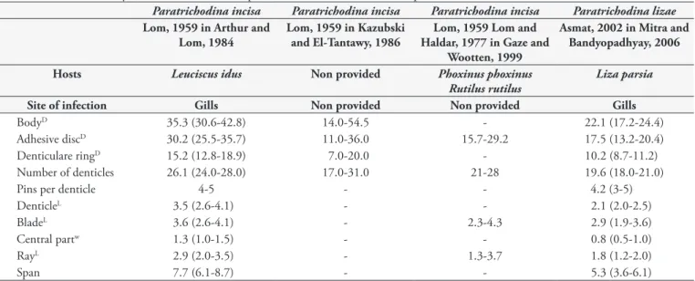
Paratrichodina Lom, 1963 comprises approximately 14 species, all characterized by small body dimensions and occurrence in the gills of the host (except for Paratrichodina phoxini, P. alburni, and P. degiustii - all endoparasites of the urinary tract). Paratrichodina incisa, the genus type, shows great similarity with P. africana in respect to some morphological features (Table 2). However, it differs from the latter by presenting a smaller and less rounded spine-like processus continuous with the central part. In addition, the distal surface of the blade of P. incisa is clearly straight, whereas it tends to be rounded in P. africana. Paratrichodina lizae also Figure 1. Photomicrographs of Paratrichodina africana from Oreochromis niloticus in the State of Amapá, North of Brazil. Silver impregnated specimens. A. Aboral view. B. Macronucleus horse-shoe shaped. Bars: 5 µm.
presents biometrical data similar to the present species (Table 2). However, as reported by Mitra and Bandyopadhyay (2006), the species contains one to eight non-impregnable round particles in the central part of the adhesive disc, which are absolutely absent in P. africana.
Lom (1963) reported “If there is an anterior projection near the base of the blade, it does not communicate with a notch in the blade of the preceding denticle”. Later, Gaze and Wootten (1999) illustrated the distinct interlocking mechanism of the denticles of P. incisa and better elucidated the positioning of the anterior projection, describing it as a well-defined structure continuous with the central part.
Gaze and Wootten (1999) suggested the absence of ray apophysis in P. incisa as an additional discriminating feature of the genus. Our observations support this idea since it was also not recorded in the specimens of P. africana herein observed.
Paratrichodina africana reported in this study fall within the ranges for P. africana presented in the original description by Kazubski and El-Tantawy (1986) with respect to measures of body diameter, number of denticles, denticulate ring, size of adoral spiral, and wide border membrane. The subsequent report provided by Mitra and Bandyopadhyay (2006) also presents similar measures of the denticulate ring, number of denticles, pins per denticle, and some dimensions of denticle components, but it differs in body diameter (Table 1). Additionally, all other Paratrichodina species biometrically differed from the present studied specimens, as presented in Table 2.
Conclusion
This is the fourth register of P. africana parasitizing a fish host and the first report of the genus in eastern Amazonia. By the fact that tilapia is one of the most popular and cultured freshwater fish, fish farmers must be careful with the water quality and feeding rate in ponds and especially in cages when the fish stocking density are higher than normal predisposing the fish for parasitism.
Acknowledgments
The authors thank to CAPES for Doctoral scholarship to Natalia Marchiori. Maurício Laterça Martins and Marcos Tavares-Dias were supported by a Research Fellowship from CNPq (Conselho Nacionalde Pesquisa e Desenvolvimento Tecnológico, Brazil).
References
Arthur JR, Lom J. Trichodinid Protozoa (Ciliophora: Peritrichida) from freshwater fishes of Rybinsk Reservoir, USSR. J Eukaryot Microbiol 1984; 31(1): 82-91. http://dx.doi.org/10.1111/j.1550-7408.1984. tb04294.x
Basson L, Van As JG. Trichodinidae and other ciliophorans (Phylum Ciliophora). In: Woo PTK. Fish Diseases and Disorders. 2nd ed. Cab International; 2006. vol. 1: Protozoan and Metazoan infections, p. 154-182.
Braccini GL, Vargas L, Ribeiro RP, Alexandre Filho L, Digmayer M. Ectoparasitos de tilápia do Nilo (Oreochromis niloticus) cultivados em tanques-rede nos rios do Corvo e Guairacá, Paraná, Brasil. Rev Bras Parasitol Vet 2008; 17(S1): 24-29. PMid:20059810.
Dias RJ, Fernandes NM, Sartini B, Silva-Neto ID, D’Agosto M. Occurrence of Trichodina heterodentata (Ciliophora: Trichodinidae) infesting tadpoles of Rhinella pombali (Anura: Bufonidae) in the Neotropical area. Parasitol Int 2009; 58(4): 471-474. PMid:19580885. http://dx.doi.org/10.1016/j.parint.2009.06.009
Evans JJ, Klesius PH, Pasnik DJ, Shoemaker CA. Influence of natural
Trichodina sp. parasitism on experimental Streptococcus iniae or
Streptococcus agalactiae infection and survival of young channel catfish
Ictalurus punctatus (Rafinesque). Aquac Res 2007; 38(6): 664-667. http:// dx.doi.org/10.1111/j.1365-2109.2007.01710.x
Gaze WH, Wootten R. An SEM study of adhesive disc skeletal structures isolated from trichodinids (Ciliophora: Peritrichida) of the genera Trichodina Ehrenberg, 1838 and Paratrichodina Lom, 1963.
Syst Parasitol 1999; 43(3): 167-174. PMid:10619066. http://dx.doi. org/10.1023/A:1006149318264
Table 2. Measurements (µm) of the most similar species of Paratrichodina with the present material from Northern Brazil.
Paratrichodina incisa Paratrichodina incisa Paratrichodina incisa Paratrichodina lizae
Lom, 1959 in Arthur and Lom, 1984
Lom, 1959 inKazubski and El-Tantawy, 1986
Lom, 1959 Lom and Haldar, 1977 in Gaze and
Wootten, 1999
Asmat, 2002 in Mitra and Bandyopadhyay, 2006
Hosts Leuciscus idus Non provided Phoxinus phoxinus Rutilus rutilus
Liza parsia
Site of infection Gills Non provided Non provided Gills
BodyD 35.3 (30.6-42.8) 14.0-54.5 - 22.1 (17.2-24.4)
Adhesive discD 30.2 (25.5-35.7) 11.0-36.0 15.7-29.2 17.5 (13.2-20.4)
Denticulare ringD 15.2 (12.8-18.9) 7.0-20.0 - 10.2 (8.7-11.2)
Number of denticles 26.1 (24.0-28.0) 17.0-31.0 21-28 19.6 (18.0-21.0)
Pins per denticle 4-5 - - 4.2 (3-5)
DenticleL 3.5 (2.6-4.1) - - 2.1 (2.0-2.5)
BladeL 3.6 (2.6-4.1) - 2.3-4.3 2.9 (1.9-3.6)
Central partw 1.3 (1.0-1.5) - - 0.8 (0.5-1.0)
RayL 2.9 (2.0-3.5) - 1.3-3.7 1.8 (1.2-2.0)
Span 7.7 (6.1-8.7) - - 5.3 (3.6-6.1)
Ghiraldelli L, Martins ML, Jerônimo GT, Yamashita MM, Adamante WB. Ectoparasites communities from Oreochromis niloticus raised in the State of Santa Catarina, Brazil. J Fish Aquat Sci 2006a; 1: 181-190. http://dx.doi.org/10.3923/jfas.2006.181.190
Ghiraldelli L, Martins ML, Adamante WB, Yamashita MM. First record of Trichodina compacta Van As and Basson, 1989 (Protozoa: Ciliophora) from cultured Nile tilapia in the state of Santa Catarina, Brazil. Int J Zool Res 2006b; 2(4): 369-375. http://dx.doi.org/10.3923/ijzr.2006.369.375 Jerônimo GT, Marchiori NC, Pádua S B, Dias Neto J, Pilarski F, Ishikawa MM, et al. Trichodina colisae (Ciliophora: Trichodinidae): new parasite records for two freshwater fish species farmed in Brazil. Rev Bras Parasitol Vet 2012: 21(4): 366-371. PMid:23207983. http://dx.doi.org/10.1590/ S1984-29612012005000008
Kattar MR. Sobre Trichodina steini Claparède & Lachmann (Protozoa: Urceolariidae) encontrada em girino de Bufo ictericus do Brasil. Braz J Biol 1975; 35(2): 253-258.
Kazubski SL, El-Tantawy AM. The Ciliate Paratrichodina africana sp. n. (Peritricha, Trichodinidae) from Tilapia fish (Cichlidae) from Africa.
Acta Protozool 1986; 25: 433-438.
Lom J. A contribution to the systematics and morphology of endoparasitic trichodinids from amphibians, with a proposal of uniform specific characteristics. J Eukaryot Microbiol 1958; 5(4): 251-263. http:// dx.doi.org/10.1111/j.1550-7408.1958.tb02563.x
Lom J. The ciliates of the family Urceolariidae inhabiting gills of fishes (Trichodinella group). Acta Soc. Zool. Bohemoslov 1963; 27: 7-19.
Madsen HCK, Buchmann K, Mellergaard S. Association between trichodiniasis in eel (Anguilla anguilla) and water quality in recirculation systems. Aquacult 2000; 187(3-4): 275-281. http://dx.doi.org/10.1016/ S0044-8486(00)00323-9
Martins ML, Ghiraldelli L. Trichodina magna Van As and Basson, 1989 (Ciliophora: Peritrichia) from cultured Nile tilapia in the state of Santa Catarina, Brazil. Braz J Biol 2008; 68(1): 169-72. PMid:18470393. http://dx.doi.org/10.1590/S1519-69842008000100024
Martins ML, Marchiori N, Nunes G, Rodrigues MP. First record of Trichodina heterodentata (Ciliophora: Trichodinidae) from channel catfish, Ictalurus punctatus cultivated in Brazil. Braz J Biol 2010; 70(3): 637-644. PMid:20730352. http://dx.doi.org/10.1590/ S1519-69842010000300022
Martins ML, Shoemaker CA, Xu D, Klesius PH. Effect of parasitism on vaccine efficacy against Streptococcus iniae in Nile tilapia.
Aquacult 2011: 314(3-4): 18-23. http://dx.doi.org/10.1016/j. aquaculture.2011.01.022
Mitra AK, Bandyopadhyay K. Trichodina haldari n. sp. and Paratrichodina bassonae n. sp. (Ciliophora: Peritrichida) from Indian fresh water fishes.
Acta Protozool 2006; 45(3): 289-294.
Moraes FR, Martins ML. Condições predisponentes e principais enfermidades de teleósteos em piscicultura intensiva. In: Cyrino JEP, Urbinati EC, Fracalossi DM, Castagnolli N, editores. Tópicos especiais em piscicultura de água doce tropical intensiva. São Paulo: TecArt; 2004. p. 343-383.
Pádua SB, Martins ML, Carraschi SP, Cruz C, Ishikawa MM. Trichodina heterodentata (Ciliophora: Trichodinidae): a new parasite for Piaractus mesopotamicus (Pisces: Characidae). Zootaxa 2012; 3422: 62-68. Pantoja WMF, Neves LR, Dias MKR, Marinho RGB, Montagner D, Tavares-Dias M. Protozoan and metazoan parasites of nile tilapia Oreochromis niloticus cultured in Brazil. Rev. MVZ Córdoba 2012; 17(1): 2659-266.
Piazza RS, Martins ML, Guiraldelli L, Yamashita MM. Parasitic diseases of freshwater ornamental fishes commercialized in Florianópolis, Santa Catarina, Brazil. BolInst Pesca 2006; 32(1): 51-57.
Pinto HA, Wieloch AH, Melo AL. Uma nova espécie de Trichodina
Ehrenberg, 1838 (Ciliophora: Trichodinidae ) em Biomphalaria schrammi
(Crosse, 1864) (Mollusca: Planorbidae ). Lundiana 2006; 7(2): 121-124.
Pinto E, Garcia AM, Figueiredo HCP, Rodrigues MP, Martins ML. Primeiro relato de Tripartiella sp. (Ciliophora: Peritrichia) em
Pseudoplatystoma corruscans (Osteichthyes: Pimelodidae) cultivado no Estado de Mato Grosso do Sul, Brasil, com descrição de nova espécie.
Bol Inst Pesca 2009; 35(1): 91-97.
Silva WM, Roche KF, Vicente FS, Delben AST. First record of the peritrich