BRAZILIAN KEFIR: STRUCTURE, MICROBIAL COMMUNITIES AND CHEMICAL COMPOSITION
Karina Teixeira Magalhães1, Gilberto Vinícius de Melo Pereira1, Cássia Roberta Campos1, Giuliano Dragone2, Rosane Freitas Schwan1*
1
Departamento de Biologia, Universidade Federal de Lavras, Lavras, MG, Brasil; 2Institute for Biotechnology and Bioengineering,
Centre of Biological Engineering, University of Minho, Campus de Gualtar, 4710-057, Braga, Portugal.
Submitted: October 30, 2009; Approved: January 13, 2011.
ABSTRACT
Microbial ecology and chemical composition of Brazilian kefir beverage was performed. The
microorganisms associated with Brazilian kefir were investigated using a combination of phenotypic and
genotypic methods. A total of 359 microbial isolates were identified. Lactic acid bacteria (60.5%) were the
major isolated group identified, followed by yeasts (30.6%) and acetic acid bacteria (8.9%). Lactobacillus
paracasei (89 isolates), Lactobacillus parabuchneri (41 isolates), Lactobacillus casei (32 isolates),
Lactobacillus kefiri (31 isolates), Lactococcus lactis (24 isolates), Acetobacter lovaniensis (32 isolates),
Kluyveromyces lactis (31 isolates), Kazachstania aerobia (23 isolates), Saccharomyces cerevisiae (41
isolates) and Lachancea meyersii (15 isolates) were the microbial species isolated. Scanning electron
microscopy showed that the microbiota was dominated by bacilli (short and curved long) cells growing in
close association with lemon-shaped yeasts cells. During the 24 h of fermentation, the protein content
increased, while lactose and fat content decreased. The concentration of lactic acid ranged from 1.4 to 17.4
mg/ml, and that of acetic acid increased from 2.1 to 2.73 mg/ml. The production of ethanol was limited,
reaching a final mean value of 0.5 mg/ml.
Key words:Lactobacillus; Saccharomyces; fermented beverage; Brazilian kefir grains
INTRODUCTION
Kefir is a known culture employed to produce the
traditional Russian beverage “kefir” from milk, which has low
alcohol content (8, 13). It is a mixed culture of various yeast
species of the genera Kluyveromyces, Candida, Saccharomyces
and various lactic acid bacteria of the genus Lactobacillus, are
contained in a matrix of proteins and polysaccharide ‘kefiran’,
which are formed during cell growth under aerobic conditions
(6, 19, 24). Kefir grains are small irregularly shaped,
yellowish-white, hard granules which resemble miniature
cauliflower blossoms (10).
The beverage is a self-carbonated that owes its distinctive
flavor to a mixture of lactic acid, ethanol, carbon dioxide and
other flavor products, such as acetaldehyde. The unique flavor
is the result of the symbiotic metabolic activity of a number of
lactic bacteria and yeast species (9, 13). The typical yoghurt
flavor kefir is caused by lactic bacteria that produce lactic acid,
*Corresponding Author. Mailing address: Departamento de Biologia, Universidade Federal de Lavras, 37.200-000 Lavras-MG, Brazil.; Te.: +55 35 38291614
which imparts an acidic and refreshing taste, and the mixture of various carbonyl compounds like acetone, diacetyl, and
acetaldehyde of which the latter is considered the major flavour
component (5, 20).
Kefir grains are added to different types of milk. It can be
made from any type of milk; cow, goat or sheep, coconut, rice
and soy but commonly cow milk is used. The grains cause its
fermentation that results in the production of numerous
components in the kefir, including lactic acid, acetic acid, CO2,
alcohol (ethyl 2 alcohol) and aromatic compounds. That
provides kefir's unique sensory characteristics: fizzy, acid taste,
tart and refreshing flavor (9, 22). The beverage contains
vitamins, minerals and essential amino acids that help the body
with healing and maintenance functions and also contains
easily digestible complete proteins (9, 13, 22). The benefits of
consuming kefir in the diet are numerous (19): antitumor
activity (28), antimicrobial activity (25), antiinflammatory, and
antiallergic activity (15).
In Brazil, kefir grains are used in private household for
fermentation of milk and while microbial and chemical
information is available concerning Irish (23), Turkish (10, 31),
and Spanish kefir grains (13), there are no reports concerning
the microbial or chemical characterization of Brazilian kefir.
Therefore, the aim of this study was to assess the microbial
diversity of Brazilian kefir beverage. For this purpose, a
combined approach of phenotypic and genotypic identification
using sequencing of portions of the 16S rRNA gene and Iternal
Tanscribed Spacer region (ITS) was performed. Also,
physicochemical and Scanning Electron Microscopy (SEM)
characterizations were used for the beverage and kefir grains
respectively. The microbial and chemical composition of
Brazilian kefir beverage is one of the prerequisites for the
successful future implementation of industrial-scale
production.
MATERIALS AND METHODS
Milk Brazilian kefir production
Brazilian kefir grains were obtained from a private
household in the city of Lavras, which is located in the
southern State of Minas Gerais, Brazil. The grains (250 g) were
washed with distilled water and inoculated in 2.250 ml of
substrate (Pasteurized whole milk, Ipê - Cooperativa Agrícola
Alto Rio Grande Ltda. Lavras, Minas Gerais, Brazil) and were
statically incubated for 24 h at 25°C. Samples of the beverage
were aseptically taken every 6 h. Four fermentations were
performed in the same conditions described above.
Enumeration of mesophilic bacteria, acetic acid bacteria, lactic acid bacteria and yeasts
Bacteria and yeasts were enumerated by the surface spread
technique, plating in triplicate 100 l of each diluted sample.
Enumeration of microorganisms was carried out using 7
different culture media. Lactic acid bacteria’s (LAB) were
enumerated on Nutrient Agar medium (Oxoid, S/P, Brazil), De
Man, Rogosa and Sharpe Agar (MRS) (Oxoid, S/P, Brazil),
M17 agar (Oxoid, S/P, Brazil), Edwards modified medium
(Oxoid, S/P, Brazil) and LUSM medium (1.0% glucose, 1.0%
Bacto Peptone (Difco, S/P, Brazil), 0.5% yeast extract (Difco,
S/P, Brazil), 0.5% meat extract (Difco, S/P, Brazil), 0.25%
gelatin (Difco, S/P, Brazil), 0.5% calcium lactate, 0.05% sorbic
acid, 75 ppm of sodium azide (Sigma, St. Louis, USA), 0.25%
sodium acetate, 0.1% (vol/vol) Tween 80, 15% tomato juice,
30 micrograms of vancomycin (Sigma, St. Louis, USA) per ml,
0.20 microgram of tetracycline (Sigma, St. Louis, USA) per
ml, 0.5 mg of cysteine hydrochloride per ml, and 1.5% agar
(Difco, S/P, Brazil). Acetic acid bacteria’s (AAB) were
enumerated on 135 medium (DSMZ, Deutsche Sammlung von
Mikroorganismen und Zellkulturen GmbH, Germany). All
media for bacterial enumeration were supplemented with 0.4
mg/ml nystatin (Sigma, St. Louis, USA). Yeasts were
enumerated on YEPG agar containing 100 mg chloramphenicol
(Sigma, St. Louis, USA) and 50 mg chlortetracycline (Sigma,
St. Louis, USA) to inhibit bacterial growth. After spreading,
plates were incubated at 28°C for 48 h for bacteria, and 5 days
for yeasts; and colony forming units (log10 c.f.u./ml) were
colonies, the square root of the number of colonies was taken at
random for identification (12).
Phenotypic identification of microorganisms
Bacterial isolates were Gram-stained. Gram-negative
bacteria were identified using Bac-Tray Kits I, II and III
(Difco, S/P, Brazil) according to the manufacturer instructions.
Gram-positive bacteria were subdivided into sporeformers and
non-spore-formers by heating at 80°C for 10 min to kill the
vegetative cells. Subsequent identification was performed
using biochemical and motility tests as recommended in
Bergey’s Manual of Determinative Bacteriology (12) and The
Prokaryotes (11), and results were confirmed by using the API
50 CHB galleries (Bio-Merieux, S/P, Brazil). Isolates were
examined for colony and cell appearance, catalase activity,
Gram staining, motility and production of CO2 from glucose in
MRS broth with a Durham tube. Biochemical characterizations
of the strains were performed with API ID 32 for Lactococcus
and Enterococcus and API 50 CHL (BioMerieux, S/P, Brazil)
for Lactobacillus and Leuconostoc. All Lactobacillus were
recognized as catalase-negative, oxidase-negative, regular
fermentative rods. They were classified into obligately
homofermentative, facultatively heterofermentative and
obligately heterofermentative by their ability to produce CO2
from glucose and gluconate. Phenotypic characteristics of all
yeast isolates was determining by their morphology, spore
formation, assimilation and fermentation of different carbon
sources (3) and yeast identities were verified using the keys of
Yeast (3, 14).
Molecular identification of microorganisms
Representatives of each species of microorganisms
identified by traditional methods were selected ( n = |n| for
numbers of microorganisms identified of each species) for
sequencing. DNA from pure cultures of the bacteria and yeasts
isolated was extracted according to the method described by
Wang et al. (29) and Makimura et al. (17), respectively.
Sequencing of portions of the 16S rRNA gene and Iternal
Tanscribed Spacer region (ITS) was used for identification of
representative bacteria and yeast isolates to species level. The
primers 27f (5’–AGAGTTTGATCCTGGCTCAG–3’) and
1512r (5’– CGGCTACCTTGTTACGACT – 3’) were used to
amplify 16S rRNA gene while the primers ITS1
(5’-TCCGTAGGTGAACCTGCGG-3’) and ITS4 (5’-TCCTCCGC
TTATTGATATGC-3’) were used to amplify ITS region. PCR
was performed according to Wang et al. (29) (bacteria) and
Naumova et al. (21) (yeast). The amplicons were analysed by
electrophoresis on agarose gels at 60-65V. The sequencing of
portions of the 16S rRNA gene and ITS region was
accomplished by the (Applied Biosystems Company, Foster
City, CA, USA). GenBank searches (http://www.ncbi.nlm.nih.
gov/BLAST/) were performed to determine the closest known
relatives of the partial ribosomal DNA sequences obtained.
Analysis by Scanning Electron Microscopy (SEM)
Brazilian kefir grains were sliced for Scanning Electron
Microscopy (SEM) (18). Samples were collected from the
outer and inner parts of the grains. The grains were fixed
(Karnovisk's fixative solution) at pH 7.2 for 24 h. The samples
were then transferred to 30% glycerol for 30 min and immersed
in liquid nitrogen for subsequent fracture in the metal surface.
Then, grains were post-fixed in 10 g/l osmium tetroxide in
phosphate buffer for 1 h at 25°C and dehydrated in acetone: 15,
30, 50 and 70%, three times. After dehydrating, samples were
critical-point dried and coated with gold using a Bal-tec SDC
050(Capovani Brothers Inc. Scotia, NY, USA). The
preparations were observed using a scanning electron
microscope (LEO EVO 040) (A Carl Zeiss SMT AG
Company, Germany).
Chemical analysis
At 0 and 24 h, the kefir beverage samples were
characterized in relation to total titratable acidity and pH,
matter according to the AOAC methodology (1). Calcium was
determined by atomic absorption spectrophotometry using a
Varian (Model SpectrAA 100/200) spectrophotometer
equipped with an air-acetylene flame. Ethanol, organic acids
(acetic acid and lactic acid) and lactose were obtained from
sample extracts every 6 h and analysed according Schwan et al.
(26) and Duarte et al. (7). All samples were examined in
triplicate.
RESULTS AND DISCUSSION
Microbial enumeration
Descriptions of yeast and bacteria present in different
batches of milk kefir grains have been reported by different
authors (27, 30, 31). However, these studies were restricted to
the grains, and none of them analysed the beverage. Using
conventional culture techniques, we have monitored the
development of bacterial and fungal communities during
24h fermentation of Brazilian kefir beverage. Previous results
showed that two groups of microorganisms co-exist in milk
kefir grains: lactic acid bacteria and yeast (10, 23). In order to
establish the different species of bacteria and yeast present
during fermentation, a representative number of isolates from
each culture medium were identified (Table 1). Lactic acid
bacteria was the most frequently found microorganism group,
showing an initial population of around 3.51 log10 c.f.u./ml that
reached 12.41 log10 c.f.u./ml. Acidification of the substratum
was mainly stimulated by the presence of lactic acid bacteria
(9). Acetobacter showed also growth, ranging from 5.92 log10
c.f.u./ml to 7.72 log10 c.f.u./ml. In general, lactic acid bacteria
were more numerous than yeast and acetic acid bacteria in milk
kefir grains, although fermentation conditions can affect this
pattern (10).
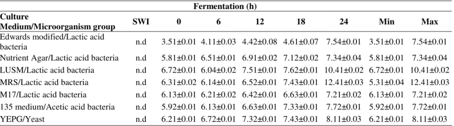
Table 1. Microbial enumeration (log10 c.f.u./ml) during fermentation of Brazilian kefir grains.
Fermentation (h) Culture
Medium/Microorganism group SWI 0 6 12 18 24 Min Max
Edwards modified/Lactic acid
bacteria n.d 3.51±0.01 4.11±0.03 4.42±0.08 4.61±0.07 7.54±0.01 3.51±0.01 7.54±0.01
Nutrient Agar/Lactic acid bacteria n.d 5.81±0.01 6.51±0.01 6.91±0.02 7.12±0.02 7.34±0.04 5.81±0.01 7.34±0.04 LUSM/Lactic acid bacteria n.d 6.72±0.01 6.04±0.02 7.51±0.01 7.62±0.01 10.41±0.02 6.72±0.01 10.41±0.02 MRS/Lactic acid bacteria n.d 6.31±0.02 6.14±0.01 6.52±0.01 7.43±0.01 12.41±0.03 5.31±0.04 12.41±0.03 M17/Lactic acid bacteria n.d 6.13±0.01 6.21±0.02 6.42±0.01 6.63±0.01 7.21±0.02 6.13±0.01 7.21±0.02 135 medium/Acetic acid bacteria n.d 5.92±0.01 6.13±0.01 6.63±0.01 7.33±0.01 7.72±0.01 5.92±0.01 7.72±0.01
YEPG/Yeast n.d 6.21±0.01 6.72±0.01 7.32±0.01 7.43±0.01 8.11±0.03 6.21±0.01 8.11±0.03
SWI = Substrate without inoculum, n.d. = not detected. Data are average values of triplicate ± standard deviation.
Identification of microbial isolates
Yeasts and bacteria were identified by phenotypic
methods (Table 2). Representatives of each species of
microorganisms identified by phenotypic methods were
selected for sequencing of the ITS region and 16S rRNA gene.
Table 2 shows the accession number and the percentage of
similarity between the sequences of the isolates from kefir
beverage and the reference sequences from GenBank. Isolates
showed 97% or higher similarities with respect to the
sequences available in the NCBI database.
A total of 359 isolates were obtained from Brazilian kefir
beverage (Table 2). Among the isolates, 249 were bacteria and
110 were yeast. During the fermentative process, the
predominant microorganisms identified were lactic acid
bacteria (60.5%), followed by yeast (30.6%) and acetic acid
Lactobacillus paracasei represented the largest and most
commonly identified LAB isolates, with 89 of a total of 249
isolates, followed by Lactobacillus parabuchneri (41 isolates),
Lactobacillus casei (32 isolates), Lactobacillus kefiri (31
isolates) and Lactococcus lactis (24 isolates)
Our data indicated that the Brazilian kefir beverage
contained a diverse spectrum LAB group including
Lactobacillus and Lactococcus. Lactobacillus kefiri is another
important bacterium found during kefir fermentation. There are
reports on the presence of Lactobacillus kefiri as a prevailing
member of the lactic acid microbiota in milk Kefir (8, 10, 16).
In our study, Lactobacillus kefiri fixed on the grain surface
might be easily freed from kefir grains into the substrate of
milk, thus resulting in the increased cell counts. Lactobacillus
kefiri is also important for production of kefiran polymer
present in the kefir grains structure (6). Kefiran has frequently
been claimed to be effective against a variety of complaints and
diseases. Several studies have investigated the antitumor
activity (6), antibacterial and antifungal activities (25).
Lactococcus lactis (24 isolates) was also identified in Brazilian
kefir beverage. Previous studies showed that a variety of
different species of Lactobacillus and Lactococcus have been
isolated and identified in milk kefir grains from around the
world (10, 16). The acetic acid species, Acetobacter
lovaniensis, was also identified (32 isolates). Acetobacter
lovaniensis species belongs to the Acetobacter pasteurianus
group. The species Acetobacter pasteurianus consists of five
subspecies, and Acetobacter pasteurianus subsp. lovaniensis
has been also described in fermented food from Indonesia and
the Philippines (16) and sugary Brazilian kefir (16).
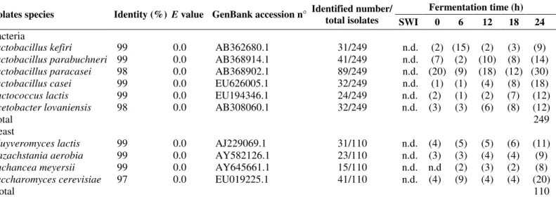
Table 2. Identification of representative bacterial and yeast isolates by sequencing of portions of the 16S rRNA and ITS, respectively.
Fermentation time (h) Isolates species Identity (%) E value GenBank accession n° Identified number/
total isolates SWI 0 6 12 18 24 Bacteria
Lactobacillus kefiri 99 0.0 AB362680.1 31/249 n.d. (2) (15) (2) (3) (9)
Lactobacillus parabuchneri 99 0.0 AB368914.1 41/249 n.d. (7) (2) (10) (8) (14)
Lactobacillus paracasei 98 0.0 AB368902.1 89/249 n.d. (20) (9) (18) (12) (30)
Lactobacillus casei 99 0.0 EU626005.1 32/249 n.d. (1) (1) (4) (8) (18)
Lactococcus lactis 99 0.0 EU194346.1 24/249 n.d. (2) (1) (2) (7) (12)
Acetobacter lovaniensis 98 0.0 AB308060.1 32/249 n.d. (3) (3) (6) (8) (12)
Total 249
Yeast
Kluyveromyces lactis 99 0.0 AJ229069.1 31/110 n.d. (4) (5) (5) (6) (11)
Kazachstania aerobia 99 0.0 AY582126.1 23/110 n.d. (3) (3) (4) (4) (9)
Lachancea meyersii 99 0.0 AY645661.1 15/110 n.d. n.d (2) (3) (2) (8)
Saccharomyces cerevisiae 97 0.0 EU019225.1 41/110 n.d. (4) (9) (4) (4) (20)
Total 110
SWI = Substrate without inoculum; n.d. = not detected; ( ) = numbers of microrganism identified.
The lactose-fermenting yeast, Kluyveromyces lactis, was
found in the Brazilian kefir beverage together with
non-lactose-fermenting yeast: Saccharomyces cerevisiae, Lachancea
meyersii and Kazachstania aerobia (Table 2). The yeast
isolates of Brazilian kefir was dominated by lactose-negative
strains. Among them, Saccharomycescerevisiae predominated
(41isolates), followed by Kazachstania aerobia (23 isolates)
and Lachancea meyersii (15 isolates). Saccharomyces
cerevisiae represented the largest and most commonly
identified yeast isolates. This species, which exhibits strong
fermentative metabolism and tolerance to ethanol, is known to
alcohol fermentation, regarding spontaneous fermented sugar
cane (26). The presence of Saccharomyces cerevisiae
contributes to the enhancement of the sensory quality of the
kefir beverage, promoting a strong and typically yeasty aroma,
as well as its refreshing, pungent taste (16). This yeast also
reduces the concentration of lactic acid, removes the hydrogen
peroxide and produces compounds that stimulate the growth of
other bacteria, thus increasing the production of kefiran
exopolysaccharides (6). It is worth noting that the yeast
species, Kazachstania aerobia and Lachancea meyersii whose
presence in milk kefir has not been reported previously, was
detected in this study. Its presence in Brazilian kefir beverage
could be connected with the assimilation of some acids
produced by lactic acid bacteria.
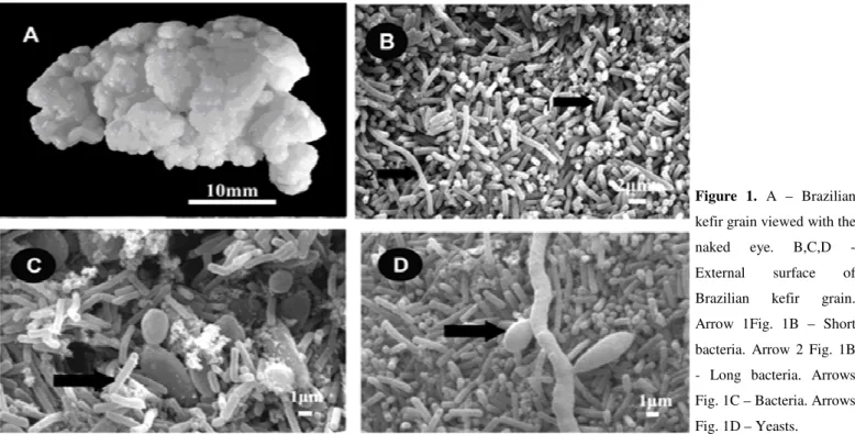
Scanning electron microscopy of milk Brazilian kefir grains As described previously by some authors (10, 23), a
complex and tightly packed biofilm could be observed around
the grains, while the interior was comprised mainly of
unstructured material. Figure 1 and 2 show the association of
the kefir microbiota through scanning electron microscopy
(SEM). The Brazilian kefir grains showed a smooth surface
(Figure 1A) and its outer portion was covered by an
agglomerate of microorganisms (Figure 1B,C and D). The
microbiota in the outer portion of the grain was dominated by
bacilli (Short and curved long) cells growing in association
with lemon-shaped yeast cells (Figure 1B,C and D). The
microbial cells on the inner portion were less than that on the
outer portion (Figure 2). Fibrillar material (probably the
polysaccharide kefiran) was observed on the outer portion, as
well as in the inner portion of the grains (Figure 1C and 2B).
Similar results for Turkish kefir grains (10) and sugary kefir
grains (16) were found.
Based on the results from plating, Lactococcus lactis
species were identified as member of the lactic acid bacteria
population (Table 2). However cocci bacteria could not be seen
on the SEM graph in our study, thus suggesting the weak
adherence of these two species on Brazilian kefir grains. This
weak adherence may have resulted in the bacteria falling into
the milk substrates, a possibility that is in agreement with the
results reported by Guzel-Seydim et al. (10) for Turkey kefir
grains and Magalhães et. al. (16) for sugary Brazilian kefir.
Figure 1. A – Brazilian
kefir grain viewed with the
naked eye. B,C,D -
External surface of
Brazilian kefir grain.
Arrow 1Fig. 1B – Short
bacteria. Arrow 2 Fig. 1B
- Long bacteria. Arrows
Fig. 1C – Bacteria. Arrows
Chemical characterization of fermented kefir beverage Table 3 shows the pH and total titratable acidity (TTA)
values over a 24 h fermentation period. The mean pH and TTA
values of the pasteurized whole milk prior to inoculation with
kefirr grains were 6.61 and 26°D, respectively. The pH
decreased to 4.42 and the TTA increased to 93°D during the
24h fermentation of milk by Brazilian kefir grains. The main
reason for pH decrease and TTA increase is the production of
certain organic acids, ethanol, CO2, and other volatile
compounds by the microbial population in grains and kefir
beverage (2, 9). Calcium content was not modified during the
fermentation and Vitamin C was not found (Table 3). The
content of fat decreased, due to the lipases production by the
microorganisms present in kefir (13). The protein content
increased, due to the increase of the microbial biomass;
however the dry matter contents of kefir grains remained
constant in the final fermentation process (Table 3).
Table 3. Values of total titratable acidity, pH, proteins, fat,
vitamin C, Calcium in Brazilian kefir beverage and dry matter
in Brazilian kefir grains.
Chemical characteristics Fermentation time (h)
0 24
Beverage
Total titratable acidity (TTA) (°D) 26±1 93±1
pH 6.61±0.02 4.42±0.01
Proteins (%) 2.12±0.02 3.91±0.02
Fat (%) 3.63±0.03 2.34±0.01
Vitamin C (%) n.d. n.d.
Calcium (Ca) (%) 0.21±0.01 0.22±0.01
Grains
Dry matter (%) 9.84±0.02 9.62±0.03
n.d. = not detected
Data are average values of triplicate ± standard deviation.
High performance liquid chromatography (HPLC) was
used to analyze organic acids, ethanol e sugars in the kefir
beverages produced. Figure 3 shows the concentration of Figure 2. Internal surface of Brazilian
kefir grain. Arrows
Fig. 3A and 3C –
Bacteria. Arrow 1 Fig.
3B – Polysaccharide.
Arrow 2 Fig. 3B –
Yeasts. Arrow Fig. 3D
sugars, organic acids and ethanol obtained through milk
fermentation. Lactose, lactic acid, acetic acid and ethanol were
quantified by HPLC. Organic acids may occur in dairy
products as a result of hydrolysis of butterfat (fatty acids),
biochemical metabolic processes, or bacterial metabolism (9).
The lactose consumption was observed after 24h of milk
fermentation (Figure 3). Lactose is readily degraded to
galactose and glucose by some strains of Streptococcus and
Kluyveromyces. Similar results were reported by Irigoyen et al.
(13) during milk fermentation with 5% inoculum of kefir
grains. According to Irigoyen et al. (13), the percentage of
kefir grains inoculated in the substrate, as well as the
cultivation method employed affects the lactose consumption.
In the present work, the lactic acid content increased
during the 24 h of the fermentation process in Brazilian kefir
beverage, reaching maximum value of 17.4 mg/ml. This result
is of great importance, since lactic acid acid provides pleasant
taste and inhibits the development of undesirable or pathogenic
microorganisms, due to the substrate acidity increase (16).
Acetic acid was also formed during the fermentation process of
Brazilian kefir beverage (Figure 3), probably by heterolactic
bacteria (16) which were identified in the present work (Table
2). Similar to lactic acid, accumulation of acetic acid turns the
beverage more acidic and inhibitory for pathogens
microorganisms (16).
Ethanol concentration increased during the kefir beverage
fermentation process (Figure 3); reaching maximum
concentration of 0.5mg/ml. Saccharomyces cerevisiae
identified in kefir beverages in the present work is primarily
responsible for the alcohol production. However, some
Lactobacillus strains also have the ability to produce ethanol,
since they possess alcohol-dehydrogenase activity, an enzyme
able to convert acetaldehyde to ethanol (4). The content of
alcohol should be enough to give kefir the flavour of a light
alcoholic beverage that is typical of traditional (ancient) kefir
of the Caucasus and the yeast aroma ensures the specificity of
this type of fermented beverage (4).
0
0,5
1
1,5
2
2,5
3
0
6
12
18
24
Time (h)
E
th
a
n
o
l
a
n
d
a
c
e
ti
c
a
c
id
(m
g
/m
l)
0
5
10
15
20
25
30
35
40
45
50
55
L
a
c
to
s
e
a
n
d
l
a
c
ti
c
a
c
id
(m
g
/m
l)
CONCLUSIONS
Three distinct microbial populations were identified
during the fermentation of Brazilian kefir beverage: lactic acid
bacteria were the predominant, followed by the yeasts and
Gram-negative bacteria from the Acetobacter genus.
Lactobacillus paracasei was the most abundant bacterium,
while Saccharomyces cerevisiae was the predominant yeast
strain. The distinct microorganism groups identified in this
beverage performed three different kinds of fermentations,
including lactic, alcoholic and acetic fermentations. The
increase in lactic bacteria population caused an increase in the
lactic acid concentration in the beverage, whereas the increase
in yeast population favored the ethanol formation. To the best
of our knowledge, this is the first study evaluating chemical
and microbiological composition of Brazilian kefir beverage.
Interestingly, two microbial species that had not been described
as belonging to the microbiota of milk kefir were found in
Brazilian kefir: Lachancea meyersii and Kazachstania aerobia.
The present study contributes for a better knowledge of the
microbiological and chemical constitution of the kefir beverage
consumed in Brazil, which have not been elucidated up till
now. Future work involving nutritional and therapeutic benefits
will improve the characterization of Brazilian kefir.
ACKNOWLEDGEMENTS
The authors acknowledge Coordenação de
Aperfeiçoamento de Pessoal de Nível Superior (CAPES) for
scholarshipsand and CAARG - Cooperativa Agrícola Alto Rio
Grande Ltda. (Lavras/MG, Brazil) for the milk supply.
REFERENCES
1. Association of Official Analytical Chemist. (2000). Official Methods of Analysis of the Association of Official Analytical Chemist. 17thed.
Washington.
2. Athanasiadis, I.; Paraskevopoulou, A.; Blekas, G.; Kiosseoglou, V. (2004). Development of a novel whey beverage by fermentation with
kefir granules. Effect of various treatments. Biotech. Progr. 20 (4), 1091-1095.
3. Barnett, J.A.; Payne, R.W.; & Yarrow, D. (2000). Yeast. Characteristic and Identification. 3rd ed. Cambridge, UK: Cambridge Univ. Press. p.
1150.
4. Beshkova, D.M.; Simova, E.D.; Frengova, G.I.; Simov, Z.I.; Dimitrov, Z.H.P. (2003). Production of volatile aroma compounds by kefir stater cultures. Int. Dairy J., 13 (7), 529-535.
5. Chaves, A.C.S.D.; Ruas-Madiedo, P.; Starrenburg, M.; Hugenholtz, J.; Lerayer, A.L.S. (2003). Impact of engineered Streptococcus thermophilus trains overexpressing glyA gene on folic acid and acetaldehyde production in fermented Milk. Braz. J. Microbiol. 34, 17-20.
6. Cheirsilp, B.; Shoji, H.; Shimizu, H.; Shioya, S. (2003). Interactions between Lactobacillus kefiranofaciens and Saccharomyces cerevisiae in mixed culture for kefiran production. J. Biosci. Bioeng., 96 (3), 279-284. 7. Duarte, W.F.; Dias, D.R.; Pereira, G.V.M.; Gervasio, I.M.; Schwan, R.F. (2008) Indigenous and inoculated yeast fermentation of gabiroba (Campomanesia pubescens) pulp for fruit wine production. J. Ind. Microbiol. Biotechnol. 36, 557–569.
8. Garrote, G.L.; Abraham, A.G.; de Antoni, G.L. (1997). Preservation of kefir grains, a comparative study. Lebensm.-Wiss. U.-Technol., 30 (1), 77-84.
9. Güzel-Seydim, Z.B.; Seydim, A.C.; Greene, A.K.; & Bodine, A.B. (2000). Determination of organic acids and volatile flavor substances in kefir during fermentation. J. Food Compos. Anal., 13 (1), 35-43. 10. Güzel-Seydim, Z.; Wyffels, J.T.; Seydim, A.C.; & Greene, A.K. (2005).
Turkish kefir and kefir grains: microbial enumeration and electron microscobic observation. Int. J. Dairy Technol., 58 (1), 25-29.
11. Hammes, W.P.; Hertel, C. (2003). The genera Lactobacillus and
Carnobacterium. In: Dworkin, M. (Eds.). The prokaryotes. New York: Springer-Verlang. New York, USA, p. 1535-1594.
12. Holt, J.G.; Krieg, N.R.; Sneath, P.H.A.; Staley, J.T.; Williams, S.T. (1994). Bergey’s manual of determinative bacteriology. 9th ed.
Baltimore: Williams & Wilkins, Baltimore, USA.
13. Irigoyen, A.; Arana, I.; Castiella, M.; Torre, P.; Ibáñez, F.C. (2005). Microbiological, physicochemical, and sensory characteristics of kefir during storage. Food Chem., 90 (4), 613-620.
14. Kurtzman, C.P.; Boekhout, T.; Robert, V.; Fell, W.J.; Deak, T. (2003). Methods to identify yeasts. In: V. Robert (Ed.). Yeasts in food: beneficial and detrimental aspects. Hamburg, Germany: B. Beh’s Verlag GmbH., p. 69-117.
15. Lee, M.Y.; Ahn, K.S.; Kwon, O.K.; Kim, M.J.; Kim, M.K.; Lee, I.Y.; Oh, S.R.; Lee, H.K. (2007). Anti-inflammatory and anti-allergic effects of kefir in a mouse asthma model. Immunobiol., 212 (8), 647-654. 16. Magalhães, K.T.; Pereira, G.V.M.; Dias, D.R.; Schwan, R.F.; (2010).
sugary Brazilian kefir. World J. Microbiol. Biotechnol., 33, 1-10. 17. Makimura, K.; Tamura, Y.; Mochizuki, T.; Hasegawa, A.; Tajiri, Y.;
Hanazawa, R.; Uchida, K.; Saito, H.; Yamaguchi, H. (1999). Phylogenetic classification and species identification of dermatophyte strains based on DNA sequences of nuclear ribosomal internal transcribed spacer 1 regions. J.Clin. Microbiol., 37 (4), 920-924. 18. Marques, S.C.; Rezende, J.G.O.S.; Alves, L.A.F.; Silva, B.C.; Alves, E.;
Abreu, L.R.; Piccoli, R.H. (2007). Formation of biofilms by
Staphylococcus aureus on stainless steel and glass surfaces and its resistance to some selected chemical sanitizers. Braz. J. Microbiol.,.38 (3), 538-543 .
19. Medrano, M.; Pérez, P.F.; Abraham, A.G. (2008). Kefiran antagonizes cytopathic effects of Bacillus cereus extracellular factors. Int. J. Food Microbiol., 122 (1), 1-7.
20. Moreira, S.R.; Schwan, R.F.; Carvalho, E.P.; Wheals, A.E. (2001). Isolation and identification of yeasts and filamentous fungi from yoghurts in Brazil. Braz. J. Microbio.l, 32 (2), 117-122.
21. Naumova, E.S.; Ivannikova, Yu.V.; Naumov, G.I. (2004). Genetic differentiation of the sherry yeasts Saccharomyces cerevisiae. Appl. Biochem. Microbiol., 41 (6), 578-582.
22. Otles, S.; Cagindi, O. (2003). Kefir: a probiotic dairy-composition, nutritional and therapeutic aspects. Pak. J. Nutr., 2 (1), 54-59.
23. Rea, M.C.; Lennartsson, T.; Dilon, P.; Drinan, F.D.; Reville, W.J.; Heapes, M.; Cogan, T.M. (1996). Irish kefir like grains: their structure, microbial composition and fermentation kinetics. J. Appl. Microbiol., 81 (1), 83–94.
24. Rimada, P.S.; Abraham, A.G. (2006). Kefiran improves rheological properties of glucono- -lactone induced skim milk gels. Int. Dairy J., 16 (1), 33-39.
25. Rodrigues, K.L.; Caputo, L.R.G.; Carvalho, J.C.T.; Evangelista, J.; Schneedorf, J.M. (2005). Antimicrobial and healing activity of kefir and kefiran extract. Int. J. Antimicrob. Ag., 25 (5), 404-408.
26. Schwan, R.F.; Mendonça, A.T.; Silva Jr, J.J.; Rodrigues, V.; Wheals, A.E. (2001). Microbiology and physiology of cachaça (aguardente) fermentations. Anton. Leeuw. Int. J. G., 79, 89–96.
27. Simova, E.; Beshkova, D.; Angelov, A.; Hristozova, Ts.; Frengova, G.; Spasov, Z. (2002). Lactic acid bacteria and yeasts in kefir grains and kefir made from them. J. Ind. Microbiol. Biotechnol., 28 (1), 1-6. 28. Vinderola, C.G.; Duarte, J.; Thangavel, D.; Perdigón, G.; Farnworth, E.;
Matar, C. (2005). Immunomodulating capacity of kefir. J. Dairy Res., 72 (2), 195-202.
29. Wang, X.; Haruta, S.; Wang, P.; Ishii, M.; Igarashi, Y.; Cui, Z. (2006). Diversity stable enrichment culture which is useful for silage inoculant and its succession in alfalfa silage. FEMS Microbiol. Ecol., 57 (1), 106-115.
30. Witthuhn, R.C.; Schoeman, T.; Britz, T.J. (2005). Characterisation of the microbial population at different stages of kefir production and kefir grain mass cultivation. Int. Dairy J., 15 (4), 383-389.
31. Yüksekdag, Z.N.; Beyatli, Y.; Aslim, B. (2004). Determination of some characteristics coccoid forms of lactic acid bacteria isolated from Turkish kefirs with natural probiotic. Lebensm.-Wiss. U.-Technol., 37 (6), 663-667.