* Corresponding author. Mailing address: Universidade Federal Rural de Pernambuco, Departamento de Agronomia, Área de Fitossanidade. 52171-900; Recife, PE, Brasil. E-mail: [email protected]
ANTAGONISM OF TRICHODERMA SPECIES ON CLADOSPORIUM HERBARUM
AND THEIR ENZIMATIC CHARACTERIZATION
Maria Angélica G. Barbosa1*; Kurt Georg Rehn2; Maria Menezes2; Rosa de Lima R. Mariano2
1Departamento de Agronomia/Fitossanidade; Universidade Federal Rural de Pernambuco, Recife, PE, Brasil.
2Departamento de Bioquímica, Universidade Federal de Pernambuco, Recife, PE, Brasil.
Submitted: August 21, 2000; Returned to authors for corrections: November 16, 2000; Approved: February 02, 2001
ABSTRACT
The verrucose caused by Cladosporium herbarum reduces production and quality of Passion fruit (Passiflora
edulis Sims.), a largely consumed tropical fruit. This work aimed to investigate the antagonism of Trichoderma
species (T. polysporum, T. koningii, T. viride and T. harzianum) against Cladosporium herbarum, and to study the
production of extracellular hydrolytic enzymes by the pathogen and the antagonists. The results showed considerable
antagonistic potential for the biocontrol of C. herbarum isolates by all Trichoderma species, except T. koningii.
The most distinguished effect was observed for T. polysporum. In relation to the pattern of esterase obtained by
electrophoresis in poliacrylamyde gel, the major activity presented by the isolates was observed after five days of
incubation. The C. herbarum isolates produced extracellular enzymes, lipase, pectinase, cellulase, and protease, all
possibly related to the infection process. Amylase excretion, not known to be associated with phytopathogens,
was detected in Trichoderma species, but not in C. herbarum. In addition to amylase, all Trichoderma species
tested produced also extracellular cellulase and pectinase, except T. koningii in relation to the latter enzyme. The
demonstration of various esterase isoenzymes in zymograms of the Trichoderma species and C. herbarum isolates
was markedly improved by washing the mycelia with detergents or EDTA. This fact suggested that a major fraction of extracelular enzymes may remain attached to outside fungal cell wall after being excreted.
Key words: passion fruit, biological control, extracellular enzymes, fungi
fungicide in relation to verrucose disease of passion fruit. Preliminary studies testing the possibility to use biological agents,
such as antagonistic species from the genus Trichoderma are
reported in the literature(10,43). These species are well studied
and have shown efficience on biocontrol of different phytopathogens (8,20,31,36,37), including some from phyloplane,
such as Botrytis (Pers. Ex Fr.), Cladosporium, Sclerotinia Fuckel
and Alternaria Nees (4). Nowdays a biofungicide formuled with T. harzianum, named Trichodex, is used to control soilborne and
phyloplane pathogens and other formulations, such as Binab-T, GlioGard and RootShield, are used to control several soilborne pathogens which cause damping-off and root rot (24,43).
Considering the importance of both pathogen and host, the purpose of this work was to study the antagonistic potential of INTRODUCTION
Cladosporium herbarum (Pers: Fr.) Link is considered a
saprophyte that turns into a plant pathogen in various stress conditions (15,16,21,40). It produces rot diseases in a number of important fruit crops, e.g. pear, grape and cherry, surface mold on raisins and figs, and also damages corn and rye. Its teleomorph,
Mycosphaerella tassiana (De Not.) Johans is responsible for the
brown leaf spot disease of date palm (9). Cladosporium herbarum
occurs in Brazil (3) as an important pathogen on passion fruit (Passiflora edulis Sims.) causing the verrucose symptoms,
reducing the production and fruit quality (Fig. 1).
Figure 1. Symptoms induced by Cladosporium herbarum on
passion fruits.
Trichoderma species against C. herbarum, and the capacity of
pathogen and antagonist to produce extracellular enzymes that could be related to the mechanism of pathogenicity or antagonistic activity.
MATERIALS AND METHODS Fungal isolates
Four isolates of C. herbarum designated Ch-1, Ch-2, Ch-3,
and Ch-5, obtained from passion fruits showing verrucose
symptoms, and four Trichoderma species, T. polysporum (T-11),
T. koningii (T-15), T. harzianum (T-25)and T. viride (TR-2) from
the collection of Laboratory of Phytopathogenic Fungi, Agronomy Department from Universidade Federal Rural de Pernambuco, were studied.
All isolates were grown on standard PDA (42), at 26ºC and
60% of relative humidity, for three days (Trichoderma) and ten
days (C. herbarum), in order to obtain young colonies for the
studies on antagonistic and enzimatic activities.
Antagonistic activity
The effect of non-volatile metabolites from Trichoderma species
against C. herbarum was tested by the method described by
Lundberg and Unestan (33), and Dennis and Webster (12). Initially, mycelial agar plugs (6 mm diameter) removed from the edge of a
young culture of Trichoderma species were transferred to the center
of Petri dishes (10 mm diameter) containing PDA and a sterilized cellophane disc (10 cm diameter) adjusted on the medium surface, where the antagonist grown for 60 h. Then the cellophane containing the Trichoderma growthwasremoved, and on the same medium a
disc of each pathogen isolate was placed. The control treatments had C. herbarum growing similarly on PDA medium where
previously there was a cellophane disc without antagonist.
The effect of volatile metabolites from Trichoderma species
against C. herbarum was tested in the assemblage described by
Dennis and Webster (13). Two bottons of Petri dishes containing PDA were individually inoculated with a disc of pathogen and antagonist, and bottons were adjusted and attached by tape. The control sets did not contain the antagonist. The cultures were incubated at 26ºC and 60% relative humidity under alternating luminosity (12 h light/12 h darkness). The studies of non-volatile and volatile metabolites were conducted in four replications arranged in a randomized design.
Growth rates, in both assays, were recorded daily by measuring colony diameter according to Lilly and Barnett (32). The inhibition percent was obtained using the formula: I% = [ (C2
– C1) / C2) ] x 100 (19), where C1 means growth of C. herbarum and
C2 means growth of control.
The sporulation was evaluated using the Neubauer chamber, and the results were expressed in spore by milliliter of distilled water.
Extracellular Enzymes
Esterase profile was obtained by electrophoresis in
polyacrylamide gel. The isolates of Trichoderma and C.
herbarum weregrowninpotato dextrose broth under continuous
luminosity, at 26ºC, for five and ten days, respectively. After these periods, 300 mg of wet mycelium were suspended in 3 mL of 2 mM EDTA, pH 7.4 and left for 20 minutes at 26ºC. Cells were spun down for 1 minute, in an Eppendorf centrifuge and the supernatant was used.
For comparison, the fluid was also tested, after filtering cells on a Whatman #1 filter. Samples of extracts were applied in wells on the top of the gel. Bromophenol blue was added as a marker dye and then electrophoresis was carried out at 10 mA for 4 hr at 4ºC. Gel was removed and stained with fast blue RR for 1 hr under dark conditions at 37ºC. A relative mobility
(Rf) value was assigned to each band of enzyme activity using
the formula: Rf = (d/D) x 100, where “d” is the distance runned by the molecules in the gel, and “D” is the distance runned by the marker dye (1).
The activities of other extracellular hydrolytic enzymes were
detected on solid media by placing mycelial discs of Trichoderma
species or C. herbarum isolates on the medium containing the
enzyme substrate and measuring the zone of degraded substrate formed around the colony an after incubation period. Monoacyl esterase activity was studied using Tween 20 as substrate (26); amylase, using soluble starch (26); protease, using milk agar (44); and cellulase, using microcrystalline cellulose (39). Pectinolytic enzymes were assayed by isolates growing induction medium with citrus pectin during 72 h (35). In all these
experiments, isolates of Trichoderma species and C. herbarum
RESULTS Growth and fungal antagonism
As showed in Fig. 2, no significant difference of mycelial
growth was observed between individual isolates of Trichoderma
species or between isolates of Cladosporium herbarum,
respectively (p > 0.05). However, all species of Trichoderma grew
considerably faster on PDA than the isolates of C. herbarum, in
the same conditions of culture (p > 0.01).
When C. herbarum was cultivated in the same Petri dish,
where previously Trichoderma was grown, antagonism was readily
observed. Non-volatile substances were responsible for most of the growth inhibition and sporulation (Fig. 3 A, B). Considering
growth inhibition by non-volatile toxins, our isolates of T.
polysporum (T11) were more efficient than T. harzianum (T25)
and T. viride (TR2) in retarding growth (p > 0.01) and sporulation
(p > 0.05) of C. herbarum. Of all Trichoderma species tested, T.
polysporum was also the only one to produce some growth
inhibition by volatile substances (Fig. 3 C). Most of the tests (Fig.
Figure 3. Inhibition of mycelial growth and sporulation of Cladosporium herbarum by non-volatile (A, B) and volatile (C, D) substances
produced by Trichoderma species.Isolates of C. herbarum: 1 = Ch-1; 2 = Ch-2; 3 = Ch-3; 4 = Ch-5. Isolates of Trichoderma spp. a = T.
polysporum; b = T. viride; c = T. harzianum; d = T. koningii.
0
10
mm/day
20
30
40
Figure 2. Mycelial growth of Trichoderma species and Cladosporium herbarum isolates on PDA. From left to right: T. harzianum; T. koningii; T. polysporum; T. viride, and C. herbarum
isolates Ch-1, Ch-2, Ch-3 and Ch-5.
Mycelial
Growth
Inhinition
(%)
Sporulation
Inhibiotoin
(%)
Mycelial
Growth
Inhinition
(%)
Sporulation
Inhibiotoin
(%)
A
B
C
D
1 2
3 4
a b c d 0
25 50 75 100
1 2
3 4
a b c d 0
25 50 75 100
1 2
3 4
a b c d 0
5 10
1 2
3 4
a b c d 0
3 A, B, C), showed an inferior performance of T. koningii (T15) as
compared with the other species (p > 0.01). On the other hand, when antagonism was observed for longer periods, all species of
Trichoderma but T. polysporum produced inhibition halos and
sporulated over the colonies of C. herbarum, at least in some of
its four isolates. Interestingly, some remarkable differences in the
interactions of the four isolates of C. herbarum withthe species
of Trichoderma attested the genuine differences between them.
Inrelationtosporulation, Ch2 was much more resistant against T.
harzianum than the other isolates and, in the same test, Ch1
demonstrated a rather selective sensitivity against dissolved
metabolites of T. koningii (Fig. 3 B), In another assay, only Ch3
and Ch4 were affected by volatile substances of the four species of Trichoderma (Fig. 3 D).
Microscopic studies of antagonized Cladosporium isolates
most frequently revealed wilt of mycelium and coagulated protoplasm, fragmented hyphae, shorter cells and thicker septa.
Trichoderma spores within Cladosporium hyphae were also seen
(Fig. 4).
Extracellular enzymes
All isolates of C. herbarum produced much less cellulolytic
activity (p < 0.01) than the four species of Trichoderma (Fig. 5 A).
Otherwise, pectinolytic enzymes were secreted in similar
concentrations both by Trichoderma and C. herbarum isolates,
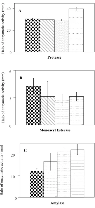
with exception to T. koningii (Fig. 5 B). Protease and monoacyl
esterase were exclusively found in filtrates of C. herbarum isolates
(Fig. 6 A, B). On the other hand, only the species of Trichoderma
secreted amylase (Fig. 6 C).
The latter findings are possibly related to the observation, that naphtyl esterase zymograms, after electrophoresis of extracellular enzymes, showed bands of different mobility in
0
A
B
5 10 15
Cellulase
0 10 20 30
Pectinase
Halo
of
enzymatic
activity
(mm)
Halo
of
enzymatic
activity
(mm)
Figure 5. Production of plant cell wall degrading enzymes in semisolid media by Trichoderma spp. and Cladosporum herbarum
isolates. From left to right: T. harzianum; T. koningii; T. polysporum; T. viride; isolates Ch-1, Ch-2, Ch-3 and Ch-5 of C. herbarum. The
halo of enzymatic activity does not include the halo of the mycelium disc.
Figure 4. Morphologicals characteristics between Trichoderma
spp. and Cladosporium herbarum: hyphal shrinking (A); cell
shortening and septa thickening (B) of C. herbarum, in the presence
Trichoderma species and C. herbarum isolates, respectively. In
the zymogram, considerably more activity was seen after washing the mycelia in 2mM EDTA, as compared to the untreated culture fluid (Fig. 7 A, B).
DISCUSSION
This study has shown that colonies of Trichoderma species
grew always faster than C. herbarum, in single or mixed culture. Its
rapid growth gives Trichoderma an important advantage in the
competition for space and nutrients with plant pathogenic fungi, even before it deploys its arsenal of mycotoxins (10,11). Toxic action
was evident in various alterations of the hyphal structure of C.
herbarum growntogether with Trichoderma species, similar to
the effects described in other systems of mixed cultures (14,30).
The species of Trichoderma are known to produce a number of
antibiotics, such as trichodermin, trichodermol, harzianum A and harzianolide (7,17). These compounds were responsible for most
of the inhibition of C. herbarum seen in this work, and observed in
other experiments involving biocontrol of fungal phytopathogens (5,14,25,33). Volatile antifungals, among which 6-pentyl-2-pyrone is the best known substance (6) were much less effective against this particular phytopathogen, although other species seem to be rather susceptible (27,44). In any case, the pattern of highly specific
inhibition caused by the four Trichoderma species, that permitted
differentiate individual isolates of C. herbarum shows the intricacies
of antagonistic action, considering complex phenomena such as growth and sporulation.
The method of Hankin and Anagnostakis (26) was used to detect the secretion of extracellular cell wall degrading enzymes,
both by the isolates of C. herbarum and Trichoderma species.
Figure 6. Production of extracellular hydrolytic enzymes in
semisolid media produced by Cladosporum herbarum isolates
(A, B) and Trichoderma species (C). In (A) and (B) from left to
right, isolates Ch-1, Ch-2, Ch-3 and Ch-5 of C. herbarum. In (C)
from left to right T. harzianum; T. koningii; T. polysporum; T.
viride. The halo of enzymatic activity does not include the halo of
the mycelium disc.
Figure 7. Esterase patterns of Cladosporium herbarum isolates
(Ch1, Ch2, Ch3, Ch5), and Trichoderma species (T11= T.
polysporum, T15= T. koningii, T25= T. harzianum, TR2= T. viride),
after five days of incubation, in PD medium (A) and mycelial mass washed with EDTA (B).
0 10 20
Halo
of
enzymatic
activity
(mm)
Amylase
C
0 3 6
Halo
of
enzymatic
activity
(mm)
Monoacyl Esterase
B
0 20 40
Halo
of
enzymatic
activity
(mm)
Protease
These assays measured, the combined activity of various enzymes upon pectin and cellulose, respectively (2). Since both pectinolytic and cellulolytic enzymes would be needed for pathogenic action as well as for saprophytic activity, it is difficult to explain the
apparent lack of pectinase in the T. koningii isolate. On the other
hand, the difference in cellulolytic activity found between
Trichoderma species and C. herbarum, though statistically
significant, has probably no important biological meaning.
The very different production of amylase by Trichoderma
and Cladosporium was already observed by Hankin and
Anagnostakis (26), and it may be speculated, that ready expression of amylolytic enzymes is partially responsible for the rapid growth of Trichoderma species in the standard potato-dextrose broth. In
contrast, there is no immediate explanation for the fact that the
isolates of C. herbarum produced much more protease and
monoacyl esterase than Trichoderma species, as demonstrated
by the agar plate assay. No was investigate a possible attachment of enzymes to the mycelium as the reason for obtaining a negative result, as was suggested with esterases. Nevertheless, the same
discrepancy was detected between Trichoderma species and C.
herbarum, usinga differentkind of lipase assay (results not
shown) than the method employed for monoacyl esterase, because it has been argued that soluble substrates are possibly not a good
assay for lipase activity (29,41). Since C. herbarum is known to
spoil butter and to damage oil paintings (16) it is quite possible that this organism secrets lipase. On the other hand, there is evidence that at least part of the activity measured in all acyl esterase assays is in fact cutinase, an enzyme responsible for
pathogenicity of Colletotrichum gloeosporioides on Carica
papaya and other plants (18,28). Even so, in the case of C. herbarum, the monoacyl esterase detected in fungal culture
certainly contains insufficient cutinase activity to allow the
pathogen to attack undamaged leaves or fruits of Passiflora edulis.
Zymograms of esterase isoenzymes are useful as additional parameters to distinguish microbial isolates. An apparent lack of
extracellular esterases in C. herbarum was in fact due to enzymes
that could be recovered by washing the mycelium in EDTA or various detergents (results not shown). As can be seen in Fig. 7, the effect is not limited to the pathogen. The hypothesis of absorved rather than eluted enzymes is favored, because the cells remained viable after the extraction procedure. Addition of EDTA to filtered culture fluid did not reactivate eventually denatured enzymes. However, experiments to test for enzymatic protein in the culture fluid were not performed in order to know if the enzymes lose activity after having been diffused away from the cells that secreted them.
RESUMO
Antagonismo de espécies de T r i c h o d e r m a
s o b r e C l a d o s p o r i u m h e r b a r u m
e suas caracterização enzimática
O maracujá (Passiflora edulis Sims.), um fruto tropical
amplamente consumido, tem sua produção e a qualidade dos seus
frutos reduzidos pela verrugose causada por Cladosporium
herbarum. Este trabalho objetivou investigar o antagonismo de
espécies de Trichoderma (T. polysporum, T.koningii, T. viride e
T. harzianum) contra C. herbarum, e estudar a produção de
enzimas hidrolíticas extracelulares pelo fitopatógeno e antagonistas. Os resultados mostraram considerável potencial
antagônico para o biocontrole dos isolados de C. herbarum por
todas as espécies de Trichoderma, exceto T. koningii. O efeito
mais promissor foi observado para T. polysporum. Em relação ao
padrão de esterase obtido por eletroforese em gel de poliacrilamida, a maior atividade apresentada pelos isolados foi observada cinco
dias após a incubação. Os isolados de C. herbarum produziram
enzimas extracelulares, lipase, pectinase, cellulase e protease, todas possivelmente relacionadas ao processo de infecção do hospedeiro. A excreção de amilase que parece não estar associada
com fitopatógenos foi detectada nas espécies de Trichoderma,
mas não em C. herbarum. Além disso, todas as espécies de
Trichoderma testadasproduziram também celulase e pectinase,
exceto T. koningii com relação a esta última enzima. A
demonstração de várias isoesterases no zimograma das espécies de Trichoderma e isolados de C. herbarum, foi notavelmente
melhorada através da lavagem do micélio com detergentes ou EDTA. Este fato sugere que uma grande fração de enzimas extracelulares pode permanecer presa externamente na parede celular fúngica após excreção.
Palavras-chave: Maracujá, controle biológico, enzimas extracelulares, fungos
REFERENCES
1. Alfenas, A.C.; Peters, I.; Brune, W.; Passador, G.C. Eletroforese de proteínas e isoenzimas de fungos e essências florestais. Edit.Universitária, UFV, Viçosa, 1991, 242p.
2. Annis, S.L.; Goodwin, P.H. Recent advances in the molecular genetics of plant cell wall-degrading enzymes produced by plant pathogenic fungi. European J. Plant Pathol., 103:1-14, 1997.
3. Bitancourt, A.A. Uma nova doença do maracujá. O Biológico,
1:202-204, 1935.
4. Blakeman, J.P.; Fokkema, N.J. Potential for biological control of plant disease on the phylloplane. Annu. Rev. Phytopathol., 20:167-192, 1982.
5. Callegarin, A.M.; Cugguda, L. Terreni repressivi verso Fusarium oxysporum: ricerça preliminari di microorganismi antagonisti. Anal. Fac. Sci. Agr. Stud. Torino, 13:19-33, 1984.
6. Claydon, N.; Allan, M.; Hanson, J.R.; Avent, A.G. Antifungal alkylpyrones of Trichoderma harzianum. Trans. Br. Mycol. Soc., 88:503-513, 1987.
7. Claydon, N.; Hanson, J.R.; Truneh, A.; Avent, A.G. Harzianolide, a butenolide metabolite from cultures of Trichoderma harzianum. Phytochemistry, 30:3802-3803, 1991.
8. Chet, J.; Baker, R. Indication of suppressiveness to Rhizoctonia solani
in soil. Phytopathology, 70:994-998, 1980.
10. Cook, R.J.; Baker, K.F. The nature apractice of biological control of plant pathogens. APS Press, St. Paul, 1989. 539p.
11. Deacon, J.W.; Berry, L.A. Modes of actions of mycoparasites in relation to biocontrol of soilborne plant pathogens. In: Tjamos, E.C.; Papavizas, G.C.; Cook, R.J. (eds). BiologicalControlofPlantDiseases.
Plenum Press, New York, 1992, p.157-167.
12. Dennis, C.; Webster, J. Antagonistic properties of species-groups of
Trichoderma. I. Production of non-volatile antibiotics. Trans. Br. Mycol. Soc., 84:25-39, 1971a.
13. Dennis, C.; Webster, J. Antagonistic properties of species-groups of
Trichoderma. II. Production of volatile antibiotics. Trans. Br. Mycol. Soc., 84:41-48, 1971b.
14. Dennis, C.; Webster, J. Antagonistic properties of species-groups of
Trichoderma. III. Hyfal interaction. Trans. Br. Mycol. Soc., 57:363-369, 1971c.
15. Despoulain, B.; Seigle-Murandi, F.; Steiman, R.; De Giorgis, L. Fungal flora of corn White draff. Cryptogam. Mycol., 11:79-88, 1990.
16. De Vries, G.A. Contribution to the knowledge of the genus Cladosporium, Link ex. Fr. Hollandia Press, Baarn, 1952, 121p.
17. Dickinson, J.M.; Hanson, J.R.; Truneh, A. Metabolites of some biological control agents. Pestic. Sci., 44:389-393, 1995.
18. Dickman, M.B.; Podilha, G.K.; Kolattukudy, P.E. Insertion of a cutinase gene into a wound pathogen enables it to infect intact host. Nature,
342:446-448, 1989.
19. Edington, L.V.; Khew, K.L.; Barron, G.L. Fungitoxicspectrumof benzimidazole compounds. Phytopathology, 61:42-44, 1971. 20. Elad, Y.; Chet, J.; Katan, J. Trichoderma harzianum a biocontrol
effective against Sclerotium rolfsii and Rhizoctonia solani. Phytopathology, 70:119-121, 1980.
21. Ellis, M.B. Dematiaceous Hyphomycetes. Commonwealth Mycological Institute, Kew, 1971. 608p.
22. Ellis, M.B. More Dematiaceous Hyphomycetes. Commonwealth Mycological Institute, Kew, 1976. 507p.
23. Fravel, D.Commercial biocontrol products for use against soilborne crop diseases.[on line]. Beltsville: Institute of Plant Science – USDA, 1999. [cited in 01 01 99]. Available in the web: http://www.bar.usda.gov/ psi/bpdl/bioprod.htm.
24. Gashe, B.A. Initial observations on the cellulytic activities of some fungal isolates. Mircen J., 4:491-494, 1988.
25. Gupta, V.P.; Govindaiah, A.K.B.; Datta, R.K. Antagonistic potential of
Trichoderma and Gliocladium species to Botryodiplodia theobromae
infecting mulberry. Indian J. Mycol. and Pl. Pathol., 25:125, 1995. 26. Hankin, L.; Anagnostakis, S.L. The use of solid media for detection of
enzyme production by fungi. Mycologia, 67:597-607, 1975. 27. Hutchinson, S.A. Biological activities of volatile fungal metabolites.
Ann. Rev. Phytopathology, 11:223-246, 1973.
28. Köller, W.; YAO, C.; Trial, F.; Parker, D.M. Role of cutinase in the invasion of plants. Can. J. Bot., 73 (Suppl.): 1109-1118, 1995.
29. Kouker, G.; Jaeger, K.E. Specific and sensitive plate assay for bacterial lipases. Appl. Environ. Microbiol., 53:211-213, 1987.
30. Lee, Y.A.; Wu, W.S. The antagonisms of Trichoderma spp. And
Gliocladium virens against Sclerotinia sclerotiorum. Plant Prot. Bull.,
26:293-304, 1984.
31. Lifshitz, R.; Witingham, M.T.; Baker, R. Mechanisms of biological control of pre-emergence damping-off of pea by seed treatment with
Trichoderma spp. Phytopathology, 76:720-725, 1986.
32. Lilly, V.G.; Barnett, N.L. Physiology of Fungi. McGraw Hill, New York, 1951, 463p.
33. Lundberg, A.; Unestan, T. Antagonism against Fomes annosus.
Comparison between different test methods “in vitro” and “in vivo”.
Mycopathologia., 70:107-115, 1980.
34. Lynch, J.H. “In vitro” identification of Trichoderma harzianum asa
potential antagonistofplant pathogens. Curr. Microbiol., 16:49-53, 1987.
35. Manachini, P.L.; Fortina, M.G.; Parini, C. Purification and properties of an endo-polygalacturonidase produced by Rhizopus stolonifer. Biotech. Lett., 9:219-224, 1987.
36. Mehta, R.D.; Patel, K.A.; Roy, K.K.; Mehta, M.H. Biological control of soilborne plant pathogens with Trichoderma harzianum. Indian J. Mycol. Pl. Pathol., 25:126, 1995.
37. Melo, I.S. Trichoderma e Gliocladium como bioprotetores de plantas.
Revs. Anu. Patol. Plantas, 4:261-295, 1996.
38. Montenecourt, B.S.; Eveleigh, D.E. Semi-quantitative plate assay for determination of cellulase production by Trichoderma viride. Appl. Environ. Microbiol., 33:178-183, 1977.
39. Neirotti, E.; Azevedo, J.L. Técnica semiquantitativa de avaliação de produção de celulases em Humicola sp. Rev. Microbiol., 19:78-81, 1988.
40. O’Donnell, J.; Dickinson, C.H. Pathonicity of Alternaria and
Cladosporium isolates on Phaseolus. Trans. Br. Mycol. Soc.,
74:335-342, 1980.
41. Rapp, P.B.; Backhaus, S. Formation of extracellular lipases by filamentous fungi yeasts, and bacteria. Enzyme Microb. Technol., 14:938-943, 1992.
42. Riker, A.J.; Riker, R.S. Introduction to Research on Plant Diseases. John S. Swift Co., St. Louis, 1936. 117p.
43. Samuels, G.J. Trichoderma: a review of biology and systematics of the genus. Mycol. Res., 100:923-935, 1996.
44. Sarath, G.; De la Motte, R.S.; Wagner, F.W. Protease assay methods.