BIOLOGICAL CONTROL OF RICE BROWN SPOT WITH NATIVE ISOLATES OF THREE TRICHODERMA SPECIES
Elham Khalili1*; Mehdi Sadravi2;Shahram Naeimi3; Vahid Khosravi4
1
Department of Plant Pathology, Gorgan University of Agriculture Science and Natural Resources, Gorgan, Golestan, Iran; 2
Department of Plant Pathology, Yasouj University, Yasouj, Iran; 3Amol Biological Control Research Laboratory, Iranian Research Institute of Plant Protection, Tehran, Iran; 4Deputy of Rice Research Institute in Mazandaran, Amol, Iran.
Submitted: January 09, 2011; Returned to authors for corrections: March 13, 2011; Approved: June 06, 2011.
ABSTRACT
Brown spot caused by Bipolaris oryzae is an important rice disease in Southern coast of Caspian Sea, the major rice growing region in Iran. A total of 45 Trichoderma isolates were obtained from rice paddy fields in Golestan and Mazandaran provinces which belonged to Trichoderma harzianum, T. virens and T. atroviride species. Initially, they were screened against B. oryzae by antagonism tests including dual culture, volatile and nonvolatile metabolites and hyperparasitism. Results showed that Trichoderma isolates can significantly inhibit mycelium growth of pathogen in vitro by producing volatile and nonvolatile metabolites Light microscopic observations showed no evidence of mycoparasitic behaviour of the tested isolates of Trichoderma spp. such as coiling around the B. oryzae. According to in vitro experiments, Trichoderma isolates were selected in order to evaluate their efficacy in controlling brown spot in glasshouse using seed treatment and foliar spray methods. Concerning the glasshouse tests, two strains of T. harzianum significantly controlled the disease and one strain of T. atroviride increased the seedling growth. It is the first time that the biological control of rice brown spot and increase of seedling growth with Trichoderma species have been studied in Iran.
Key words: Rice brown spot, Biocontrol, Trichodermaharzianum, T. virens,T. atroviride
INTRODUCTION
Rice (Oryza sativa L.) is among the most important cereals in Iran. On the basis of nutrition value, it is comparatively rated more than other cereals and plays a key role in nutrition (1). Rice brown spot caused by Bipolaris oryzae Breda de Hann (formerly, Helminthosporium oryzae) (Teleomorph: Cochliobolus miyabeanus) is occurred in all rice-growing areas of the world. The pathogen causes infection on all growth stages of rice plant from nursery to field and results in significant yield and grain quality losses. Rice brown spot was
a major factor for the ‘‘Great Bengal Famine’’ during 1942– 1943 (27). In 1956, the disease was reported by Petrak at the first time from Iran and then it was reported by Sharif and Ershad from coastal area of Caspian sea in 1966 (16). The disease is prevalent in northern provinces of Iran and under environmental conditions conducive to disease; it can cause severe yield loss (30, 22).
Continuous, inappropriate and non-discriminative use of chemicals is known to cause undesirable effects such as residual toxicity, development of pathogen resistance to fungicides, environmental pollution, health hazards to humans
and animals and increased expenditure for plant protection. Instead, plant pathologists have focused their attention to develop environmentally safe, long-lasting and effective biocontrol methods for the management of plant diseases. Among various fungal and bacterial biocontrol agents, Trichoderma spp. was most frequently used against various plant diseases. Research during the previous two decades has led to the possibility of biological control as an increasingly realistic option for rice disease management (32). Trichoderma spp. has been shown to be effective for the control of brown spot disease and the increase of plant growth on rice (17). Rice plants sprayed with spore suspension of T. harzianum obtained a significant reduction in the severity of disease under greenhouse conditions (2). Also, Trichoderma species are able to colonize the root surface and rhizosphere from the treated seeds, protecting them from fungal diseases and stimulate plant growth and productivity (4). This study was accomplished to obtain indigenous Trichoderma isolates from paddy fields and to examine their biocontrol activities against B. oryzaein vitro as well as in vivo and also to evaluate their effects on rice growth parameters.
MATERIALS AND METHODS
Fungal isolates
In order to isolate Trichoderma spp., phyllosphere and soil samples were collected from paddy fields in Mazandaran and Golestan provinces from March to August 2007. Soil samples were taken at 0-30 cm depth. Isolation of Trichoderma from soil samples was performed by the soil dilution method (21). Ten grams of each soil sample was added to 90 mL distilled water on a shaker for one hour. Then, 10 mLaliquot from this suspension was transferred aseptically to 90 mL distilled water. This dilution process was repeated to get dilutions up to 10-5. In the case of phyllosphere samples, rice stems and leaves were cut into 5 mm pieces and added to 150 mL distilled water on a shaker for 30 minutes. From each dilution suspension, one millilitre of aliquots (from soil and phyllosphere sample) was spread on RB-S-F selective medium
(9) in 9 cm Petri dishes. The dishes were incubated at 26±1°C in the dark for 3-4 days. Putative Trichoderma isolates were purified using single spore technique.
For isolation of pathogen, naturally diseased leaves of different rice cultivars infected with varying degrees of brown spot were collected from nurseries and fields at different geographic locations in Mazandaran province, Iran. Eight B. oryzae isolates were obtained and purified using single spore technique (15, 30). For pathogenicity test 21-day-old rice seedlings of the most common cultivar in north of Iran i. e. Tarom, were grown under greenhouse conditions, and sprayed with spore suspension (4 × 10⁵ spore mL-1) of each B. oryzae isolate. Seven days after inoculation, the severity of brown spot disease was assessed following the standard evaluation system of International Rice Research Institute (20). The most aggressive B. oryzae isolate was selected for in vitro and in vivo tests of this study.
Identification of Trichoderma isolates
For morphological identification, Trichoderma isolates were grown on 2% malt extract agar under ambient laboratory conditions of light and temperature (about 21°C). Microscopic examination was carried out by mounting the culture in lactic acid 25%. Individual isolates were identified at the species level using morphological keys and fungal species descriptions (5, 6, 13, 35). For molecular identification, a nuclear rDNA region, containing the internal transcribed spacers 1 and 2 (ITS1 and 2) and the 5.8S rRNA gene was amplified using the primers ITS1 and ITS4 (34). PCR amplifications were performed as described previously (19). Amplicons were purified with the GenElute PCR Clean-up Kit (Sigma, USA) and sequenced at Macrogen Inc., South Korea. In order to identify the isolates at the species level, ITS sequence analysis was carried out with the aid of the program TrichOkey 2.0 available online at http://www.isth.info/.
In vitro antagonism tests
mycelial disks of either B. oryzae and Trichoderma isolates 10 mm away from the edge of the plate opposite to each other. Plates inoculated with B. oryzae alone served as control. Plates were incubated at 26 ± 1°C for seven days (12 h light/12 h darkness). The linear growth was measured. Three replicate plates were done for each treatment. The percentage of growth inhibition was calculated using the equation RI= 100 x (R2 - R1) / R2. Where RI was the percentage of reduction in mycelial growth, R1 was the averaged growth of pathogen in treated plates and R2 was the averaged growth of pathogen in control plates (12).
Production of volatile compounds: The bottoms of Petri dishes containing PDA were separately inoculated with mycelial plugs of pathogen and Trichoderma isolates. Then two bottoms held together with pathogen at top, sealed with adhesive tape and incubated at 26 ± 1°C in the dark for seven days. The radial growth of the pathogens was measured. Three replicates of each treatment were used (11).
Production of non-volatile compounds: A piece of autoclaved cellophane was placed on the surface of PDA medium in a 9 cm plate then a 7 mm disk of Trichoderma isolate was inoculated on the cellophane. The plates were incubated at 26 ± 1°C for 72 h. After the incubation period, the cellophane and the adhering Trichoderma mycelia were aseptically removed and the centre of each dish was inoculated with a 5 mm diameter disk of B. oryzae taken from an actively growing colony. The plates were incubated at 26 ± 1°C for a further five days. The control treatment was B. oryzae grown on PDA plate where previously there was a cellophane disc without antagonist. Three replicate plates were done for each treatment. The percentage of growth inhibition was calculated (12).
Mycoparasitism test: The slide culture method (28) was used to investigate the mycoparasitic nature of Trichoderma isolates against B. oryzae. A microscope glass slide covered with a thin layer of PDA in the agar plate was inoculated with
5 mm mycelial disks of Trichoderma spp. and pathogen,1 cm apart from each other. All paired cultures incubated at 26 °C and regions where the hyphae of Trichoderma isolates met the hyphae of the pathogen were periodically observed under a light microscope.
Evaluation of Trichoderma isolates on wet filter paper: Based on in vitro antagonism test, 20 Trichoderma isolates were selected. Rice seeds were soaked in spore suspension of B. oryzae (105 spores mL-1) containing 0.05% Tween 20 for 24 h. Then, seeds were air-dried and soaked in spore suspension of Trichoderma isolates (108 spores mL-1) containing 0.05% Tween 20 for 2 h and then placed on the sterile wet filter paper in a seedling tray with 100 seeds per treatment (7). Plant height, root length and the percentage of disease control were determined. The number of infected seedlings was counted for each treatment and then compared to infected control with B. oryzae.
Glasshouse experiments
Seed treatment: According to wet filter paper test, 11 Trichoderma isolates were selected and used to inoculate infested rice seeds with pathogen and were sown in plastic pots containing autoclaved rice field soil. The pots were kept in glasshouse with the condition of 21 to 30°C temperature and 90% relative humidity. Three pots were used for each treatment. Rice seedlings growth parameters namely stem height, root length, stem and root wet weight, stem and root dry weight and percentage of disease control were determined 45 days after inoculation. Rice stem and root were dried in an oven at 80°C for 24 hours to a dry weight.
Foliar spray
inoculation of pathogen and the percentage of disease control was calculated for each individual treatment (12). The completely randomized design with three replications (pots) and five seedlings per pot was used.
Statistical analysis
The completely randomized design with three replications per treatment was used. Data were analyzed with statistical analysis software (6.0 SAS, 1999). All data were first subjected to analysis of variance (ANOVA). Means were compared using Duncan’s multiple range test at P = 0.01.
RESULTS
A total of 45 Trichoderma isolates were obtained which
belonged to three species namely Trichoderma harzianum, T. virens and T. atroviride. The percentage of the total Trichoderma isolates obtained from the rice phyllosphere and soil samples were 63 and 38 percent, respectively. The most frequent species was T. harziunum with 31 isolates (69%). Eleven and three isolates belonged to T. virens and T. atroviride, respectively.
Based on the pathogenicity test, B. oryzae 3 (from a rice field in Amol, Mazandaran) showed highest disease severity, and this most aggressive isolate was used for all subsequent studies.
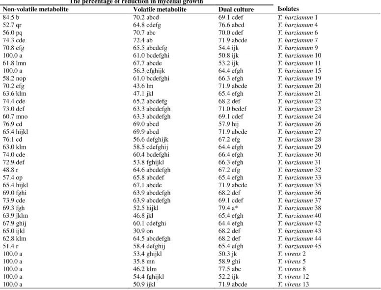
Data from dual culture tests showed that although all 45 Trichoderma isolates inhibited the mycelium growth ofpathogen, there were significant differences among them at P = 0.01. T. harzianum isolates 38, 4, 27, 35, 20 and 7; T. virens isolates 8 and 13 and T. atroviride isolate 3 substantially reduced the growth of the pathogen (Table 1).
Table 1.In vitro effects of Trichoderma isolates on mycelial growth of Bipolaris oryzae.
The percentage of reduction in mycelial growth
Non volatile metabolite Volatile metabolite Dual culture Isolates
84.5 b 70.2 abcd 69.1 cdef T. harzianum 1
52.7 qr 64.8 cdefg 76.6 abcd T. harzianum 4
56.0 pq 70.7 abc 70.0 cdef T. harzianum 6
74.3 cde 72.4 ab 71.9 abcde T. harzianum 7
70.8 efg 65.5 abcdefg 54.4 ijk T. harzianum 9
100.0 a 61.0 bcdefghi 50.8 ijk T. harzianum 10
61.8 lmn 67.7 abcde 53.2 ijk T. harzianum 11
100.0 a 56.3 efghijk 64.4 efgh T. harzianum 15
58.2 nop 61.0 bcdefghi 66.3 efgh T. harzianum 19
70.2 efg 43.6 lm 71.9 abcde T. harzianum 20
63.6 klm 47.1 jkl 65.4 efgh T. harzianum 21
74.4 cde 65.2 abcdefg 68.2 def T. harzianum 22
73.0 def 63.3 abcdefgh 71.0 bcdef T. harzianum 23
60.7 mno 63.3 abcdefgh 69.1 cdef T. harzianum 24
76.9 cd 69.0 abcd 57.9 hij T. harzianum 26
65.4 hijkl 69.9 abcd 71.9 abcde T. harzianum 27
76.1 cd 56.6 defghijk 67.2 efg T. harzianum 28
63.0 klm 58.5 cdefghij 64.4 efgh T. harzianum 29
74.0 cde 60.4 bcdefghi 66.4 efgh T. harzianum 30
72.9 def 53.8 fghijkl 66.3 efgh T. harzianum 31
48.8 r 64.6 abcdefgh 67.2 efg T. harzianum 32
57.4 op 65.8 abcdef 65.4 efgh T. harzianum 33
65.4 hijkl 67.1 abcde 71.9 abcde T. harzianum 35
69.0 fghi 63.9 abcdefgh 68.2 def T. harzianum 36
73.9 cde 63.9 abcdefgh 69.1 cdef T. harzianum 37
69.3 fgh 52.5 hijkl 79.4 a* T. harzianum 38
63.9 jklm 46.8 jkl 65.4 efgh T. harzianum 40
67.9 ghij 60.1 cdefghi 64.4 efgh T. harzianum 42
65.0 ijkl 30.9 on 68.2 def T. harzianum 43
62.8 klm 64.5 abcdefgh 68.2 def T. harzianum 44
51.4 r 58.4 defghij 65.4 efgh T. harzianum 45
100.0 a 53.4 ghijkl 50.3 jk T. virens 2
100.0 a 35.8 mn 58.9 ghi T. virens 5
100.0 a 46.2 klm 77.5 abc T. virens 8
100.0 a 54.4 fghijkl 52.2 ijk T. virens 12
77.6 c 54.1 fghijkl 62.6 fgh T. virens 14
76.9 cd 66.7 abcde 50.8 ijk T. virens 16
69.7 fg 56.6 efghijk 53.2 ijk T. virens 18
76.6 cd 56.6 efghijk 66.3 efgh T. virens 25
66.6 ghijk 22.1 o 59.0 ghi T. virens 39
100.0 a 62.6 bcdefghi 66.3 efgh T. virens 41
42.6 s 21.8 o 78.4 ab T. atroviride 3
64.9 ijklm 66.7 abcde 48.5 k T. atroviride 17
100.0 a 75.0 a 69.1 cdef T. atroviride 34
* Values within a column followed by the same letter(s) are not significantly different according to Duncan’s multiple range test (P = 0.01) * Each value represents the mean of three replicates.
Volatile metabolites of Trichoderma isolates also had inhibitory effects on the growth of B. oryzae. The most effective isolates were T. harzianum 4, 27, 35, 7, 23, 6, 24, 37, 1, 22, 44, 36, 32, 33, 26, 9 and 11; T. virens 16 and T. atroviride 34 and 17 (Table 1).
All tested Trichoderma isolates released non-volatile compounds that diffused through the cellophane membrane onto the agar medium, resulting inhibition growth ofpathogen. The highest reduction in the mycelial growth of the pathogen (100%) was recorded for T. harzianum 15 and 10;T. virens 8, 13, 41, 5, 12, 2 and T. atroviride 34 (Table 1).
Light microscopic observations showed no evidence of mycoparasitic behaviour of the tested isolates of Trichoderma spp. such as coiling around the B. oryzae.
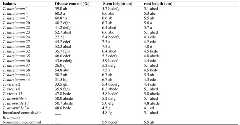
In wet filter paper test, significant differences were observed in plant height, root length and the percentages of disease control between treated and untreated rice seeds. The highest disease control was recorded for T. harzianum 7, 4, 1, 43, 38, 23, and 28; T. atroviride 17 and 3. Furthermore, T. harzianum isolates 7, 4, 43, 38, 23, 28, 27, 22, 20, 32, andT. virens isolate 8 showed increasing effects on the rice seedling growth (Table 2).
Table 2. Effect of Trichoderma isolates in controlling brown spot as well as on growth factors of rice seedlings in wet filter paper
test.
Isolates Disease control (%) Stem height(cm root length (cm
T. harzianum 1 59.0 ab 5.7 bcdefg 5.1 abcd
T. harzianum 4 60.3 a 6.6 abc 5.3 abc
T. harzianum 7 60.8* a 6.6 ab 5.5 ab
T. harzianum 20 40.2 efgh 6.7 ab 5.8 a
T. harzianum 22 41.2 defgh 6.4 abcd 5.7 a
T. harzianum 23 52.7 abcd 6.6 abc 5.1 abcd
T. harzianum 24 22.2 j 5.4 bcdefg 4.3 cde
T. harzianum 27 45.2 cdef 7.5 a 4.2 cde
T. harzianum 28 52.2 abcd 7.3 a 4.0 e
T. harzianum 32 35.7 fghi 6.4 abcd 4.5 bcde
T. harzianum 35 46.0 cdef 5.3 cdefg 4.8 abcde
T. harzianum 36 43.6 cdefg 5.9 bcdef 4.4 cde
T. harzianum 37 26.0 ij 5.2 defg 5.1 abcd
T. harzianum 38 54.8 abc 7.5 a 4.5 bcde
T. harzianum 43 58.2 ab 6.7 ab 5.5 ab
T. harzianum 44 31.5 hij 6.7 ab 4.3 cde
T. virens 2 33.5 ghi 5.4 bcdefg 4.4 cde
T. virens 8 35.9 fghi 6.2 abcde 5.1 abcd
T. virens 13 47.8 bcde 5.8 bcdef 5.0 abcde
T. atroviride 3 50.0 abcde 5.2 defg 5.1 abcd
T. atroviride 17 50.7 abcde 5.0 efg 4.8 abcde
T. atroviride 34 48.0 bcde 4.5 g 4.1 ed
Inoculated control(with
B. oryzae)
___ 4.8 fg 5.1 abcd
Non-inoculated control ___ 5.9 bcdef 5.5 ab
Seed treatment with Trichoderma isolates significantly decreased rice brown spot in glasshouse. T. harzianum 1 and 20 were the most effective isolates. In the case of growth factors, highest values were recorded for T. atroviride isolate 3 (Table 3).
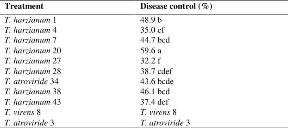
Foliar spray with selected Trichoderma isolates in glasshouse showed that all treatments were significantly different in controlling the disease and T. harzianum 20 was the most effective isolate (Table 4).
Table 3. Effects of treatment of rice seeds by selected Trichoderma isolates on brown spot and growth characteristics of rice seedlings in glasshouse *.
Root dry weight (g)
Stem dry weight (g)
Root wet weight (g)
Stem wet weight (g)
Root length (cm)
Stem height (cm)
Disease control (%)
Isolate
0.2 e 0.2 f 0.8 ghi 1.1 fgh 16.7 a 48.4 de 61.2 a T. harzianum 1 0.5 bc 0.4 bcd 2.0 ab 2.3 b 18.1 a 65.4 abcd 32.3 dce T. harzianum 4 0.4 c 0.3 bcde 1.2 defg 1.4 defg 19.8 a 53.2 abcde 45.3 bc T. harzianum 7 0.2 e 0.2 f 0.7 hi 0.9 gh 16.6 a 46.5 e 58.1 a T. harzianum 20 0.4 cd 0.4 bc 1.3 def 1.9 bcd 20.6 a 67.1 abc 20.8 e T. harzianum 27 0.3 e 0.3 bcde 0.5 i 0.8 h 17.8 a 54.7 abcde 50.3 ab T. harzianum 28 0.4 cd 0.4 ab 1.6 bcd 2.1 bc 17.9 a 68.0 ab 34.7 cd T. atroviride 34 0.3 de 0.3 def 1.2 defg 1.2 efgh 17.3 a 50.3 cde 44.7 bc T. harzianum 38 0.6 a 0.3 bcde 1.8 bc 1.7 cde 17.5 a 60.5 abcde 34.6 cd T. harzianum 43 0.3 de 0.3 bcde 1.1 efgh 1.4 defg 19.6 a 53.2 abcde 39.4 bc T. virens 8 0.6 ab 0.5 a 2.3 a 3.0 a 20.4 a 69.6 a 24.0 de T. atroviride 3 0.2 e 0.2 fe 1.0 fgh 1.2 efgh 15.8 a 50.3 cde ___ Infected check
(with B. oryzae) 0.4 c 0.3 bcde 1.5 cde 1.6 cdef 17.0 a 59.2 abcde Healthy check * Values within a column followed by the same letter(s) are not significantly different according to Duncan’s multiple range test (P = 0.01)
* Each value represents the mean of three replicates.
Table 4. Control of rice brown spot after foliar spray of Trichoderma isolates on Tarom cultivar *.
* Values within a column followed by the same letter(s) are not significantly different according to Duncan’s multiple range test (P = 0.01)
*Each value represents the mean of three replicates.
DISCUSSION
The majority of isolates (90%) recovered from the soil identified as T. harzianumandT. virens which is in agreement with the findings of Naeimi et al. (25). In addition, fungal species belonging to the genus Trichoderma are worldwide in
occurrence and they have been widely reported from rice fields in other countries such as Philippine, China and Bangladesh (23, 26, 36, 29). The results of the present study showed that the Trichoderma isolates could easily adopt and survive in irrigated rice ecosystems of northern Iran and this gives hope for future application of the promising native strains in paddy
Treatment Disease control (%)
T. harzianum 1 48.9 b
T. harzianum 4 35.0 ef
T. harzianum 7 44.7 bcd
T. harzianum 20 59.6 a
T. harzianum 27 32.2 f
T. harzianum 28 38.7 cdef
T. atroviride 34 43.6 bcde
T. harzianum 38 46.1 bcd
T. harzianum 43 37.4 def
T. virens 8 T. virens 8
fields in order to control brown spot and other rice diseases. In vitro antagonism tests revealed that the native isolates of Trichoderma spp.significantly inhibited the mycelial growth of B. oryzae in several ways. The most effective isolates were belonged to T. harzianum. It is clear from the previous results that competition plays an important role in the T. harzianum and B. oryzae interaction (2, 17). Trichoderma isolates inhibited the growth of the target organisms through its ability to grow much faster than the pathogenic fungi thus competing efficiently for space and nutrients. Angelica et al. (3) reported that inhibition of pathogen growth is due to production of amylase by Trichoderma spp., which is partially responsible for the rapid growth of antagonists on potato dextrose agar medium. In addition to amylase, it has been reported that Trichoderma species also produce extracellular cellulose and pectinase enzymes that are capable of hydrolyzing the cell walls of other fungi (24). A comparison between the inhibition effects of volatile and nonvolatile metabolites of our Trichoderma isolates revealed that the nonvolatile metabolites seemed to be more effective. In a similar study, Abdel-Fattah et al. (2) also reported that the antifungal metabolites of T. harzianum strains completely inhibited the linear growth of B. oryzae. Gary et al. (14) identified five classes of volatile compounds, such as alcohols, esters, ketones, acids and lipids, produced by some fungi and bacteria. A large number of volatile and non-volatile antifungal substances such as diterpenes, peptaibols, butenolides, furanones, pyrones, and pyridones are produced by T. harzianum which have stimulus influence on plant growth and development. Trichoderma antifungal substances are also able to arrest the hyphal growth of different fungal pathogens. It is believed that these enzymes and antibiotics act synergistically on the host (8).
Light microscopic observations revealed that mycoparasitism did not appear to contribute to the aggressive nature of our Trichoderma isolates against B. oryzae which is in agreement with the findings of Abdel-Fattah et al. (2).
Results from seed treatments and foliar spray, demonstrated that significant reduction in disease severity is observed when T. harzianum 1 and 20 were applied in
glasshouse. In addition, Trichoderma atroviride isolate 3 had substantial influence on plant growth and development. Similarly, Harish et al. (17) stated that spraying of spore suspension of Trichoderma isolates on rice plant significantly inhibited the growth and spore germination of B. oryzae besides increasing seedling growth. There are high levels of nutrients in the rhizosphere and pathogen and the introduced biocontrol agent (antagonist) compete for the availability of space and nutrients. Trichoderma spp. has been reported to be more rhizosphere competent than most soil fungi (32). Abundant production and germination of conidia by Trichoderma spp. and their rapid mycelial growth may suppress the growth of the pathogen population in the rhizosphere and thus reduce disease development. Promotion of plant growth induced by Trichoderma spp. appeared to be due to both the control of minor pathogens and production of growth-regulating factors. A number of plant-associated microbes were found to produce poisonous compound cyanide which causes reduction of the plant growth. Trichoderma spp. could produce enzymes which detoxify waste cyanide and this process lead to increase in plant growth. Recent advances demonstrate that the effects of Trichoderma on plants, including induced systemic or localized resistance, are also very important. These fungi colonize the root epidermis and outer cortical layers and release bioactive molecules that cause to induction of pathways for resistance in plants, increased plant growth and nutrient uptake occurs (4, 188).
This study has demonstrated that a number of naturally occurring fungal isolates can inhibit growth of B. oryzae. Furthermore, this research raises some interesting possibilities for future research. These include testing whether the antagonist can show the same level of efficacy under natural field conditions; testing whether mixtures of biocontrol agents are more effective than a single strain on control of disease severity and testing for preparing the best formulation of promising Trichoderma isolates.
ACKNOWLEDGMENTS
Department of Microbiology, Faculty of Sciences and Informatics, University of Szeged, Hungary for molecular identification of Trichoderma isolates.
REFERENCES
1. Alinia, F.; Bahrami, M.; Amoogholi-Tabari, M. (2002). Rice Significant Pests and Diseases and Their Control Methods. Jihad Agriculture Organization of Mazandaran Publications, Iran.
2. Abdel-Fataah, G.M.; Shabana, Y.M.; Ismail, A.E.; Rashad, Y.M. (2007). Trichoderma harzianum: a biocontrol agent against Bipolaris oryzae. Mycopathol. 164, 81-99.
3. Angelica, M.; Barbosa, G.; Rehn, K.G.; Menzes, M.; Mariano, R.R. (2001). Antagonism of Trichoderma sp. on Cladosporium herbarum and their enzymatic characterization. Braz. J. Microbiol. 32, 98–104. 4. Baker, R. (1988). Trichoderma spp. as plant-growth stimulants.
Biotechnol. 7(2), 97–106.Benitez, T.; Rincon, A.M.; Limon, M.C.; Codon, A.C. (2004). Biocontrol mechanisms of Trichoderma strains. Int. Microbiol. 7(4), 249–260.
5. Bisset, J. (1991a). A revision of the genus Trichoderma. A. Infrageneric classification. Can. J. Bot. 69, 2357-2372.
6. Bisset, J. (1991b). A revision of the genus Trichoderma. B. Additional notes on section Longibrachiatum. Can. J. Bot. 69, 2418-2420.
7. Burgess, D.R.; Hepworth, G. (1996). Biocontrol of sclerotinia stem rot (Sclerotinia minor) in sunflower by seed treatment with Gliocladium virens. Plant Pathol. J. 45(3), 583-592.
8. Calistru, C.; Mclean, M.; Berjak, P. (1997). In vitro studies on the potential for biological control of Aspergillus flavus and Fusarium moniliforme by Trichoderma species. Mycopathol. 137, 115–24. 9. Davet, P.; Rouxel, F. (2000). Detection and Isolation of Soil Fungi.
Science Publishers Inc, Enfield, USA.
10. Dennis, L.; Webster, J. (1971 a). Antagonistic properties of species groups of Trichoderma. I. Production of non–volatile antibiotics. Trans. Brith. Mycol. Soc. 57, 25-29.
11. Dennis, L.; Webster, J. (1971 b). Antagonistic properties of species groups of Trichoderma II. Production of non-volatile antibiotics. Trans. Br. Mycol. Soc. 57, 41-48.
12. Desai, S., Reddy, M.S., and Kloepper, J.W. (2002). Comprehensive testing of biological agents. In: Gnanamanickam, S.S. (Ed.). Biological Control of Crop Diseases. Marcel Dekker Inc, New York, USA, p. 387– 420.
13. Gams,W.; Bisset, J. (1998). Morphology and identification of Trichoderma. In:Kubicek, C.P.; Harman, G.E. (eds). Trichoderma and Gliocladium Basic Biology, Taxonomy and Genetics. Taylorand Francis Ltd, London, UK, p. 57–74.
14. Gary, A.; Dirkse, E.; Sears, J.; Markworth, C. (2001). Volatile antimicrobials from Muscodor albus, a novel endophytic fungus. Microbiol. 147, 2943–2950.
15. Ellis, M.B. (1971). Dematiaceous Hyphomycetes. Common-wealth Mycological Institute, KEW, England.
16. Ershad, D. (1995). Fungi of Iran. Iranian Ministry of Agriculture, Tehran, Iran.
17. Harish, S.; Saravavakumar, D.; Radjacommar, R.; Ebenezar, E.G.; Seetharaman, K. (2007). Use of plant extracts and biocontrol agents for the management of brown spot disease in rice. Biocontrol 53(3), 555-567. 18. Harman, G.E. (2006). Overview of mechanisms and uses of Trichoderma
spp.Phytopathol. 96, 190–194.
19. Hermosa, M.R., Grondona, I., Iturriaga, E.A., Diaz-Minguez, J.M., Castro, C., Monte, E., Garcia-Acha, I. 2000. Molecular characterization of biocontrol isolates of Trichoderma. Appl. Environ. Microbiol. 66:1890–1898.
20. International Rice Research Institute. (2002). Standard Evaluation System for Rice (SESR). Available at: http://www.irri.org.
21. Johnson, L.; Curl, E.; Bond, J.; Fribourg, H. (1959). Methods for Studying Soil Mycoflora. – Plant Disease Relationships. Burgess Publishing Company, Minneapolis.
22. Khosravi, V.; Zand, J.; Hedjaroude, GH-A.; Okhovvat, S.M. (2007). Seed-borne fungi of seed component in Mazandaran provinces. Abstract Book of 59th International Symposium on Crop Protection, Iran, p. 295. 23. Kubicek, C.P.; Bisset, J.; Druzhinina, I.; Kullnig-Gradinger, C.; Szakacs,
G. (2003). Genetic and metabolic diversity of Trichoderma: a case study on South-East Asian isolates. Fungal Genet. Biol. 38(3), 310-319. 24. Marco, J.L.; Inglis, M.C.; Felix, C.R. (2003). Production of hydrolytic
enzymes by Trichoderma isolates with antagonistic activity against Crinipellis perniciosa, the causal agent of witches, broom of cocoa. Braz. J. Microbiol. 34(1), 148–54.
25. Naeimi, S.; Okhovvat, S.M.; Javan-Nikkhah, M.; Khosravi, V. (2008). Introducing Trichoderma spp. isolated from rice fields in Mazandaran Province. 18th Iranian Plant Protection Congress, Hamedan, p. 626. 26. Nagamani, A.; Mew, T.W. (1987). Survey of Trichoderma species in rice
field soils of the Philippines. Philippine Phytopathol, 23- 35.
27. Ou, S.H. (1985). Rice Diseases. Commonwealth Mycological Institute, London, USA.
28. Riddell, R.W. (1950). Permanent stained mycological preparation obtained by slide culture. Mycologia 42, 265–70.
29. Shovan, L.R.; Bhuiyan, M. K. A.; Begum, J. A.; Pervez, Z. (2008). In vitro Control of Colletotrichum dematium causing anthracnose of soybean by fungicides, plant extracts and Trichoderma harzianum. J. Sustain. Crop Prod. 3(3), 10-17.
30. Sivanesan, A. (1987). Graminicolous Speciesof Bipolaris, Curvularia, Drechslera, Exserohilum and TheirTeleomorphs. Mycol. Pap. 158, 1-261.
31. Torabi, M. (1984). Comparison few isolation methods of Drechslera oryzae from contaminated rice seeds Iran. J. of Plant Pathol. 20, 1-7. 32. Tsahouridou, P.C.; Thanassoulopoulosh, C.C. (2002). Proliferation of
33. Vasudevan, P.; Kavitha, S.; Priyadarsini, V.B.; Babuje, L.; Gnanamanickam, S.S. (2002). Biological Control of Rice Diseases. In: Gnanamanickam S.S.(ed). Biological Control of Crop Diseases. Marcel Dekker, Inc, New York, Basel, p. 11–32.
34. White, T.J.; Bruns, T.; Lee, S.; Taylor, J. (1990). Amplification and direct sequencing of fungal ribosomal RNA genes for phylogenetics. In:Innes, M.H., Gelfand, D.H., Sninsky, J.J., White, T.J. (eds), PCR protocols.
Academic Press, San Diego, CA, pp. 315–322.
35. Zafari, D.; Zare, R.; Ershad, D.; Alizadeh, A. (2004). Three new species of Trichoderma for the mycoflora of Iran. Rostaniha 5, 159-169. 36. Zhang, C.; Druzhinina, I. S.; Kubicek, C. P.; XU, T. (2005).
Trichoderma biodiversity in China: Evidence for a North to South distribution of species in East Asia. FEMS Microbiol. Lett., 251-257.